588 Copyright © 2018 Korean Neurological Association
JCN
Open AccessCerebral Large-Vessel Vasculitis in Sjogren’s Syndrome:
Utility of High-Resolution Magnetic Resonance Vessel Wall Imaging
Dear Editor,
Sjogren’s syndrome is characterized by the involvement of exocrine glands, predominantly the salivary and lacrimal glands. The commonly reported central nervous system (CNS) pre- sentations include a multiple sclerosis-like presentation with focal or multifocal white matter lesions, cranial nerve palsies, and myelitis.1 Small-vessel vasculitis has often been postulated as a pathogenic mechanism of the CNS involvement in Sjogren’s syndrome,2,3 but the in- volvement of major cerebral vessels in Sjogren’s syndrome has only rarely been reported.4-6
A 50-year-old female without vascular risk factors presented with recurrent episodes of right-hemispheric transient ischemic attacks involving left upper limb paresthesia and weak- ness, with complete recovery of symptoms after several minutes, and two episodes of right- hemispheric minor stroke with recovery after 2 days. The patient also reported dryness of the eyes with no joint or cutaneous symptoms. An examination revealed no focal neurolog- ical deficits. The routine blood parameters, ESR, and findings of a CSF study were normal.
The antinuclear antibody profile revealed strong positivity for anti-Sjogren’s-syndrome-re- lated antigen A(SSA/Ro), anti-Sjogren’s-syndrome-related antigen B (SSB/La), and PM-Scl.
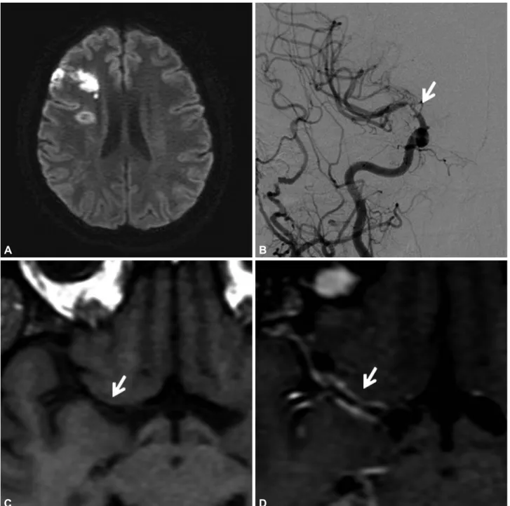
Schirmer’s test was positive for dry-eye syndrome. Head CT revealed infarcts in the right middle cerebral artery (MCA) and anterior cerebral artery (ACA) deep watershed territory, and in the MCA and posterior cerebral artery cortical watershed territory. CT angiography revealed stenosis of the right supraclinoid and communicating segments of the intracranial internal carotid artery (ICA). Digital-subtraction angiography showed 80% stenosis of the right supraclinoid ICA with normal distal smaller vessels. High-resolution MR vessel wall im- aging (HRVWI) revealed smooth homogeneous concentric wall thickening with enhance- ment involving the communicating segment of the right ICA, the proximal M1 segment of the MCA, and the A1 segment of the ACA, without any significant positive or negative re- modeling, suggestive of vasculitis of the terminal right ICA and its branches. Large-vessel vas- culitis secondary to Sjogren’s syndrome was diagnosed. The patient was treated for 5 days with intravenous methylprednisolone followed by oral prednisolone, along with mycophenolate mofetil and hydroxychloroquine. The patient had not experienced any further cerebral isch- emic events at a 3-month follow-up.
This case demonstrates vasculitis of cerebral large-vessels in a patient with primary Sjogren’s syndrome and provides further insight into the pathogenesis of the CNS involvement in Sjo- gren’s syndrome. Our patient had dry-eye syndrome, based on a positive Schirmer’s test, and strong positivity for SSA/Ro and SSB/La, suggestive of Sjogren’s syndrome. The presence of right supraclinoid ICA stenosis in a setting of primary Sjogren’s syndrome suggested a vascu- litic etiology. The alternative possibilities considered were intracranial atherosclerotic disease and moyamoya disease. The homogenous concentric thickening and enhancement involv- Gopikrishnan Unnikrishnan
Nikhil Hiremath
Kesavadas Chandrasekharan Sapna E Sreedharan
Padmavathy Narayanan Sylaja
Comprehensive Stroke Care Program, Department of Neurology,
Sree Chitra Tirunal Institute for Medical Sciences and Technology, Trivandrum, Kerala, India
pISSN 1738-6586 / eISSN 2005-5013 / J Clin Neurol 2018;14(4):588-590 / https://doi.org/10.3988/jcn.2018.14.4.588
Received June 11, 2018 Revised July 7, 2018 Accepted July 10, 2018 Correspondence
Padmavathy Narayanan Sylaja, MD, DM Comprehensive Stroke Care Program, Department of Neurology,
Sree Chitra Thirunal Institute for Medical Sciences and Technology, Trivandrum 695011, India
Tel +91-471-2524482 E-mail [email protected]
cc This is an Open Access article distributed under the terms of the Creative Commons Attribution Non-Com- mercial License (https://creativecommons.org/licenses/by-nc/4.0) which permits unrestricted non-commercial use, distribution, and reproduction in any medium, provided the original work is properly cited.
LETTER TO THE EDITOR
www.thejcn.com 589
Unnikrishnan G et al.
JCN
ing the communicating segment of the right ICA and a few millimeters of the proximal M1 segment of the MCA and the A1 segment of the ACA, without any significant positive or negative remodeling, is characteristic of vasculitis. Eccentric wall thickening and the juxtaluminal T2-weighted hyperin- tensity that is evident in intracranial atherosclerotic disease was not seen in our patient. The absence of collateral vessels
on angiography as well as the absence of negative remodeling in HRVWI excluded the possibility of moyamoya disease.
Vasculitis is one of the mechanisms underlying CNS involve- ment in primary Sjogren’s syndrome. Vasculitis involving small and medium-size vessels has been demonstrated in histopath- ological examinations of brain and spinal cord specimens, and in angiography of patients with Sjogren’s syndrome.3,7 Large-ar-
Fig. 1. A: MR diffusion-weighted image showing an acute infarct in the right middle cerebral artery and anterior cerebral artery cortical watershed territory, and a subacute infarct in the deep watershed territory. B: Cerebral digital-subtraction angiography with injection into the right common carotid artery showing 80% stenosis of the terminal right internal carotid artery and the proximal M1 segment of the middle cerebral artery (arrow), with sig- nificant attenuation of the A1 segment of the right anterior cerebral artery. C and D: High-resolution MR vessel-wall pre- and postcontrast T1-weighted images showing smooth homogeneous contrast enhancement along the middle cerebral artery (arrows).
A
C
B
D
590 J Clin Neurol 2018;14(4):588-590
Large Vessel Vasculitis in Sjogren's Syndrome
JCN
tery involvement in Sjogren’s syndrome is rare, with only three cases reported in the literature.4-6 We did not perform a histo- pathological confirmation of vasculitis and follow-up HRVWI after treatment to look for resolution of the vessel-wall en- hancement. Our case highlights the presence of vasculitic in- volvement of the large cerebral vessels in primary Sjogren’s syndrome, which can be confirmed by HRVWI.
Conflicts of Interest
The authors have no financial conflicts of interest.
REFERENCES
1. Ramos-Casals M, Brito-Zerón P, Sisó-Almirall A, Bosch X. Primary Sjogren syndrome. BMJ 2012;344:e3821.
2. Alexander EL. Neurologic disease in Sjögren’s syndrome: mononucle- ar inflammatory vasculopathy affecting central/peripheral nervous system and muscle. A clinical review and update of immunopathogen-
esis. Rheum Dis Clin North Am 1993;19:869-908.
3. Alexander EL, Ranzenbach MR, Kumar AJ, Kozachuk WE, Rosen- baum AE, Patronas N, et al. Anti-Ro(SS-A) autoantibodies in central nervous system disease associated with Sjögren’s syndrome (CNS-SS):
clinical, neuroimaging, and angiographic correlates. Neurology 1994;
44:899-908.
4. Sakata H, Fujimura M, Sato K, Shimizu H, Tominaga T. Efficacy of extracranial-intracranial bypass for progressive middle cerebral artery occlusion associated with active Sjögren’s syndrome: case report. J Stroke Cerebrovasc Dis 2014;23:e399-e402.
5. Matsuki Y, Kawakami M, Ishizuka T, Kawaguchi Y, Hidaka T, Suzuki K, et al. SLE and Sjögren’s syndrome associated with unilateral moy- amoya vessels in cerebral arteries. Scand J Rheumatol 1997;26:392-394.
6. Hasiloglu ZI, Albayram S, Tasmali K, Erer B, Selcuk H, Islak C. A case of primary Sjögren’s syndrome presenting primarily with central nervous system vasculitic involvement. Rheumatol Int 2012;32:805-807.
7. Alexander EL, Craft C, Dorsch C, Moser RL, Provost TT, Alexander GE. Necrotizing arteritis and spinal subarachnoid hemorrhage in Sjögren syndrome. Ann Neurol 1982;11:632-635.