Ⅰ. INTRODUCTION
Periodontal disease is the inflammatory disor- ders of the periodontium ranging from the rela- tively benign form of gingivitis, in which the inflammation is confined to the marginal tissues, to more aggressive forms such as rapidly progres-
The effect of neuropeptides on secretion of Interleukin-8(IL-8)
Kyung-Jun Kim1, Sang-Hyuk Park1,2, Kyoung-Kyu Choi1,2, Sang-Jin Park2*
1Department of Conservative Dentistry, Division of Dentistry, Graduate School, Kyunghee University
2Oral Biology Research Institute, College of Dentistry, Kyunghee University
We investigated the secretion of Interleukin-8 (IL-8) from ginviva and periodontal ligament stimu- lated with Substance P (SP) and Calcitonin Gene-related Peptide (CGRP). Gingiva (GF), periodon- tal ligament (PDLF) and pulp (PF) tissues were collected from extracted intact 3rd molars.
Cultured cells were stimulated with different concentrations of SP for 4 hrs, and stimulated with SP, CGRP and Tumor Necrosis Factor-α(TNF-α) for 8 hrs. Then RNase Protection Assay was car- ried out. ELISA was performed using supernatants of stimulated cells for quantitative analysis of IL-8. Results were assessed using student t-test with significance of P < 0.05.
According to this study, the results were as follows:
1. IL-8 mRNA was detected in all type of cells studied (PF, GF and PDLF).
2. IL-8 mRNA expression was not increased after stimulating 4 hrs with SP (10-5M) and SP (10-8M) compared with Mock stimulation in all type of cells studied.
3. IL-8 mRNA expression was not increased after stimulating 8 hrs with SP (10-4M) and CGRP (10-6M) compared with Mock stimulation in all type of cells studied.
4. TNF-α(2 ng/㎖) increased the expression of IL-8 mRNA in all kind of cells studied.
5. The secretion of IL-8 from GF was increased 8 hrs after the stimulation with CGRP (10-6M) (p < 0.05).
6. The secretion of IL-8 from PDLF was increased 8 hrs after the stimulation with SP (10-4M) (p < 0.05).
Calcitonin Gene-related Peptide (CGRP) increased Interleukin-8 (IL-8) which plays an important role in chemotaxis of neutrophil in Calcitonin Gene-related Peptide (CGRP) gingival tissue, whereas Substance P increased the secretion of IL-8 from periodontal ligament.[J Kor Acad Cons Dent 31(3):153- 160, 2006]
Key words : Interleukin-8 (IL-8), Substance P (SP), Tumor Necrosis Factor-α(TNF-α), pulp fibroblast (PF), gingival fibroblast (GF), periodontal ligament fibroblast (PDLF)
- Received 2005.8.23., revised 2006.2.6., accepted 2006.3.9.
ABSTRACT
* Corresponding Author: Sang-Jin Park
Professor of Division of Dentistry, Graduate school, KyungHee University
1, Hoegi Dong, Dongdaemun Gu, Seoul, Korea, 130-702 Tel: 82-2-958-9335
E-mail: [email protected]
sive periodontitis, in which the disease process leads to loss of connective tissue attachment to the root surfaces, loss of alveolar support, and impaired function of the dentition. Although intense microbiological, immunological, and bio- chemical studies about the pathogenesis of the various periodontal disease were performed, pathogenesis of several diseases were too compli- cated to understand. Recently it has come to know that nervous system can regulate immuno- logic and inflammatory responses1). One of the potent reasons of individually different response to same irritation, is that neurologic system can modulate immunologic system2). The nervous sys- tem affects the development of inflammatory processes through local release of various neu- ropeptides3).
Neutrophils are the most predominant cell pop- ulation that migrate into inflammatory lesions.
Besides their protective role, they are involved in tissue damage under pathologic conditions because of their high contents in neutral and acid proteases and their ability to generate superox- ides and other reactive oxygen derivatives4). Inflammatory neutrophils are recruited into affected tissues through the process of multiple activation events that eventually lead to the release of antimicrobial and inflammatory prod- ucts.
The recent discovery of a family of mediators called chemokines, which play pivotal roles in regulating inflammatory reactions, has provided an insight into the molecular mechanisms under- lying the kinetics of inflammation5,6). However, the characterization of chemokine expression in the cells of dental pulp, gingiva and periodontal ligament during inflammation and infection has been sparse7,8).
Interleukin-8 (IL-8) is a neutrophil-stimulating factor and displays a wide range of biological effects including chemotaxis and activation of neutrophils8). It was originally described as a 72 amino acid peptide produced by human blood monocytes stimulated in culture with E. coli lipopolysaccharide4). IL-8 is a member of the chemokine C-X-C subfamily that displays potent
chmotactic activities for human neutrophils and T lymphocytes4,7,9). In addition to chemotaxis, IL-8 induces neutrophil degranulation, resulting in the release of enzymes that cause tissue destruc- tion10).
Substance P (SP) and Calcitonin Gene-related Peptide (CGRP) are major sensory neuropeptides, released from the peripheral nerve endings of sen- sory nerves during inflammation1,6,11). Although the primary function of these two neuropeptides is to induce vasodilatation and consequently to increase blood flow, they can play more direct roles in infiltrations of local inflammatory cells12). In addition, they can modulate local inflammatory responses by modifying the secretion of various cytokines1).
The cellular elements inside the periodontal lig- ament are fibroblasts, endothelial cells, cemento- blasts, osteoblasts, and osteoclasts and neu- trophiles can be seen predominantly in gingival connective tissues and periodontal ligament space as well14). Fibroblast which is predominant cell in gingival connective tissue plays an important role in wound healing following the injury such as gin- gival surgery or pathologic processes through renewal of collagen fibers and other chemical con- stituents13,14).
In the present study, whether these neuropep- tides such as SP and CGRP could induce the release of IL-8 in gingiva, periodontal ligament, and pulp during inflammation was evaluated. We investigated responses of these cells cultured from the gingiva, periodontal ligament, and pulp in releasing IL-8 after stimulation with SP or CGRP.
Ⅱ. MATERIALS AND METHODS Sample Collection and Cell Culture
Freshly extracted, intact, caries-free third molars (n = 10) were obtained from the patients (15 - 25 years old) in the Department of Oroma- xillofacial Surgery at Kyung-Hee Medical Center.
Immediately after extraction, teeth were stored in phosphate buffered saline (PBS) and transferred to the laboratory. Gingival tissues were cut
around the cemento-enamel junction area, and the periodontal ligaments were obtained by scrap- ing the root surface. Each tooth was grooved lon- gitudinally with a fissure bur at high speed. The tooth was then split with the screw driver, and the entire pulp (coronal and radicular) elevated with cotton pliers, maximum pulpal tissues were obtained.
Repeated washing with PBS, all kinds of tissues obtained from dental pulp, gingiva and periodon- tal ligament were placed in 48-well plate culture dishes containing Dulbecco’s modified Eagle medium (DMEM; Life Technologies/ GIBCO BRL, Gaithersburg, MD, USA) supplemented with 10%
fetal bovine serum (FBS) and 100 units/㎖
Penicillin-G, 100 ㎍/㎖ Streptomycin, and 0.25 ㎍/
㎖ Fungizone (Gemini Bio-Products, Inc., Woodland, CA, USA). The cells from pulp tissue (PF), gingival tissue (GF), and periodontal liga- ment (PDLF) were grown to confluence and passed at 1 : 2 ratio until used for experiments (passages 5-8 were used).
Once all kind of cells obtained were confluent at passage 5, they were pooled in 100 ㎜ diameter culture dish and cultured until it became conflu- ent for the stimulation with SP and CGRP for RNase Protection Assay. In addition, GF and PDLF were seeded on the 24-well plates, and grown into confluent for Enzyme-Linked Immunosorbent Assay (ELISA).
Stimulation of cells with SP and CGRP
Synthetic human SP and CGRP (Sigma, St.
Louis, MO, USA) were prepared in sterile water with 0.1% low-endotoxin bovine serum albumin (BSA, Sigma, St Louis, MO, USA). Cells were seeded in 100 ㎜ culture dish and 24-well plates and grown to confluence before used for experi- ments. Alpha minimal essential medium contain- ing L-glutamine (Life Technologies/GIBCO BRL, Gaithersburg, MD, USA) without FBS was used 24 hrs before cells were stimulated with neu- ropeptiedes. Cells seeded in 100 ㎜ culture dish were stimulated with SP (10-8M) or SP (10-5M) or only with medium (Mock stimulation) for 4 hrs,
and stimulated with SP (10-4M) or CGRP (10-6M) or TNF-α(2 ng/㎖) or only with medium (Mock stimulation) for 8 hrs which will be subjected to RNase Protection Assay.
GF and PDLF seeded on 24-well plate were stimulated SP (10-4M) or CGRP (10-6M) or TNF-α (2 ng/㎖) or only with medium (Mock stimula- tion) for 8 hrs for the ELISA. Experiments with pulp cells after stimulation with SP and CGRP were excluded, because those data with PF was already known in the previous study15,16).
The serum was not used in the medium to pre- vent the increase in the base-line level of IL-8 secretion from these cells. Based on the prelimi- nary experiments, the serum induced significant levels of IL-8 and from pulp. Varying doses of neuropeptides or 0.1% BSA (Mock stimulation) were added to the cell cultures. Recombinant human TNF-α(R&D Systems, Minneapolis, MN, USA), a known IL-8 agonist, was used as a posi- tive control for IL-8 induction.
Extraction of Total RNA
After cells were stimulated with SP for 4 hrs, and with SP and CGRP, and TNF-αfor 8 hrs, supernatants were discarded. All kinds of cells stimulated were collected scraping with cell scraper (Fisher Scientific, Pittsburgh, PA, USA).
5 - 10㎍ of total RNA was extracted from all kinds of cells stimulated using the RNeasy mini kit following the manufacturer’s instructions (Qiagen, LRS Laboratories, Inc., Seoul, Korea).
RNase Protection Assay
The total RNA extracted were subjected to RNase protection assay as specified by the manu- facturer, using the human cytokine-5 probe set (BD Biosciences, San Diego, CA, USA) and BD RiboQuant Ribonuclease Protection Assay Systems (BD Biosciences, San Diego, CA, USA).
The resulting protected RNAs were resolved on 5% denaturing polyacrylamide gels and exposed to X-ray films.
Enzyme Linked Immunosorbent Assay (ELISA) for IL-8
After GF and PDLF were stimulated with SP (10-4M), CGRP (10-6M) and TNF-α(2 ng/㎖) and only with medium(Mock stimulation), whose total volume were 300 ㎕ in each well for 8 hrs, super- natants were collected and stored at -80℃.
Standard ELISA for IL-8 was performed as described previously7) using 2 ㎍/㎖ of polyclonal goat anti-human IL-8 (R&D Systems, Minnea- polis, MN, USA) as capturing antibodies, 1 g/㎖
polyclonal rabbit anti-human IL-8 (Endogen Inc., Cambridge, MA, USA) as detecting antibodies, and 0.1 g/㎖ horseradish peroxidase (HRPO)- labeled polyclonal goat anti-rabbit immunoglobu- lin G (Biosource International, Camarillo, CA, USA) as a secondary antibody. Subsequently fresh developing buffer containing substrate of optimal concentrations of TMB (3,3’,5,5’- Tetramethyl benzidine; Sigma, St. Louis, MO, USA), H2O2 and sodium acetate, pH 6.0 was added and the developing reaction was stopped with 1.2 M/L sulfuric acid. Absorbance at 450 ㎚ was determined with a microplate reader(Bio-Tek Instrument, Inc., Laguna Hills, CA, USA) and concentrations were derived using the Delta Soft III software (Bio-Tek Instrument, Inc., Laguna Hills, CA, USA). Known concentrations of recom- binant human (rh) IL-8 (Endogen Inc., Cambridge, MA, USA) was used to establish a standard curve for determining the concentrations of experimental samples.
Statistical Analysis
The Student t-test was used to assess the sig- nificance of differences between data. The differ- ences were considered significant when the proba- bility (P) value was < 0.05.
Ⅲ. RESULTS RNase Protection Assay
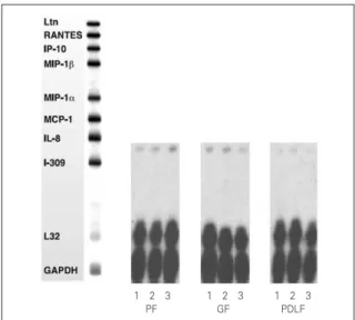
IL-8 mRNA was detected in all kinds of cells
studied (PF, GF, PDLF) 4hrs after stimulation with Mock, SP (10-5M), and SP (10-8M) as shown in Figure 1. But, other cytokines (Ltn, RANTES, IP-10, MIP-1β, MIP-1α, MCP-1, and I-309) included in the human cytokine-5 probe set (BD Biosciences, San Diego, CA, USA) were not detected after Mock stimulation and after SP stimulation as well. The expression of IL-8 mRNA was not increased 4 hrs after stimulation with SP (10-5M) and SP (10-8M) compared with Mock stimulation in all kinds of cells studied (Figure 1).
IL-8 mRNA was detected in all kinds of cells studied (PF, GF, PDLF) 8hrs after stimulation with Mock, SP (10-4M), and CGRP (10-6M) as shown in Figure 2. The expression of IL-8 mRNA was not increased 8 hrs after stimulation with SP (10-4M) or CGRP (10-6M) compared with Mock stimulation in all type of cells studied.
On the other hand, TNF-α(2 ng/㎖) remarkably increased the expression of IL-8 mRNA in all kind of cells studied (Figure 2). In addtion, I-309
Figure 1. RNase Protection Assay 4hrs after stimulation of the cells with MOCK and various dose of Substance P.
1. MOCK stimulation with medium only 2. Stimulation with Substance P (10-5M) 3. Stimulation with Substance P (10-8M) PF: Pulp Fibroblasts
GF: Gingival Fibroblasts
PDLF: Periodontal Ligament Fibroblasts
1 2 3 1 2 3 1 2 3 PF GF PDLF
which was not detected 8 hrs after Mock stimula- tion was distinctively expressed 8 hrs after stimu- lation with TNF-α(2 ng/㎖) in all kinds of cells studied (Figure 2).
Enzyme Linked Immunosorbent Assay for IL-8
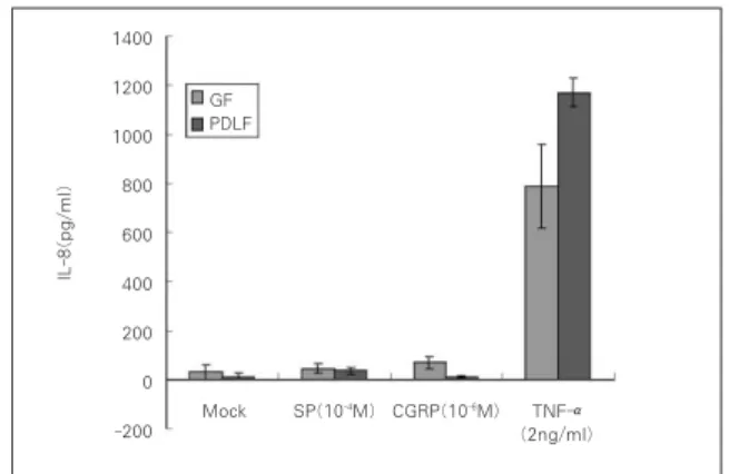
As a result of quantitative analysis from the supernatent of the cells after the stimulation with SP (SP10-4M) and CGRP (10-6M), and TNF-α(2 ng/㎖) as a positive control, The secretion of IL-8 from GF was increased mildly up to 44.2 pg/㎖
(1.5-fold) 8 hrs after stimulation with SP (10-4M) and was increased significantly to 68.5 pg/㎖ (2.3- fold) 8 hrs after stimulation with CGRP (10-6M) compared with Mock stimulation (30.1 pg/㎖) as shown in Figure 3 (p < 0.05). IL-8 from GF was significantly increased (788.3 pg/㎖) 8 hrs after stimulation with TNF-α(2 ng/㎖) (p < 0.05).
PDLF stimulated with SP (10-4M) for 8 hrs increased IL-8 (35.7 pg/㎖) compared with Mock stimulation (12.3 pg/㎖) significantly (p < 0.05).
There was no induction of IL-8 from PDLF (10.2 pg/㎖) after the stimulation with CGRP (10-6M) in comparison with Mock stimulation. As same as in GF, the secretion of IL-8 from PDLF was signifi- cantly increased up to approximately 100-fold (1170.7 pg/㎖) after TNF-αstimulation(2 ng/㎖) (p < 0.05).
Ⅳ. DISCUSSION
In teeth and tooth supporting tissues, SP and CGRP are shown to exert vasoactive functions including increase in blood flow and interstitial tissue pressur15,16), regulation of local inflamma- tion17), guiding of immune cells18) and enhance- ment of cell proliferation19).
Other cell types from different tissues release IL-8 during stimulation with neuropeptides20-22) and substantial evidence has shown that neu- ropeptides can stimulate the production of proin- flammatory cytokines in various cell types. SP induces skin keratinocytes to release IL-1 and mast cells to produce tumor necrosis factor-α (TNF-α). CGRP stimulates human dermal microvascular endothelial cells to secrete IL-8, whereas SP induces human dermal microvascular Figure 2. RNase Protection Assay 8hrs after
stimulation of the cells with Mock and Substance P, CGRP, and TNF-α.
1. MOCK stimulation with medium only 2. Stimulation with Substance P (10-4M)
3. Stimulation with Calcitonin Gene Related Peptides (CGRP, 10-6M)
4. Stimulation with TNF-α(2 ng/㎖) PF: Pulp Fibroblasts
GF: Gingival Fibroblasts
PDLF: Periodontal Ligament Fibroblasts
1 2 3 4 1 2 3 4 1 2 3 4 PF GF PDLF
Figure 3. IL-8 secretion from the cells 8hrs after stimulation with SP (10-4M), CGRP (10-6M) and TNF-α(2 ng/㎖).
GF: Gingival Fibroblasts
PDLF: Periodontal Ligament Fibroblasts
GF PDLF
Mock SP(10-4M) CGRP(10-6M) TNF-α (2ng/ml) 1400
1200 1000 800 600 400 200 0 -200
IL-8(pg/ml)
endothelial cells to express vascular cell adhesion molecule-121). Furthermore, SP induces IL-8 pro- duction by osteoarthritis fibroblasts, whereas CGRP increases IL-8 and IL-6 secretion from rheumatoid arthritis fibroblasts22).
Neutrophil recruitment depends on chemotactic agonists that are synthesized and released at the site of inflammation. Chemotactic agonists may be derived from the host or infecting microorgan- isms.
IL-8 stimulates neutrophils to transport CD11 /CD18 (LFA-1 or Mac) integrin from the neu- trophil cytoplasm to cell surface23). This facilitates neutrophil migration into the tissue. Neutrophil migration may be induced by a haptotactic gradi- ent of IL-8 either on the endothelial cell surface or in the extracellular matrix23,24).
IL-8 can be induced by IL-1α, IL-1βand tumor necrosis factor-α(TNF-α) in many different cells4). It was also derived in vitro from dermal fibrob- lasts, keratinocytes, endothelial cells, hepatocytes and synovial cells in response to inflammatory mediators such as IL-1βand TNF-α25).
As mentioned above, various cell types from dif- ferent tissues release IL-8 during stimulation with neuropeptides. In some cells, both SP an CGRP increase the secretion of IL-8. Cultured human pulp cells stimulated with SP have been reported to increase IL-8 secretion in the previous study12).
Our data demonstrated that cultured primary human gingival cells having a fibroblast-like phe- notype increased the secretion of IL-8 significant- ly in response to CGRP, but mildly to SP, where- as cultured periodontal ligament cells increased the secretion of IL-8 significantly in response to SP, but mildly to CGRP. Our findings suggest that SP and CGRP have a more direct role in the initiation of inflammatory cell infiltration in gingi- va and periodontal ligament. Our report indicated that SP and CGRP stimulate the chemotaxis of leukocytes, which may have been in part a result of this IL-8 induction effect.
In the previouse study20), the basal level of IL-8 from the cultured primary pulp cells were between 100 pg/㎖ and 200 pg/㎖ after Mock
stimulation, nevertheless endothelial cells (ECV 304 cells) were around 20 pg/㎖. In this study, the basal level of IL-8 from the cultured primary gingival cells (30.1 pg/㎖) and periodontal liga- ment cells (10.2 pg/㎖) were lower than pulp cells after Mock stimulation.
From the immunohistochemical studies, in healthy gingival tissues SP was found in the con- nective tissues between the collagenous elements, whereas in inflamed tissue SP was markedly increased particularly around the blood vessels as well as in close association with much of the inflammatory cell infiltrate2). IL-8 was elevated in chronically inflammed gingiva, and the major IL-8 was detected only in the epithelial cell layer, which means that IL-8 may play a crucial role in the recruitment and activation of neutrophils and T lymphocytes in periodontitis8). The neural mod- ulation of inflammatory events in the gingiva, a site in which continuous tissue reactivity is gen- erated in response to the accumulation of dental plaque, is of particular interest in light of the rec- ognized potential for stress to serve as modifying factor in inflammatory lesions which affect the periodontium, which is accumulating evidence that interactions between the nervous system, immune cells, and cells such as fibroblasts are critical to the development and persistence of a variety of inflammatory disorders.
Ⅴ. CONCLUSION
According to this study, the results were as follows:
1. IL-8 mRNA was detected in all type of cells studied (PF, GF and PDLF).
2. IL-8 mRNA expression was not increased after stimulating 4 hrs with SP (10-5M) and SP (10-8M) compared with Mock stimulation in all type of cells studied.
3. IL-8 mRNA expression was not increased after stimulating 8 hrs with SP (10-4M) and CGRP (10-6M) compared with Mock stimulation in all type of cells studied.
4. TNF-α(2 ng/㎖) increased the expression of IL-
8 mRNA in all kind of cells studied.
5. The secretion of IL-8 from GF was increased 8 hrs after the stimulation with CGRP (10-6M) (p < 0.05).
6. The secretion of IL-8 from PDLF was increased 8 hrs after the stimulation with SP (10-4M) (p < 0.05).
The present study has demonstrated that Calcitonin Gene-related Peptide (CGRP) increased Interleukin-8 (IL-8) which plays an important role in chemotaxis of neutrophil in gingival tissue, whereas Substance P increased the secretion of IL-8 from periodontal ligament.
REFERENCES
1. Kiss M, Kemeny L, Gyulai R, Michel G, Husz S, Kovacs R, Dobozy A, Ruzicka T. Effects of the neu- ropeptides substance P, calcitonin gene-related peptide and α-melanocyte-stimulating hormone on the IL-8/
IL-8 receptor system in a cultured human keratinocyte cell line and dermal fibroblasts. Inflammation 23:557- 567, 1999.
2. Bartold PM, Kylstra A, Lawson R. Substance P. An Immunohistochemical and Biochemical Study in Human Gingival Tissues. A Role for Neurogenic Inflammation? J Periodontol 65:1113-1121, 1994.
3. Rameshwar P, Gascon P. Induction of negative hematopoietic regulators by neurokinin-A in bone mar- row stroma. Blood 88:98-106, 1996.
4. Baggiolini M, Walz A, Kunkel SL. Neutrophil-activat- ing peptide-1 /Interleukin 8, a novel cytokine that activates neutrophils. J Clin Invest 84:1045-1049, 1989.
5. Kent LW, Dyken RA, Rahemtulla F, Allison AC, Michalek SM. Effects of in vitro passage of healthy human gingival fibroblasts on cellular morphology and cytokine expression.Archs oral Biol 41:263-270, 1996.
6. Guo CJ, Lai JP, Luo HM. Douglas SD, Ho WZ.
Substance P up-regulates macrophage inflammatory protein-1βexpression in human T lymphocytes. J Neuroimmunol 131:160-167, 2002.
7. Huang GTJ, Potente AP, Kim JW, Chugal N, Zhang X.
Increased interleukin-8 espression in inflamed human dental pulps. Oral Surg Oral Med Oral Pathol Oral Radiol Endod 88:214-220, 1999.
8. Fitzgerald JE, Kreutzer DL. Localization of Interleukin-8 in human gingival tissues. Oral Microbiol and Immunol 10:297-303, 1996.
9. Larsen CG, Anderson AO, Appella E, Oppenheim JJ, Matsushima K. The neutrophil-activating protein (NAP-1) is also chemotactic for T lymphocytes. Science 243:1464-1466, 1989.
10. Peveri P, Walz A, Dewald B, Baggiolini M. A novel neutrophil activating factor produced by human mononuclear phagocytes. J Exp Med 167:1547-1559, 1988.
11. Cuesta MC, Quintero L, Pons H, Suarez-Roca H.
Substance P and calcitonin gene-related peptide increase IL-1β, IL-6 and TNFαsecretion from human peripheral blood mononuclear cells. Neurochemistry Int 40:301-306, 2002.
12. Park SH, Hsiao GYW, Huang GTJ. Role of substance P and calcitonin gene-related peptide in the regulation of interleukin-8 and monocyte chemotactic protein-1 expression in human dental pulp. Int Endod J 37:185- 192, 2004.
13. Bordin S, Narayanan S, Reddy J, Cleveland D, Page RC. Fibroblast subtypes in the periodontium. J Periodont Res 19:642-644, 1984.
14. McCulloch CAG, Bordin S. Role of fibroblast subpopu- lations in periodontal physiology and pathology. J Periodont Res 26:144-154, 1991.
15. Heyeraas KJ, Berggreen E. Interstitial fluid pressure in normal and inflamed pulp. Crit Rev Oral Biol Med 10(3):328-336, 1999.
16. Berggreen E, Heyeraas KJ. Effect of the sensory neu- ropeptide antagonists h-CGRP((8-37)) and SR 140.33 on pulpal and gingival blood flow in ferrets. Arch Oral Biol 45(7):537-542, 2000.
17. Fistad I, Kvinnsland IH, Jonsson R, Heyeraas KJ.
Effect of intermittent long-lasting electrical tooth stim- ulation on pulpal blood flow and immunocompetent cells: a hemodynamic and immunohistochemical study in young rat molars, Exp Neurol 146(1):230-239, 1997.
18. Fristad I, Heyeraas KJ, Kwinnsland IH, Jonsson R.
Recruitment of immunocompetent cells after dentinal injuries in innervated and denervated young rat molars: an innunohistochemcal study. J Histochem Cytochem 43(9):871-879, 1995.
19. Bongenhielm U, Haegerstrand A, Theodorsson E, Fried K. Effect of neuropeptides on growth of cultivated rat molar pulp fibroblasts. Regul Pept 60(2-3):91-8, 1995.
20. Patel T, Park SH, Lin LM, Chiappelli F, Huang GTJ.
Substance P induces interleukin-8 secretion from human dental pulp cells. Oral Surg Oral Med Oral Pathol Oral Radiol Endod 96:478-485, 2003.
21. Ansel JC, Armstrong CA, Song I, Quinlan KL, Olerud JE, Caughman SW, Bunnett NW. Interaction of the skin and nervous system. J Investig Dermatolo Symp Proc 2(1):23-26, 1997.
22. Raap T, Justen HP, Miller LE, Cutolo M, Scholmerich J, Straub RH. Neurotransmitter modulation of inter- leukin 6(IL-6) and IL-8 secretion of synovial fibrob- lasts in patients with rheumatoid arthritis compared to osteoarthritis. J Rheumatol 27:2558-2565, 2000.
23. Detmers PA, Lo SK, Olsen-Egbert E, Walz A, Baggiolini M, Cohn ZA. Neutrophil-activating protein 1/interleukin 8 stimulates the binding activity of the leukocyte adhesion receptor CD11b /CD18 on human neutrophils. J Exp Med 117:1155-1162, 1990.
24. Rot A. Neutrophil attractant/ activation protein- 1(interleukin-8) induces in vitro neutrophil migration by haptotactic mechanism. Eur J Immunol 23:303- 306, 1993.
25. Takashiba S, Takigawa M, Takahashi K, Myokai F, Nishimura F, Chihara T, Kurihara H, Nomura Y, Murayama Y. Interleukin-8 is a major neutrophil chemotactic factor derived from cultured human gingi- val fibroblasts stimulated with interleukin-1βor tumor necrosis factor alpha. Infection and Immunity 60:5253- 5258, 1992.
Interleukin-8 (IL-8) 분비에 미치는 neuropeptides의 영향에 관한 연구
김경준1∙박상혁1,2∙최경규1,2∙박상진1,2*
1경희대학교 대학원 치의학과 치과보존학교실
2경희대학교 치과대학 구강생물학연구소
본 연구는 치수조직, 치은, 치주인대로부터 배양된 조직을 SP (Substance P)로 4시간, SP, CGRP (Calcitonin Gene-related Peptide), Tumor necrosis factor-α(TNF-α)로 8시간 자극 후 RNase Protection Assay를 시행하고, IL-8의 분비량을 측정해 다음 결과를 얻었다.
1. IL-8 mRNA는 모든세포에서 발현됐다.
2. IL-8 mRNA 발현은 SP (10-5M)와 SP (10-8M)로 4시간 자극 시 증가되지 않았다.
3. IL-8 mRNA 발현은 SP (10-4M)와 CGRP (10-6M)로 8시간 자극 시 증가되지 않았다.
4. TNF-α(2 ng/㎖) 자극 시, IL-8 mRNA 발현이 증가됐다.
5. 치은 세포를 CGRP (10-6M)로 8시간 자극 시, IL-8 분비량이 증가했다 (p < 0.05).
6. 치주인대 세포를 SP (10-4M)로 8시간 자극 시 IL-8 분비량이 증가했다 (p < 0.05).
주요어: Interleukin-8 (IL-8), SP (Substance P), Tumor necrosis factor-α(TNF-α), 치수세포, 치은세포, 치주인대세포
국문초록