INTRODUCTION
Despite recently published randomized trials suggesting no survival benefit for routine lymphadenectomy in endometrial
cancer [1,2], full pelvic and paraaortic lymphadenectomy is still recommended by many gynecologic oncologic societ- ies and guideline committees [3-5]. However, although there is ongoing controversy concerning the benefit of routine lymphadenectomy [6-8], the general consensus is that there is a certain subset of patients in which the omission of routine lymphadenectomy may be justified [9-11].
For several decades, researchers have proposed several models to predict patients at low-risk for nodal metastasis [12-15]. Most of these prediction models were designed using surgicopathological parameters, such as depth of myometrial
How low is low enough? Evaluation of various risk- assessment models for lymph node metastasis in endometrial cancer: a Korean multicenter study
Sokbom Kang1, Jong-Min Lee2, Jae-Kwan Lee3, Jae Weon Kim4, Chi-Heum Cho5, Seok-Mo Kim6, Sang-Yoon Park1, Chan-Yong Park7, Ki-Tae Kim8
1Center for Uterine Cancer, National Cancer Center, Goyang; Department of Obstetrics and Gynecology, 2Kyung Hee University School of Medicine, Seoul; 3Korea University College of Medicine, Seoul; 4Seoul National University College of Medicine, Seoul;
5Dongsan Medical Center, Keimyung University, Daegu; 6Chonnam National University Medical School, Gwangju;
7Gachon University Hospital, Incheon; 8Inje University Busan Paik Hospital, Busan, Korea
See accompanying editorial on page 210.
Received May 7, 2012, Accepted May 28, 2012
*Presented at the 2012 Annual Meeting of Society of Gynecologic Oncology.
Correspondence to Sokbom Kang
Center for Uterine Cancer, National Cancer Center, 323 Ilsan-ro, Ilsandong- gu, Goyang 410-769, Korea. Tel: 82-31-920-2382, Fax: 82-31-920-1238, E-mail: [email protected]
Objective: The aim of this study was to identify a standard for the evaluation of future models for prediction of lymph node metastasis in endometrial cancer through estimation of performance of well-known surgicopathological models.
Methods: Using the medical records of 947 patients with endometrial cancer who underwent surgical management with lymphadenectomy, we retrospectively assessed the predictive performances of nodal metastasis of currently available models.
Results: We evaluated three models included: 1) a model modified from the Gynecologic Oncology Group (GOG) pilot study;
2) one from the GOG-33 data; and 3) one from Mayo Clinic data. The three models showed similar negative predictive values ranging from 97.1% to 97.4%. Using Bayes’ theorem, this can be translated into 2% of negative post-test probability when 10%
of prevalence of lymph node metastasis was assumed. In addition, although the negative predictive value was similar among these models, the proportion that was classified as low-risk was significantly different between the studies (56.4%, 44.8%, and 30.5%, respectively; p<0.001).
Conclusion: The current study suggests that a false negativity of 2% or less should be a goal for determining clinical usefulness of preoperative or intraoperative prediction models for low-risk of nodal metastasis.
Keywords: Endometrial cancer, Low-risk group, Lymph node dissection, Lymphadenectomy, Prediction, Sensitivity and specificity
invasion or pathological grade [13,14,16,17]. Therefore, it has been frequently challenged whether we can apply frozen sec- tion results in these models due to the inaccuracy of frozen section examination [18,19], and many have claimed that rou- tine lymphadenectomy is unavoidable [20].
Although many researchers have suggested several meth- ods to identify the low-risk group of nodal metastasis before lymphadenectomy [21-25], many gynecologic oncologists are still skeptical about these results [26]. Moreover, there has been no consensus about the desirable performance of a prediction method. The American Society of Breast Surgeons recommended a false-negative rate of 5% or less in order to abandon axillary dissection [27]. Then, how should an accept- able false-negative rate of lymph node metastasis be deter- mined in endometrial cancer?
To answer these questions, we began a multi-institutional, retrospective study. If we are able to estimate the innate false- negative rate of the final pathology-based models, we may also use that as a tool for determining clinical usefulness of pre- or intra-operative prediction models which are in devel- opment.
MATERIALS AND METHODS 1. Patient selection
Using data from eight independent institutions, we retro- spectively reviewed the medical records and pathological findings of patients surgically treated for endometrial cancer between 2000 and 2006. A total of 1,298 patients were identi- fied after approval from the institutional review board. A part of the dataset has been used in previous reports; eligibility for the study and treatment strategy have been described previously [28]. Briefly, patients with histologically confirmed
endometrial cancer who underwent surgical management, including hysterectomy, were enrolled in the study. At all in- stitutions, patients were consecutively enrolled and defined using the selection criteria. Exclusion criteria were as follows:
histologic diagnosis of sarcoma including carcinosarcoma, double primary tumor, or other metastatic cancer. Our study was designed and analyzed as recommended by the Stan- dards for Reporting Diagnostic Accuracy Steering Group [29].
As an index test, three models predicting low-risk groups based on pathologic data were used. These included the following: 1) criteria modified from the GOG pilot study sug- gested by Boronow et al. [12,13] (Model A) ; 2) criteria modi- fied from the GOG-33 data suggested by Creasman et al. [14]
(Model B) ; and 3) the Mayo clinic criteria suggested by Mariani et al. [15,21] (Model C) . Detailed descriptions of these models are summarized in Table 1.
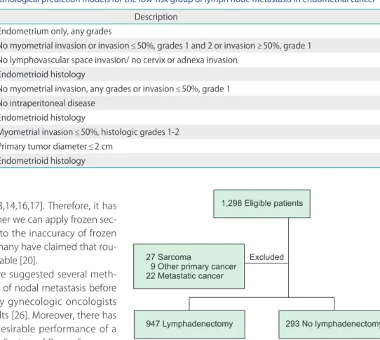
The reference standard was defined as the final pathologic diagnosis of the harvested lymph nodes. Central pathologic review was not performed, as pathologists from each par- ticipating center assessed lymph node status. No restriction of harvested lymph nodes was applied if one or more lymph Fig. 1. Flowchart of patient selection process.
Table 1. Description of three surgicopathological prediction models for the low-risk group of lymph node metastasis in endometrial cancer Model Description
Model A [11,12] Endometrium only, any grades
No myometrial invasion or invasion ≤50%, grades 1 and 2 or invasion ≥50%, grade 1 No lymphovascular space invasion/ no cervix or adnexa invasion
Endometrioid histology
Model B [13] No myometrial invasion, any grades or invasion ≤50%, grade 1 No intraperitoneal disease
Endometrioid histology
Model C [14] Myometrial invasion ≤50%, histologic grades 1-2 Primary tumor diameter ≤2 cm
Endometrioid histology
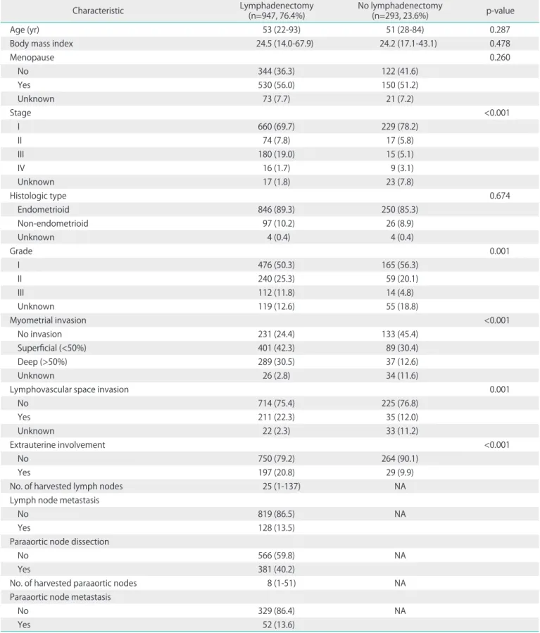
Table 2. Characteristics of the 1,240 patients included in the analysis
Characteristic Lymphadenectomy
(n=947, 76.4%) No lymphadenectomy
(n=293, 23.6%) p-value
Age (yr) 53 (22-93) 51 (28-84) 0.287
Body mass index 24.5 (14.0-67.9) 24.2 (17.1-43.1) 0.478
Menopause 0.260
No 344 (36.3) 122 (41.6)
Yes 530 (56.0) 150 (51.2)
Unknown 73 (7.7) 21 (7.2)
Stage <0.001
I 660 (69.7) 229 (78.2)
II 74 (7.8) 17 (5.8)
III 180 (19.0) 15 (5.1)
IV 16 (1.7) 9 (3.1)
Unknown 17 (1.8) 23 (7.8)
Histologic type 0.674
Endometrioid 846 (89.3) 250 (85.3)
Non-endometrioid 97 (10.2) 26 (8.9)
Unknown 4 (0.4) 4 (0.4)
Grade 0.001
I 476 (50.3) 165 (56.3)
II 240 (25.3) 59 (20.1)
III 112 (11.8) 14 (4.8)
Unknown 119 (12.6) 55 (18.8)
Myometrial invasion <0.001
No invasion 231 (24.4) 133 (45.4)
Superficial (<50%) 401 (42.3) 89 (30.4)
Deep (>50%) 289 (30.5) 37 (12.6)
Unknown 26 (2.8) 34 (11.6)
Lymphovascular space invasion 0.001
No 714 (75.4) 225 (76.8)
Yes 211 (22.3) 35 (12.0)
Unknown 22 (2.3) 33 (11.2)
Extrauterine involvement <0.001
No 750 (79.2) 264 (90.1)
Yes 197 (20.8) 29 (9.9)
No. of harvested lymph nodes 25 (1-137) NA
Lymph node metastasis
No 819 (86.5) NA
Yes 128 (13.5)
Paraaortic node dissection
No 566 (59.8) NA
Yes 381 (40.2)
No. of harvested paraaortic nodes 8 (1-51) NA
Paraaortic node metastasis
No 329 (86.4) NA
Yes 52 (13.6)
Values are presented as number (%) or median (range).
NA, not available.
nodes were harvested. Instead, we categorized optimal and suboptimal lymphadenectomy based on the number of har- vested lymph nodes. Optimal lymphadenectomy was arbi- trarily defined as more than ten harvested nodes and four or more harvested paraaortic nodes [30,31].
2. Statistical analysis
All statistical analyses were performed using STATA ver. 11.0 (STATA, College Station, TX, USA). To estimate continuous vari- ables, Student’s t-test and the Wilcoxon rank-sum test were used. For categorical variables, chi-square and Fisher exact tests were used. All p-values presented are two-sided, and as- sociations are considered significant if the p-value is <0.05.
To assess the performance of models predicting low-risk groups for lymph node metastasis, we selected the nega- tive likelihood ratio (LR) as a primary endpoint [32,33]. We concluded that the negative predictive value was not an ad- equate endpoint, as negative predictive value is vulnerable to the prevalence of events. Using Bayes’ theorem, the negative post-test probability (PTP) was derived from the negative LR based on the assumed pre-test probability of lymph node me- tastasis as 10%. PTP was calculated as: post-test odds/(post- test odds+1), where post-test odds is calculated as: preva- lence/(1-prevalence)×sensitivity/(1-specificity).
RESULTS
The records of 1,298 patients who received surgical manage- ment for uterine cancer were reviewed (Fig. 1). Of the 1,298 patients, 58 patients were excluded because of a diagnosis of non-epithelial cancer including carcinosarcoma, double primary tumor, or other metastatic cancer. Furthermore, 293 patients who did not undergo lymph node dissection were excluded. The characteristics of the remaining 947 patients are summarized in Table 2. As expected, the distribution of
stage, tumor grade, myometrial invasion, lymphovascular space invasion (LVSI), and extra-uterine involvement were significantly different between the lymphadenectomy versus non-lymphadenectomy groups, representing the tendency to avoid lymphadenectomy in cases with fewer risk factors.
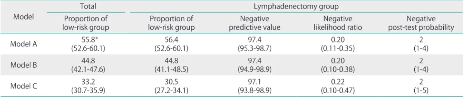
The negative predictive values (NPVs) and negative LRs were not statistically different among the three models (Table 3).
However, the proportion of patients classified as low-risk group was significantly different among the models. Model A, which included LVSI information, identified the largest num- ber of patients as a low-risk group (56.4%) without hampering the negative predictive value. Model C identified the smallest low-risk group (30.5%), although its predictive performance was similar to that of other models. In addition, using Bayes’
theorem, the negative PTP could be calculated at the 10% of assumed prevalence of lymph node metastasis (Table 3). All models indicated that false negative rate might be 2% when the prevalence of lymph node metastasis was 10%.
DISCUSSION
In the current study, we compared the predictive perfor- mance of various prediction models to identify a low-risk group in a large cohort of patients with endometrial cancer.
Several clinical implications suggested by our data are as fol- lows.
First, our study revealed that three models based on surgical pathology showed similar negative predictive powers. Our data suggest that the low-risk group can be identified with a false negativity rate of 2% by final pathologic data (Table 3), regardless of the choice of prediction model. Second, al- though the false negativity of these models was similar, the model from the Gynecologic Oncology Group (GOG) pilot study [12,13], which included LVSI as a predictor, was able to identify the largest number of patients (56%) as a low-risk
Table 3. Comparison of model performance in predicting a low-risk group of nodal metastasis Model
Total Lymphadenectomy group
Proportion of
low-risk group Proportion of
low-risk group Negative
predictive value Negative
likelihood ratio Negative post-test probability
Model A 55.8*
(52.6-60.1) 56.4
(52.6-60.1) 97.4
(95.3-98.7) 0.20
(0.11-0.35) 2
(1-4)
Model B 44.8
(42.1-47.6) 44.8
(41.1-48.5) 97.4
(94.9-98.9) 0.20
(0.10-0.38) 2
(1-4)
Model C 33.2
(30.7-35.9) 30.5
(27.2-34.1) 97.1
(93.8-98.9) 0.22
(0.10-0.47) 2
(1-5) Values are presented as percentage (95% confidence interval).
*Comparison with model B and C yielded p<0.001 for both comparisons (chi-square test).
group. The proportions of patients in the low-risk group iden- tified using those two models (A and C) were significantly dif- ferent.
In summary, even with final pathologic data, the currently available prediction identifying the low-risk group of lymph node metastasis in endometrial cancer has a false negative rate about 2% at 10% of the assumed prevalence. Therefore, future pre-/intra-operative prediction models may be regard- ed as clinically useful if the model shows a false negative rate less than 2%, when the prevalence of nodal metastasis was assumed as 10%.
CONFLICT OF INTEREST
No potential conflict of interests relevant to this article was reported.
ACKNOWLEDGMENTS
This study was funded by National Cancer Center, Korea (grant no. 1210200).
REFERENCES
1. Benedetti Panici P, Basile S, Maneschi F, Alberto Lissoni A, Signorelli M, Scambia G, et al. Systematic pelvic lympha- denectomy vs. no lymphadenectomy in early-stage endometrial carcinoma: randomized clinical trial. J Natl Cancer Inst 2008;100:1707-16.
2. ASTEC study group, Kitchener H, Swart AM, Qian Q, Amos C, Parmar MK. Efficacy of systematic pelvic lympha- denectomy in endometrial cancer (MRC ASTEC trial): a randomised study. Lancet 2009;373:125-36.
3. Creasman WT, Odicino F, Maisonneuve P, Quinn MA, Beller U, Benedet JL, et al. Carcinoma of the corpus uteri:
FIGO 26th Annual Report on the Results of Treatment in Gynecological Cancer. Int J Gynaecol Obstet 2006;95 Suppl 1:S105-43.
4. Greer BE, Koh WJ, Abu-Rustum N, Bookman MA, Bristow RE, Campos SM, et al. Uterine neoplasms: clinical practice guidelines in oncology. J Natl Compr Canc Netw 2009;7:
498-531.
5. American College of Obstetricians and Gynecologists.
ACOG practice bulletin, clinical management guidelines for obstetrician-gynecologists, number 65, August 2005:
management of endometrial cancer. Obstet Gynecol
2005;106:413-25.
6. Kitchener HC. To stage or not to stage? That is the question:
(with apologies to Shakespeare). Int J Gynecol Cancer 2010;20:S55-6.
7. Walsh CS, Karlan BY. Lymphadenectomy's role in early endometrial cancer: prognostic or therapeutic? J Natl Cancer Inst 2008;100:1660-1.
8. Uccella S, Podratz KC, Aletti GD, Mariani A. Re: Systematic pelvic lymphadenectomy vs no lymphadenectomy in early-stage endometrial carcinoma: randomized clinical trial. J Natl Cancer Inst 2009;101:897-8.
9. Chan JK, Wu H, Cheung MK, Shin JY, Osann K, Kapp DS.
The outcomes of 27,063 women with unstaged endo- metrioid uterine cancer. Gynecol Oncol 2007;106:282-8.
10. Trimble EL, Kosary C, Park RC. Lymph node sampling and survival in endometrial cancer. Gynecol Oncol 1998;
71:340-3.
11. Lee TS, Kim JW, Kim SH, Seong SJ, Song ES, Kim JH, et al.
Surgical practice patterns in endometrial cancer: results of the Korean Gynecologic Oncology Group survey. J Gynecol Oncol 2009;20:107-12.
12. Boronow RC. Surgical staging of endometrial cancer:
evolu tion, evaluation, and responsible challenge: a perso- nal perspective. Gynecol Oncol 1997;66:179-89.
13. Boronow RC, Morrow CP, Creasman WT, Disaia PJ, Silverberg SG, Miller A, et al. Surgical staging in endometrial cancer:
clinical-pathologic findings of a prospective study. Obstet Gynecol 1984;63:825-32.
14. Creasman WT, Morrow CP, Bundy BN, Homesley HD, Graham JE, Heller PB. Surgical pathologic spread patterns of endo- metrial cancer: a Gynecologic Oncology Group Study. Cancer 1987;60:2035-41.
15. Mariani A, Webb MJ, Keeney GL, Haddock MG, Calori G, Podratz KC. Low-risk corpus cancer: is lymphadenectomy or radiotherapy necessary? Am J Obstet Gynecol 2000;
182:1506-19.
16. Schink JC, Lurain JR, Wallemark CB, Chmiel JS. Tumor size in endometrial cancer: a prognostic factor for lymph node metastasis. Obstet Gynecol 1987;70:216-9.
17. Morrow CP, Bundy BN, Kurman RJ, Creasman WT, Heller P, Homesley HD, et al. Relationship between surgical- pathological risk factors and outcome in clinical stage I and II carcinoma of the endometrium: a Gynecologic Oncology Group study. Gynecol Oncol 1991;40:55-65.
18. Case AS, Rocconi RP, Straughn JM Jr, Conner M, Novak L, Wang W, et al. A prospective blinded evaluation of the accuracy of frozen section for the surgical management of endometrial cancer. Obstet Gynecol 2006;108:1375-9.
19. Neubauer NL, Havrilesky LJ, Calingaert B, Bulusu A,
Bernardini MQ, Fleming ND, et al. The role of lympha- denectomy in the management of preoperative grade 1 endometrial carcinoma. Gynecol Oncol 2009;112:511-6.
20. Chan JK, Kapp DS. Role of complete lymphadenectomy in endometrioid uterine cancer. Lancet Oncol 2007;8:831- 41.
21. Mariani A, Dowdy SC, Cliby WA, Gostout BS, Jones MB, Wilson TO, et al. Prospective assessment of lymphatic disse mination in endometrial cancer: a paradigm shift in surgical staging. Gynecol Oncol 2008;109:11-8.
22. Todo Y, Okamoto K, Hayashi M, Minobe S, Nomura E, Hareyama H, et al. A validation study of a scoring system to estimate the risk of lymph node metastasis for patients with endometrial cancer for tailoring the indication of lymphadenectomy. Gynecol Oncol 2007;104:623-8.
23. Ballester M, Dubernard G, Lecuru F, Heitz D, Mathevet P, Marret H, et al. Detection rate and diagnostic accuracy of sentinel-node biopsy in early stage endometrial cancer:
a prospective multicentre study (SENTI-ENDO). Lancet Oncol 2011;12:469-76.
24. Suh DH, Kim K, Kim JW. Major clinical research advances in gynecologic cancer in 2011. J Gynecol Oncol 2012;23:53- 64.
25. Kang S, Kang WD, Chung HH, Jeong DH, Seo SS, Lee JM, et al. Preoperative identification of a low-risk group for lymph node metastasis in endometrial cancer: a Korean gynecologic oncology group study. J Clin Oncol 2012;30:1329-34.
26. Soliman PT, Frumovitz M, Spannuth W, Greer MJ, Sharma S, Schmeler KM, et al. Lymphadenectomy during endo- metrial cancer staging: practice patterns among gyne co- logic oncologists. Gynecol Oncol 2010;119:291-4.
27. Lyman GH, Giuliano AE, Somerfield MR, Benson AB 3rd, Bodurka DC, Burstein HJ, et al. American Society of Clinical
Oncology guideline recommendations for sentinel lymph node biopsy in early-stage breast cancer. J Clin Oncol 2005;23:7703-20.
28. Lee KB, Ki KD, Lee JM, Lee JK, Kim JW, Cho CH, et al. The risk of lymph node metastasis based on myometrial inva- sion and tumor grade in endometrioid uterine cancers: a multicenter, retrospective Korean study. Ann Surg Oncol 2009;16:2882-7.
29. Bossuyt PM, Reitsma JB, Bruns DE, Gatsonis CA, Glasziou PP, Irwig LM, et al. Towards complete and accurate repor- ting of studies of diagnostic accuracy: the STARD initia- tive: Standards for Reporting of Diagnostic Accuracy. Clin Chem 2003;49:1-6.
30. Chan JK, Kapp DS, Cheung MK, Osann K, Shin JY, Cohn D, et al. The impact of the absolute number and ratio of positive lymph nodes on survival of endometrioid uterine cancer patients. Br J Cancer 2007;97:605-11.
31. Chi DS, Barakat RR, Palayekar MJ, Levine DA, Sonoda Y, Alektiar K, et al. The incidence of pelvic lymph node metastasis by FIGO staging for patients with adequately surgically staged endometrial adenocarcinoma of endometrioid histology. Int J Gynecol Cancer 2008;18:
269-73.
32. Jaeschke R, Guyatt GH, Sackett DL. Users' guides to the medical literature. III. How to use an article about a dia- gnostic test. B. What are the results and will they help me in caring for my patients? The Evidence-Based Medicine Working Group. JAMA 1994;271:703-7.
33. Jaeschke R, Guyatt G, Sackett DL. Users' guides to the medical literature. III. How to use an article about a diagnostic test. A. Are the results of the study valid?
Evidence-Based Medicine Working Group. JAMA 1994;
271:389-91.