서 론
유방의 유두상 종양은 유두종(papilloma), 유두종증(papillo- matosis), 경화 유두종(sclerosing papilloma), 비정형 유두종 (atypical papilloma), 유두상 관내암종(intraductal papillary carcinoma), 그리고 유두상 침습암종(invasive papillary car- cinoma)를 포함하는 넓은 범주의 질환군이다.(1) 초음파 유도 중 심부바늘생검은 비교적 시술이 쉽고 비침습적으로 유방 병변을 Purpose: We wanted to determine the underestimation rate
of ultrasound (US)-guided 14-gauge core needle biopsy for papillary neoplasms that were treated with subsequent sur- gical excision or vacuum-assisted biopsy (VAB) and we also wanted to evaluate the sonographic findings of papillary neo- plasms.
Methods: A retrospective review of the US-guided core needle biopsies of 984 consecutive lesions from January 2004 to April 2006 revealed 29 (3%) papillary neoplasms.
Twenty five lesions were further excised by surgery (n=16) or VAB (n=9). The remaining 4 lesions were not further excis- ed and they were excluded from this study. We evaluated the concordance between results of core needle biopsy and the final pathologic results. We reevaluate the sonographic findings of the papillary neoplasms included in our study.
Results: The pathologic results of core needle biopsy were benign in 21 and atypical in four. Of the 21 benign papillomas, none were revealed as carcinoma after further excision. Just one lesion showed focal atypical ductal hyperplasia (ADH)
after VAB. Three intraductal papillomas with ADH underwent surgical excision (n=3) or VAB (n=1), and they were proved to be the same pathologic entities with (n=1) or without (n=3) lobular neoplasia. The sonographic findings were as follows:
four intraductal masses, four intracystic masses, four solid masses with peripheral anechoic rims, five extraductal masses adjacent to dilated ducts, six pure solid masses, and two mixed masses.
Conclusion: US-guide 14-gauge core needle biopsy for papillary neoplasm showed no underestimation after surgical excision or VAB in our study, and the procedure proved to be reliable for the assessment of papillary neoplasm if the imaging and pathologic findings were concordant. Papillary neoplasms showed variable sonographic findings.
Key Words : Breast, Papilloma, Ultrasound-guided core needle biopsy, Auto- mated gun biopsy
중심단어 : 유방, 유두종, 초음파 유도 중심부바늘생검, 자동총생검
Is US-guided 14-gauge Core Needle Biopsy Valid for Papillary Neoplasm of the Breast?
Na Young Jung
1, Jaehee Lee
1,2, A Won Lee
3, Byung Joo Song
4, Sang Seol Jung
4Department of 1Radiology, The Catholic University of Korea, College of Medicine, Seoul; 2Human Medical Imaging and Intervention Center, Seoul; Departments of 3Pathology and 4Surgery, The Catholic University of Korea, College of Medicine, Seoul, Korea
Breast Cancer
O R I G I N A L A R T I C L E
정나영1ㆍ이재희1,2ㆍ이아원3ㆍ송병주4ㆍ정상설4
가톨릭대학교 의과대학1방사선과학교실, 2휴먼영상의학센터, 가톨릭대학교 의과대학3병리과학교실∙4외과학교실
유방의 유두상 종양 진단에 있어서 초음파 유도 14 게이지 중심부바늘생검의 유용성
책임저자 : 이재희
137-902 서울시 서초구 잠원동 12-25 K타워 3층, 휴먼영상의학센터 Tel: 02-512-6695, Fax: 02-512-6646
E-mail : [email protected]
접수일 : 2007년 9월 12일 게재승인일 : 2007년 12월 17일
*2006년 제62차 대한영상의학회 학술대회 구연 발표되었음.
*Scientific exhibition at 11th Congress of the World Federation for Ultrasound in Medicine and Biology (WFUMB), 2006
30
진단할 수 있어 널리 이용되는 생검 방법이다. 그러나 초음파 유 도 중심부바늘생검으로 얻은 조각난(fragmented) 제한적인 표 본으로 유두상 종양을 진단하고 양성과 악성 유두상 종양을 감별 하는 것은 매우 어려울 수 있다.(2, 3) 특히 비정형성이 동반된 유 두상 종양을 가진 환자는 양성 유두상 종양을 가진 환자보다 상대 적인 암 위험률이 7.5배 높으며, 주로 병변이 있는 유방, 특히 원 래의 유두상 종양의 인접부위에서 암이 발생하므로 비정형성이 동반된 유두상 종양은 전구 병변(precursor lesion)의 가능성이 높은 것으로 알려져 있다.(4) 이러한 비정형성이 동반된 유두상 종양은 중심부바늘생검으로 저평가(underestimation)될 가능 성이 30% 내외로 높기 때문에 수술적 절제를 해야 하는 것으로 보고되고 있다.(3, 5-11) 그에 비하여 양성 유두상 종양은 저평가 율이 0-26% 정도로 다양하게 보고되어 추가 치료에 대한 논란의 여지가 있다.(3, 5-10, 12-14)
본 연구의 목적은 유방 유두상 종양에 대한 초음파 유도 14 게이 지중심부바늘생검후수술적절제또는진공흡인생검법(vacuum- assisted biopsy)을 받은 경우의 중심부바늘생검의 저평가율을 알아보고, 초음파 유도 중심부바늘생검으로 진단받은 유두상 종 양의 초음파 소견을 평가해보고자 하는 것이다.
방 법
2004년 1월에서 2006년 4월까지 984건의 연속된 유방 병변 에 대한 초음파 유도 14 게이지 중심부바늘생검에서 29개(2.9%) 의 유두상 종양이 진단되었다. 이 중 16개의 병변은 수술적 절제 로, 9개의 병변은 초음파 유도 진공흡인생검법을 통하여 제거되었 다. 나머지 4개의 병변은 더 이상의 절제를 시행하지 않아 본 연구 에서 제외되었다.
초음파 유도 중심부바늘생검과 진공흡인생검법은 환자에게 시 술의 방법과 합병증을 설명하고 동의서를 받은 후 시행하였다. 모 든 중심부바늘생검은 환자가 똑바로 또는 비스듬하게 누운 상태 에서 초음파 유도하에 12-5 MHz의 고해상도 선형 탐촉자(HDI 5000 Sono CT [Philips medical systems, Bothell, Washing- ton, USA] and HDI 5000 ATL [Advanced Technology Labo- ratory, Bothell, Washington, USA])를 이용하여 시행되었으 며, 14 게이지 반자동 중심부생검바늘(TSK Laboratory, Tokyo, Japan)을 사용하여 적어도 4회 이상의 조직 표본을 얻었다. 초음 파 유도 진공흡인생검법은 환자가 똑바로 또는 비스듬하게 누운 상태에서 12-5 MHz 선형 탐촉자로 검사하면서, 진공흡인 조직 검사 기구(Mammotome; Ethicon-Endosurgery, Cincinnati, Ohio, USA)를 이용하여 11 게이지(n=6) 혹은 8 게이지(n=3) 바 늘을이용하여초음파로보이는범위의병변을최대한제거하였다.
중심부바늘생검의 결과와 이후 수술적 절제 또는 초음파 유도 진공흡인생검법을 통해 얻은 최종 병리 결과를 한 명의 병리과 의 사가 조직 슬라이드를 후향적으로 검토하여 재진단하였다. 각각 의 중심부바늘생검의 결과와 최종 병리 결과를 비교 분석하였다.
“조직학적 저평가(histologic underestimation)”는 중심부바늘 생검 결과는 양성 또는 비정형 유관 증식증이었으나 수술에서 암 이 나온 경우 또는 중심부바늘생검 결과는 관내암종이 나왔으나 수술에서 침습암종으로 나온 경우로 정의하였다.(15)
연구에 포함된 25명의 환자들의 의무기록을 후향적으로 검토 하였다. 환자들의 나이는 17-79세로 평균 45세였다. 25명의 환 자 중 13명은 증상이 있었는데, 혈성 유두 분비물이 있었던 경우 와 장액성 유두 분비물이 있었던 경우가 각각 3명과 2명이었고, 종괴가 만져지는 경우가 6명, 종괴와 함께 유두 분비물이 있었던 경우가 2명이었다.
각 환자의 초음파 판독지에 기술된 American College of Radiology Breast Imaging-Reporting and Data System (ACR BI-RADS�) (16)에 의한 최종 판정 카테고리를 알아보았 다. 카테고리 1은 병변이 없는 경우, 카테고리 2는 악성소견이 없 는 경우, 카테고리 3은 악성의 가능성이 희박한 양성의증의 경우, 카테고리 4는 악성의 가능성이 있어 조직검사가 필요한 경우, 카 테고리 5는 거의 확실한 악성의 경우라는 정의에 따랐다. 카테고 리 4는 악성 가능성의 정도에 따라 4A, 4B, 4C로 구분하였다. 또 한 초음파 영상을 두 명의 방사선과 의사가 합의에 의해 후향적으 로 검토하여 유두상 종양들의 크기, 위치, 경계, 유관 확장증의 동 반 여부, 방향 그리고 에코 음영에 대해 분석하였다. 각 유방 종괴 들을 종괴와 유관 사이의 관계에 따라 Han 등(17)이 보고한 여섯 가지 부류로 분류하였다; 이들은 각각 관강내(intraluminal) 종 괴, 늘어난 유관 근처에 접해있는 유관외(extraductal) 종괴, 단순 고형 종괴, 그리고 혼합형 병변이다. 관강내 종괴는 유관의 늘어난 정도와 종괴가 관을 채우고 있는 정도에 따라 유관내(intraductal) 종괴, 낭종내(intracystic) 종괴, 주변부 무에코의 띠를 갖는 고형 종괴의 세 부류로 분류되었다.
초음파 유도하 중심부바늘생검을 시행하기 전에 유방촬영(Ma- mmography; Mammomat 3000, Seimens, Solna, Sweden) 을 시행한 환자의 경우에는 유방촬영 사진을 재검토하였다.
결 과
중심부바늘생검의 병리 결과는 21예(84%)에서 양성이었고, 4예 (16%)에서 비정형성이었다. 양성 결과로는 유관내 유두종이 14예 (56%)이고 상용 유관 증식증(usual ductal hyperplasia, UDH) 을 동반한 유관내 유두종이 7예(28%)였으며, 4예(16%)의 비정형
성 결과는 모두 비정형성 유관 증식증(atypical ductal hyper- plasia, ADH)을 동반한 유관내 유두종이었다. 중심부바늘생검의 결과와 수술 또는 진공흡인생검법 후 최종 병리조직 결과를 Table 1에 비교 정리하였다. 21예의 양성 유두종 중 1예에서 진공흡인생 검법 후 비정형성 유관 증식증이 추가로 발견되었을 뿐 수술(n=13) 이나진공흡인생검법(n=8) 후악성으로진단된경우는없었다. 4예 의 비정형성 유관 증식증을 동반한 유두종들은 수술(n=3) 또는 진 공흡인생검법(n=1) 후모두같은진단명을가진병변으로확인되었 고, 수술을 했던 환자 중 한 명에서 소엽성 신생물(lobular neo- plasia)를 동반한 것으로 진단되었다. 이는 유방의 침습암종의 위 험 인자일 뿐 악성은 아니므로 중심부바늘생검에서 조직학적으로 저평가된 병변으로 분류되지는 않는다. 따라서 본 연구에서는 중 심부바늘생검시 수술이나 진공흡인생검법과 비교하여 조직학적 저평가를 보였던 증례는 없었다. 25명의 환자 중 21명이 수술 또는 진공흡인생검 후 평균 20.2개월(7-37개월)의 추적 유방 초음파 검사를 시행 받았고 이들 중 새로운 병변이 발견된 환자는 없었다.
본 연구에 포함된 병변들의 판독지를 검토한 결과 BI-RADS
카테고리는 카테고리 3 (n=2), 4A (n=19) 또는 4B (n=4)로 대 다수가 양성 가능성이 높거나 다소 의심스러운 소견이지만 악성 가능성은 낮은 병변으로 보였다(Table 2).
병변의 평균 크기는 1.1 cm이었고(0.4-2.3 cm), 병변의 위치 는 12예에서 중심성, 13예에서 말초성이었다. 병변은 16예에서 분명한 경계를 보였고 9예에서는 불분명한 경계를 보였으며, 종 괴로 보였던 경우 중 2예를 제외하고는 모두 피부와 평행한 방향 을 보였다. 유관 확장은 14예에서 동반되었다. 병변들은 다양한 에코 음영을 보였는데, 저에코성이 14예, 등에코성이 7예, 혼합 된 에코 음영을 보인 경우가 4예였다. 초음파 소견으로 병변들을 6가지 범주로 나누었는데, 유관내 종괴가 4예, 낭종내 종괴가 4예, 주변부 무에코의 띠를 갖는 고형 종괴가 4예, 늘어난 유관 근처에 접해있는 유관외 종괴가 5예, 단순 고형 종괴가 6예, 그리고 혼합 형이 2예였다(Fig 1). 중심부바늘생검에서 비정형성 유관 증식증 을 동반한 4예의 유두종은 각각 유관내 종괴, 낭종내 종괴, 늘어 난 유관 근처에 접해 있는 유관외 종괴, 단순 고형 종괴로 다양하 게 보였다. 이 중 유관내 종괴로 보였던 1예에서 수술 후 소엽성
Table 1. Histologic correlation between core needle biopsy and further excision
Histologic findings at core needle biopsy
Histologic findings at surgical excision
IP IP with
UDH
IP with ADH
IP with LN
Histologic findings at VAB
IP IP with
UDH
IP with ADH
IP with LN
Total
IP 9 1 0 0 2 1 1* 0 14
IP with UDH 0 3 0 0 0 4 0 0 7
IP with ADH 0 0 2 1* 0 0 1 0 4
Total 9 4 2 1 2 5 2 0 25
VAB=vacuum-assisted biopsy; IP=intraductal papilloma; UDH=usual ductal hyperplasia; ADH=atypical ductal hyperplasia; LN=lobular neoplasia.
*Corrected pathology after surgical excision or VAB.
Table 2. Correlation between sonographic BI-RADS categories and histologic findings at surgical excision or VAB
Histologic findings at surgical excision or VAB
BI-RADS
category N
IP IP with UDH
IP with ADH
IP with LN
C3 2 2
C4A 19 7 7 4 (1*) 1*
C4B 4 2 2
Total 25 11 9 4 1
BI-RADS=Breast imaging reporting and data system; VAB=vacuum- assisted biopsy; C3=BI-RADS category 3, probably benign; C4A=BI- RADS category 4A, suspicious abnormality with low suspicion of malig- nancy; C4B=BI-RADS category 4B, suspicious abnormality with inter- mediate concern; IP=intraductal papilloma; UDH=usual ductal hyper- plasia; ADH=atypical ductal hyperplasia; LN=lobular neoplasia.
*Corrected pathology after surgical excision or VAB.
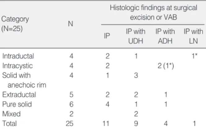
Table 3. Correlation between sonographic features and histo- logic findings at surgical excision or VAB
Histologic findings at surgical excision or VAB Category
(N=25) N
IP IP with UDH
IP with ADH
IP with LN
Intraductal 4 2 1 1*
Intracystic 4 2 2 (1*)
Solid with 4 1 3
anechoic rim
Extraductal 5 2 2 1
Pure solid 6 4 1 1
Mixed 2 2
Total 25 11 9 4 1
VAB=vacuum-assisted biopsy; IP=intraductal papilloma; UDH=usual ductal hyperplasia; ADH=atypical ductal hyperplasia; LN=lobular neoplasia.
*Corrected pathology after surgical excision or VAB.
신생물이 추가로 발견되었다(Fig 2). 중심부바늘생검에서 양성 유두종으로 나왔던 21예의 종괴들은 다양한 초음파 소견을 보였 으며, 그 이후 시행한 조직 절제술에서 낭종내 종괴로 보였던 1예 에서 비정형성 유관 증식증이 추가로 발견된 것(Fig 3) 이외에는 모두 양성 유두종으로 확진되었다. 유두상 종양의 초음파 소견에 따른 최종 조직학적 결과를 Table 3에 정리하였다.
25명의 환자 중 15명에서 수술 전 유방촬영술 사진을 얻을 수
있었는데, 정상 소견이 6예, 종괴가 보였던 경우가 6예, 비대칭 음영이 1예, 매우 치밀한 조직을 보여 초음파 검사를 권유했던 경 우가 2예였으며, 미세석회화를 동반한 경우는 없었다. 그러나 연 구에 포함된 환자 중 60% (15/25)에서만 유방 촬영 필름을 얻을 수 있었으며, 나머지 환자들은 타 병원에서 시행한 필름을 얻을 수 없었거나 환자가 젊은 나이여서 촬영하지 않은 경우로 평가가 불가능하였다.
Fig 2. A 48-yr-old woman without specific symptom. US of left outer subareolar region shows mild ductectasia with an isoechoic intraductal lesion (arrows). Histopathology of core needle biopsy revealed an intraductal papilloma with atypical ductal hyperplasia.
The lesion was surgically excised and confirmed as the same pathologic entity on core biopsy specimen with lobular neoplasia.
Fig 3. A 79-yr-old woman with a palpable breast mass. US of left mid upper breast reveals an about 1.7 cm-sized circumscribed oval cyst with an internal nodular isoechoic solid portion (arrow).
With core needle biopsy of the solid portion, the lesion was reve- aled as a benign papilloma. After 11 gauge VAB, the lesion was confirmed as papilloma with atypical ductal hyperplasia.
Fig 1. US categories of papillary neoplasms of breast. (A) Intraductal mass (n=4). (B) Intracystic mass (n=4). (C) Solid mass with peripheral anechoic rim (n=4). (D) Extraductal mass adjacent to the dilated duct (n=5). (E) Pure solid mass (n=6). (F) Mixed type (Intraductal+
Extraductal masses) (n=2).
A
D
B C
E F
고 찰
유방의 유두상 종양은 넓은 범주를 포함하는 질환군으로 초음 파 유도 중심부바늘생검으로 유두상 종양을 진단하고 양성과 악 성 유두상 종양으로 감별하는 것은 매우 어렵다. 특히 비정형성이 동반된 유두상 종양은 전암성 병변으로 여겨지며 30%에 달하는 높은 저평가율 때문에 수술적으로 절제되어야 하는 병변으로 잘 알려져 있다.(3, 5-11) 그러나 중심부바늘생검에서 양성 유두상 종양으로 진단된 경우에는 저평가율이 0-26%로 다양하게 보고 되고 있다.(3, 5-10, 12-14) 이전의 여러 연구들(3, 5, 7-9, 13) 에서 입체정위(stereotactic directional) 또는 초음파 유도 중심 부바늘생검으로 진단된 양성 유두상 종양은 절제 후 대부분 양성 으로 판명되거나 장기 추적 검사에서 변화가 없어 영상 소견과 조 직 소견이 일치한다면 중심부바늘생검은 양성 유두상 종양의 진단 에 정확하여 즉각적인 수술적 절제는 필요하지 않으나, 양성 생검 결과를 보이더라도 영상 소견과 병리 소견이 일치하지 않는 경우 에는 절제가 시행되어야 한다고 기술하였다. 그러나 최근 몇몇 연 구들에서는 중심부바늘생검에서 진단된 양성 유두상 종양도 높은 저평가율 때문에 수술적으로 절제되어야 하며, 모든 유두상 종양 은 관내암종 또는 침습암종의 배제를 위해 모두 조직 검사를 시행 하는것이바람직하다고주장하였다.(6, 10, 12, 14) 이중Mercado 등(6)이 보고한 관내암종으로 상향 진단된 유두상 종양들은 BI- RADS 카테고리 5의 병변이거나 고연령 환자에서 새로이 발견된 의심스러운미세석회화병변으로영상소견과중심부바늘생검결과 간의 불일치로 바로 수술적 절제를 했던 경우이다. 한편 Liber- man 등(10)은 경피적 유방 생검으로 양성 유두종으로 진단된 영 상-병리 일치를 보였던 35예의 증례의 추적 검사(평균 22개월) 중 5예의 악성 병변(4예의 상피내암과 1예의 임파선 전이가 없는 1.8 cm의 침습암종)과 6예의 고위험 병변이 발견되었다고 보고 하였다. 악성 증례 중 1예는 입체정위 진공흡인생검법 후 임상의 와 환자의 요구에 의해 1개월 이내 수술을 시행했던 경우였고, 나 머지는 초음파 유도 중심부바늘생검 후 7-25개월 추적검사 기간 중 종괴가 증가되거나 증상이 나타나 수술을 시행했던 경우였다.
Lam 등(12)도 중심부바늘생검으로 양성 유두종으로 진단받았으 며 영상-병리 일치를 보였던 7예 중 1예에서 수술 후 악성으로 진 단된 경우가 있어 모든 유두상 종양은 수술을 하는 것이 좋다고 주장하였다. 그러나 이 연구에서는 16 게이지의 바늘로 세 번의 중심부바늘생검 표본만을 얻었으므로 14 게이지 바늘로 네 번 이 상의 조직을 얻었던 우리의 연구보다 좀 더 검사의 정확도가 떨어 질 수 있을 것으로 보인다. 이상의 연구 결과에서 초음파 유도 중 심부바늘생검에서 양성이고 영상-병리 일치를 보이더라도 충분 하고 주의깊은 추적검사가 필요함을 알 수 있다. 그러나 본 연구
에서는 평균 20.2개월(7-37개월)의 추적검사 동안 새로운 병변 이 발견된 경우는 없었다.
또한 본 연구에서는 초음파 유도 14 게이지 중심부바늘생검에 서 양성 또는 비정형성 유두상 종양으로 진단된 환자 중 수술적 절제나 초음파 유도 진공흡인생검법으로 악성으로 진단된 경우 는 없었다. 이전 연구의 결과와 달리 비정형성 유관 증식증을 동 반한 경우에도 조직학적 저평가를 보인 예가 없었던 것은 연구에 포함된 병변의 수가 적으며 포함된 병변 중 의심스러운 미세석회 화를 포함하는 병변이 없었고 초음파에서도 대부분 악성 가능성이 비교적 낮은 BI-RADS 카테고리 4B 이하의 소견을 보여 영상- 병리 일치로 생각되었던 것과 관계가 있을 것으로 생각된다.
한편 진공흡인생검법은 14 게이지 중심부바늘생검에 비해 비교 적 많은 조직을 제거하므로 저평가율이 낮으나(18), 수술적 절제 에 비하여서는 유두상 종양을 저평가 할 수 있다.(6, 7, 10, 19) Mercado 등(6)은 입체정위 진공흡인생검법으로 유두상 관내암 종으로 진단 받은 8예의 병변 중 2예(25%)가 수술 후 침습암종으 로 진단되었으며 1예가 진공흡인생검법으로 양성 유두종으로 진 단되었다가 수술 후 유두상 관내암종으로 상향 진단되었음을 보 고하였다.(19) 또한, Rosen 등(7)의 연구에서는 11 게이지 진공 흡인생검법으로 진단받은 8예의 비정형성 유두상 종양 중 2예가 수술 후 유두상 관내암종으로 상향 진단 되었다고 보고하였다.(7) Liberman의 최근 연구(10)에서도 입체정위 진공흡인생검법 후 양성 유두종으로 진단받았던 증례에서 영상-병리 일치를 보였음 에도 불구하고 1개월 이내의 수술에서 관내암종이 동반되었음을 확인한 바 있다. 위 논문들에서 상향 진단된 증례들은 모두 의심 스러운 석회화 또는 석회화를 동반한 종괴로 보였던 것에 비해 본 연구에 포함된 중심부바늘생검 후 진공흡인생검법으로 추가로 병 변을 절제한 9예의 증례 중에서 의심스러운 석회화를 포함하는 병변은 없었다. 이들 9예의 유두상 종양 중 중심부바늘생검에서 비정형성 유관 증식증이 동반되어 있었던 2예를 포함하여 모든 경우에서 진공흡인생검법 후 추가로 관내암종 또는 침습암종이 발견된 경우는 없었으며, 모두 영상 소견과 병리 소견이 일치하므 로 병변 제거 후 추적 검사를 시행 중이다. 추적 기간(5-28개월) 중 새롭게 생긴 병변은 아직 발견되지 않았다.
연구에 포함된 25예의 유두상 종양 중 19예에서 초음파에서 유 관과의 관계를 시사할 만한 소견을 동반하고 있었다. 증례의 수가 많지 않아 통계적 분석은 어려우나 비정형성을 동반하거나 하지 않은 유두상 종양은 각각 다양한 위치에 다양한 모양으로 보였고 비정형성을 시사할 만한 초음파 소견은 발견되지 않았다. 고전적 으로 중심성 유두상 종양보다 말초성 유방 종양에서 악성의 확률 이 높다고 알려져 있으나, 근래의 연구들에서는 종양의 위치, 개 수, 모양보다는 비정형성 유관 증식증을 동반하였는가 하는 점이
악성의 확률과 더 관계가 있다고 보고하였다.(3, 4)
본 연구의 한계는 초음파 유도 중심부바늘생검으로 진단된 유두 상 종양의 증례 수가 적다는 점과 아직 충분한 추적 검사가 이루 어지지 않았다는 점이다. 중심부바늘생검의 유용성과 결과를 증 명하기 위해서는 좀 더 많은 수의 증례와 더 긴 기간의 추적 검사 를 통한 추가 연구가 필요할 것이다.
결 론
초음파 유도 14 게이지 중심부바늘생검은 영상-병리 일치 (Image-pathology concordance)를 보이는 양성 유두상 종양 의 평가에 대해서는 믿을 만한 검사법이며, 양성과 비정형성 유두 상 종양은 각각 다양한 초음파 소견을 보일 수 있다.
참고문헌
1. Tavassoli FA. Papillary lesions. In: Tavassoli FA, ed. Pathology of the breast. Norwalk Conn: Appleton & Lange; 1992;193-227.
2. Jacobs TW, Connolly JL, Schnitt SJ. Nonmalignant lesions in breast core needle biopsies: to excise or not to excise? Am J Surg Pathol 2002;26:1095-110.
3. Ivan D, Selinko V, Sahin AA, Sneige N, Middleton LP. Accuracy of core needle biopsy diagnosis in assessing papillary breast lesions:
histologic predictors of malignancy. Mod Pathol 2004;17:165-71.
4. Page DL, Salhany KE, Jensen RA, Dupont WD. Subsequent breast carcinoma risk after biopsy with atypia in a breast papilloma. Cancer 1996;78:258-66.
5. Liberman L, Bracero N, Vuolo MA, Dershaw DD, Morris EA, Ab- ramson AF, et al. Percutaneous large-core biopsy of papillary breast lesions. AJR Am J Roentgenol 1999;172:331-7.
6. Mercado CL, Hamele-Bena D, Oken SM, Singer CI, Cangiarella J.
Papillary lesions of the breast at percutaneous core-needle biopsy.
Radiology 2006;238:801-8.
7. Rosen EL, Bentley RC, Baker JA, Soo MS. Imaging-guided core needle biopsy of papillary lesions of the breast. AJR Am J Roentgenol 2002;179:1185-92.
8. Agoff SN, Lawton TJ. Papillary lesions of the breast with and without atypical ductal hyperplasia: can we accurately predict benign behavior
from core needle biopsy? Am J Clin Pathol 2004;122:440-3.
9. Ko ES, Cho N, Cha JH, Park JS, Kim SM, Moon WK. Sonographi- cally-guided 14-gauge core needle biopsy for papillary lesions of the breast. Korean J Radiol 2007;8:206-11.
10. Liberman L, Tornos C, Huzjan R, Bartella L, Morris EA, Dershaw DD. Is surgical excision warranted after benign, concordant diagnosis of papilloma at percutaneous breast biopsy? AJR Am J Roentgenol 2006;186:1328-34.
11. Lim JY, Oh SM. Usufulness of ultrasound guided core needle biopsy of breast lesions with automated gun. J Korean Breast Cancer Soc 1998;1:186-91.
12. Lam WW, Chu WC, Tang AP, Tse G, Ma TK. Role of radiologic features in the management of papillary lesions of the breast. AJR Am J Roentgenol 2006;186:1322-27.
13. Philpotts LE, Shaheen NA, Jain KS, Carter D, Lee CH. Uncommon high-risk lesions of the breast diagnosed at stereotactic core-needle biopsy: clinical importance. Radiology 2000;216:831-7.
14. Puglisi F, Zuiani C, Bazzocchi M, Valent F, Aprile G, Pertoldi B, et al. Role of mammography, ultrasound and large core biopsy in the diagnostic evaluation of papillary breast lesions. Oncology 2003;65:
311-5.
15. Burbank F. Stereotactic breast biopsy of atypical ductal hyperplasia and ductal carcinoma in situ lesions: improved accuracy with direc- tional, vacuum- assisted biopsy. Radiology 1997;202:843-7.
16. American College of Radiology. Breast imaging reporting and data system (BI-RADS). 4th ed. Reston, VA: 2003.
17. Han BK, Choe YH, Ko YH, Yang JH, Nam SJ. Benign papillary lesions of the breast: sonographic-pathologic correlation. J Ultrasound Med 1999;18:217-23.
18. Ko ES, Cho N, Yang SK, Kim DY, Moon WK. Papillary lesions of the breast: Comparison of the US-guided 14-gauge automated gun method and the 11-gause directional vacuum-assisted biopsy method.
J Korean Radiol Soc 2006;54:537-41.
19. Mercado CL, Hamele-Bena D, Singer C, Koenigsberg T, Pile-Spell- man E, Higgins H, et al. Papillary lesions of the breast: evaluation with stereotactic directional vacuum-assisted biopsy. Radiology 2001;221:650-5.