bone, including breast cancer, and are well known for the management of osteoporosis1. Medical uses of BPs began in 1977, when the U.S. Food and Drug Administration (FDA) approved the use of etidronate as a bone disease agent to treat Paget’s disease. BP uses were extended to additional diseases after the FDA recognized the clinical use of nitrogen-contain- ing BPs in 19942.
Bisphosphonate-related osteonecrosis of the jaw (BRONJ) was first reported by Marx3 in 2003 and Ruggiero et al.4 in 2004. Marx3 described patients who received intravenous BP zoledronic acid treatment and subsequently developed osteonecrosis of the jaw. Ruggiero et al.4 described clinical symptoms, radiological features and guidelines for diagnosis in 63 patients with BP oral administration who developed os- teonecrosis.
There are many hypotheses as to why BP induces osteo-
I. Introduction
Bisphosphonates (BPs), a class of drugs that inhibit os- teoclast-mediated bone resorption, are used to treat skeletal diseases that increase bone resorption, such as Paget’s disease or multiple myeloma. BPs can also counteract the hyper- calcemia of malignancies and carcinomas metastatic to the Jae-Hoon Lee
Department of Oral and Maxillofacial Surgery, College of Dentistry, Dankook University, 119 Dandae-ro, Dongnam-gu, Cheonan 31116, Korea
TEL: +82-41-550-0271 FAX: +82-41-551-8988 E-mail: [email protected]
ORCID: http://orcid.org/0000-0002-9959-4632
This is an open-access article distributed under the terms of the Creative Commons Attribution Non-Commercial License (http://creativecommons.org/licenses/by-nc/4.0/), which permits unrestricted non-commercial use, distribution, and reproduction in any medium, provided the original work is properly cited.
CC
Evaluation of the predisposing factors and involved outcome of surgical treatment in bisphosphonate-related osteonecrosis of the jaw cases
including bone biopsies
Tae-Hwan Kim, Won-Gyo Seo, Chul-Hong Koo, Jae-Hoon Lee
Department of Oral and Maxillofacial Surgery, College of Dentistry, Dankook University, Cheonan, Korea
Abstract(J Korean Assoc Oral Maxillofac Surg 2016;42:193-204)
Objectives: This study examined the statistical relevance of whether the systemic predisposing factors affect the prognosis of surgical treatment of bisphosphonate-related osteonecrosis of the jaw (BRONJ). All cases had undergone bone biopsies to determine the characteristics of the mechanisms of BRONJ by optical microscopy.
Materials and Methods: The data included 54 BRONJ cases who underwent surgery and in whom bone biopsies were performed. The results of surgery were evaluated and the results were classified into 3 categories: normal recovery, delayed recovery, and recurrence after surgery. The medical history, such as diabetes mellitus, medication of steroids, malignancies on other sites was investigated for an evaluation of the systemic predispos- ing factors in relation to the prognosis. The three factors involved with the medication of bisphosphonate (BP) were the medication route, medication period, and drug holiday of BP before surgery. The serum C-terminal cross-linking telopeptide (CTX) value and presence of microorganism colony in bone biopsy specimens were also checked. Statistical analysis was then carried out to determine the relationship between these factors and the results of surgery.
Results: The group of patients suffering from diabetes and on steroids tended to show poorer results after surgery. Parenteral medication of BP made the patients have a poorer prognosis after surgery than oral medication. In contrast, the medication period and drug holiday of BP before surgery did not have significance with the results of surgery nor did the serum CTX value and presence of microorganism colony. Necrotic bone specimens in this study typically showed disappearing new bone formation around the osteocytic lacunae and destroyed Howship’s lacunae.
Conclusion: Although many variables exist, this study could in part, predict the prognosis of surgical treatment of BRONJ by taking the patient’s medical history.
Key words: Bisphosphonate, Osteonecrosis, Jaw diseases
[paper submitted 2016. 4. 14 / revised 2016. 6. 27 / accepted 2016. 7. 22]
Copyright Ⓒ 2016 The Korean Association of Oral and Maxillofacial Surgeons. All rights reserved.
account.
II. Materials and Methods
Written informed consent was obtained from the patients for the publication of this report and any accompanying im- ages. The total of 54 BRONJ patients who received surgical therapy and a bone biopsy between January 2008 to July 2015 at Dankook University Dental Hospital (Cheonan, Ko- rea) were included. Patients who received radiation treatment in the head and neck or were diagnosed with osteopetrosis or florid osseous dysplasia after biopsy were excluded.
The following data were collected: (1) sex, (2) age, (3) osteonecrosis site, (4) BRONJ stage at first examination, (5) etiologic factors, (6) underlying disease requiring BP medica- tion, and (7) surgery type.
Classification of BRONJ stage at the first examination was done according to the position paper of the American Association of Oral and Maxillofacial Surgeons (AAOMS) released in 2007. (1) Stage 0: nonspecific clinical findings and symptoms such as jaw pain or osteosclerosis but no clini- cal evidence of exposed bone, (2) stage 1: exposed, necrotic bone or fistula that probes to bone. No symptoms or evidence of infection, (3) stage 2: exposed, necrotic bone or fistula that probes to bone, associated with infection, pain, and erythema in the regions of the exposed bone. Purulent drainage may also be present, and (4) stage 3: exposed, necrotic bone or fistula that probes to bone in patients with pain, infection, and 1 or more of the following; pathologic fracture, extraoral fistula, oral antral/oral nasal communication, and osteolysis extending to the mandibular inferior border or sinus floor.
For outcome, cases were classified after surgery according to clinical symptoms during outpatient follow-up: (1) normal tissue regeneration on surgical site, (2) delayed recovery on surgical site, and (3) recurrences on surgical site and other sites.
Delayed recovery cases included patients with wound de- hiscence and bone exposure for more than 3 months during periodic follow-up after surgery. Recurrence cases included patients with osteonecrosis on the surgical site and other sites after surgery requiring additional surgical management.
A patient scoring system was adopted to capture systemic pathology features in 3 categories according to the follow- ing criteria. If a patient had none among the three items mentioned above, then he or she was scored as 0. If a patient had a feature described above, then he or she was scored as 1. If a patient had two features, then he or she was scored as necrosis of the jaw. Impediment of osteoclast activity, de-
creased blood flow, direct toxicity to tissues, and imbalance of wound treatment, inflammation and infection have been considered5-7. However, evidence suggests that BPs have pharmacokinetic characteristics that hinder osteoclast activity and lead to ONJ from the imbalance of blood flow8.
The risk factors of BRONJ are mainly diabetes mellitus, intake of corticosteroids, malignancies, alcohol consumption, immunosuppressant treatment, autoimmune diseases, he- matopoietic diseases, peripheral vascular diseases and renal disorders9. Diabetes is a high risk factor for BRONJ because it may bring ischemic changes in microvessels, decline in endothelial cell activity, bone cells destruction, and a reduced metabolism10. Patients who take chemotherapy with a cell division suppressant, corticosteroids and stem cell transfusion are more vulnerable to developing BRONJ than others due to the decreased immunity and blood flow supply caused by bone marrow transformation10-12.
Conservative treatments are preferred to manage BRONJ, including medication to reduce pain, mouth rinsing for im- proved hygiene, and prescription of antibiotics for inflamma- tion with removal of sharp bone edges that might stimulate soft tissues13-15. Discontinuation of BP administration, hy- perbaric oxygen therapy, and low-level laser therapy have also been introduced16-18. However, those applications do not directly treat the necrosis of the jaw and surrounding soft tis- sues, and patients risk continuous bone exposure without full recovery of the mucous membrane in the jaw. Stanton and Balasanian2, on the other hand, described debridement that could bring temporary recovery of pain and infection due to BRONJ and fully solve the aggravation of osteonecrosis.
Lopes et al.19 and Stockmann et al.20 reported a success rate of more than 80% through sequestrectomy of BRONJ cases.
Carlson and Basile21 performed surgical management through the complete resection of necrotic bone in the mandible and maxilla on 95 BRONJ patients and healed them with an ac- ceptable prognosis.
This study focuses on BRONJ patients who underwent surgical therapy. All received BP medication and were di- agnosed with BRONJ. We collected and reviewed general information on the patients and assessed BRONJ risk factors for their influence on outcome after surgical management as well as the pathogenesis of BRONJ. A biopsy was performed on the extracted bones in all cases and common histological characteristics were analyzed with respect to the pathogen- esis of BRONJ. The objective was to suggest a standard for BRONJ surgical management taking systemic factors into
III. Results
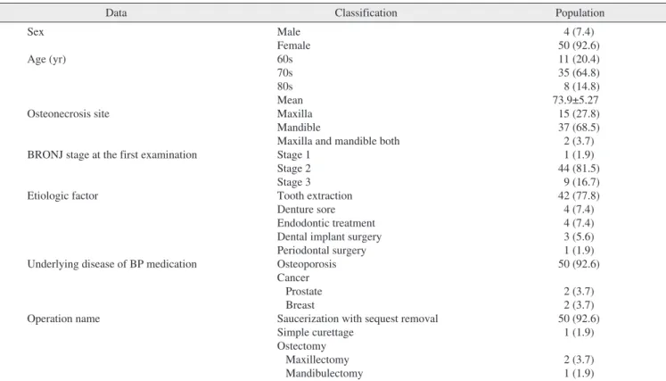
The dataset consisted of 50 female patients and 4 male pa- tients. The age distribution varied from 61 to 86 years (mean, 73.9±5.27 years). There were 11 patients in their 60s, 35 in their 70s, and 8 in their 80s. The osteonecrosis sites were di- vided into the maxilla, mandible, and maxilla and mandible simultaneously. Osteonecrosis occurred in the mandible in 37 patients, the maxilla in 15 patients and the maxilla and man- dible simultaneously in 2 patients. According to the AAOMS BRONJ categories described above, one patient was classi- fied as stage 1, 44 as stage 2, and 9 patients as stage 3. The etiology of osteonecrosis was tooth extraction in 42 patients.
Trauma due to inappropriate dentures resulted in BRONJ in four patients, dental implants in three patients and periodontal treatment in one patient. The majority of the patients, 50 out of 54, had taken BP medication for treatment or prevention of osteoporosis. Patients with malignancies (three breast cancer and one prostate cancer) received BP medication to prevent bone diseases. Most of the surgeries were saucerization with removal of sequestrations. Saucerization was performed in 50 cases, together with 1 simple curettage, 2 partial maxillecto- mies, and 1 segmental mandibulectomy.(Table 2)
Based on prognoses after surgical therapy, 32 of the 54 pa- tients (59.3%) showed normal recovery, 16 patients (29.6%) were diagnosed with delays at surgical sites, and recurrence of BRONJ at surgical and other sites was observed in 6 pa- tients (11.1%). There were two cases of recurrence at the 2. We also applied the MUCONNS (Modified University of
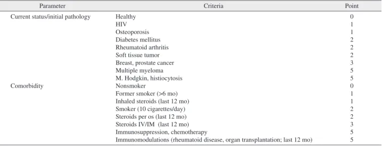
Connecticut Osteonecrosis Numerical Scale) scoring system described by Reich et al.22, which weighs each disease differ- ently. The MUCONNS scoring system is composed of sev- eral comorbidities. Depending on the assessed severity of the osteonecrosis of the jaw, each disease is scored differently.
Osteoporosis scores as 1, diabetes mellitus as 2, steroid oral administration as 2, breast cancer and prostate cancer among other malignancies as 3, and chemotherapy as 5.(Table 1) We adopted the weighted factor for each criterion and summed up all for an index score for each patient.
Patients were evaluated in several subject groupings. We looked at 36 patients for whom the complete BP medication period was confirmed as well as 44 patients who discontin- ued their BP medication before BRONJ surgery respectively.
The C-terminal cross-linking telopeptide (CTX) value, or bone resorption index, was also collected from 28 patients who received blood screening.
The presence of microorganisms in bone biopsies and im- pressions of necrotic bone and adjacent sites were evaluated by optical microscopy. The presence of microorganisms was assessed for an association with prognosis after the surgery.
Statistical analysis was performed using linear by linear as- sociation and nonparametric Kruskal-Wallis tests with IBM SPSS Statistics version 21.0 program (IBM Co., Armonk, NY, USA). A P-value <0.05 was considered statistically sig- nificant.
Table 1. MUCONNS (Modified University of Connecticut Osteonecrosis Numerical Scale) reference table
Parameter Criteria Point
Current status/initial pathology
Comorbidity
Healthy HIV Osteoporosis Diabetes mellitus Rheumatoid arthritis Soft tissue tumor Breast, prostate cancer Multiple myeloma M. Hodgkin, histiocytosis Nonsmoker
Former smoker (>6 mo) Inhaled steroids (last 12 mo) Smoker (10 cigarettes/day) Steroids per os (last 12 mo) Steroids IV/IM (last 12 mo) Immunosuppression, chemotherapy
Immunomodulations (rheumatoid disease, organ transplantation; last 12 mo)
0 1 1 2 2 2 3 5 5 0 1 1 2 2 3 5 5 (IV: intravenous, IM: intramuscular)
Data from the article of Reich et al. (J Craniomaxillofac Surg 2015;43:1809-22)22.
Tae-Hwan Kim et al: Evaluation of the predisposing factors and involved outcome of surgical treatment in bisphosphonate-related osteonecrosis of the jaw cases including bone biopsies. J Korean Assoc Oral Maxillofac Surg 2016
sites without any complications. Six patients were diagnosed with more than two diseases or received more than two types of medication. Two patients had diabetes mellitus and were taking steroid medications. Two patients had diabetes and a malignancy and two patients were taking steroid medications same surgical site and four cases of recurrence at surgical and
other sites.(Fig. 1)
A total of 19 patients took BP medication without a diag- nosis of diabetes mellitus, steroid medication, or malignancy.
Among the other 35 patients, 11 had diabetes, 22 received steroid medications and seven were diagnosed with malig- nancies. There were 8 diabetes mellitus patients without other diseases, 19 patients who took only steroid medications, and 3 patients who were diagnosed with malignancies at other
Table 2. Default data (n=54)
Data Classification Population
Sex Age (yr)
Osteonecrosis site
BRONJ stage at the first examination
Etiologic factor
Underlying disease of BP medication
Operation name
Male Female 60s 70s 80s Mean Maxilla Mandible
Maxilla and mandible both Stage 1
Stage 2 Stage 3 Tooth extraction Denture sore Endodontic treatment Dental implant surgery Periodontal surgery Osteoporosis Cancer Prostate Breast
Saucerization with sequest removal Simple curettage
Ostectomy Maxillectomy Mandibulectomy
4 (7.4) 50 (92.6) 11 (20.4) 35 (64.8) 8 (14.8) 73.9±5.27 15 (27.8) 37 (68.5) 2 (3.7) 1 (1.9) 44 (81.5)
9 (16.7) 42 (77.8) 4 (7.4) 4 (7.4) 3 (5.6) 1 (1.9) 50 (92.6)
2 (3.7) 2 (3.7) 50 (92.6)
1 (1.9) 2 (3.7) 1 (1.9) (BRONJ: bisphosphonate-related osteonecrosis of the jaw, BP: bisphosphonate)
Values are presented as number (%) or mean±standard deviation.
Tae-Hwan Kim et al: Evaluation of the predisposing factors and involved outcome of surgical treatment in bisphosphonate-related osteonecrosis of the jaw cases including bone biopsies. J Korean Assoc Oral Maxillofac Surg 2016
Fig. 1. Distribution of surgical treatment results.
Tae-Hwan Kim et al: Evaluation of the predisposing factors and involved outcome of surgical treatment in bisphosphonate-related osteonecrosis of the jaw cases including bone biopsies. J Korean Assoc Oral Maxillofac Surg 2016
10 Reccurence
Delay on recovery
Normal recovery
0 5 15 20 25 30
No. of patients (%) 6 (11.1)
16 (29.6)
35 32 (59.3)
DM+steroid (n=2)
DM+malignancy (n=2)
Steroid+malignancy (n=2) Steroid
medication (n=22)
Malignancy on other sites
(n=7) DM
(n=12)
Chemotherapy (n=5)
Fig. 2. Classification of systemic predisposing factors (medical and medication history). (DM: diabetes mellitus)
Tae-Hwan Kim et al: Evaluation of the predisposing factors and involved outcome of surgical treatment in bisphosphonate-related osteonecrosis of the jaw cases including bone biopsies. J Korean Assoc Oral Maxillofac Surg 2016
Patients with more than two factors were excluded and the difference in prognosis after surgery was compared between patients with and without each factors. For example, in the diabetes mellitus category, 8 of the 12 patients were included in the experimental group; the remaining 4 patients were ex- cluded.(Fig. 2) Patients who took steroid medication due to malignancies at other sites were also excluded.
There was a statistically significant difference in progno- sis according to the presence of diabetes mellitus (P<0.05).
Moreover, the administration of steroids was significantly associated with prognosis (P<0.05), while the presence of malignancies at other sites was not (P>0.05).(Table 3)
When each patient was scored using systemic medical his- tory criteria (the first scoring system), patients with higher scores showed significantly worse prognosis after surgical therapy (P<0.05). Prognosis after surgical therapy was also and had malignancies at other sites. All of these six patients
were distributed equally among the three categories.(Fig. 2) Four of the patients with malignancies were diagnosed with breast or prostate cancer. Two had been treated with chemo- therapy. Three other patients had received chemotherapy for carcinomas at other sites.
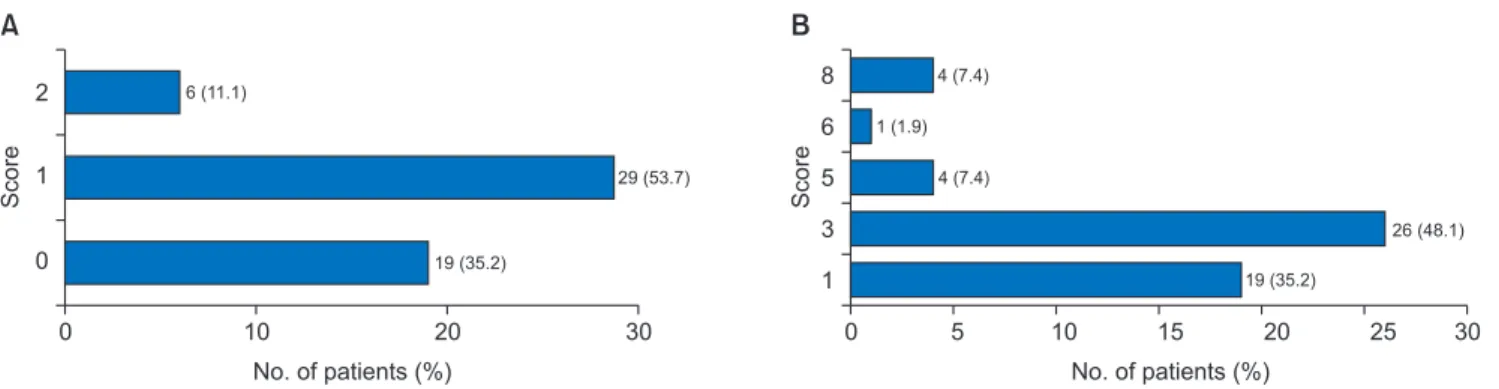
Using the first scoring system mentioned in Materials and Methods, 19 patients scored 0, 29 scored 1, and 6 patients scored 2 (Fig. 3. A), for a mean score of 0.76±0.64. Using the second scoring system, adopted from MUCONNS, the minimum value was 1 and the maximum was 8, with a mean score of 2.87±1.94.(Fig. 3. B)
In the statistical analysis, each systemic predisposing factor was assessed individually after eliminating other influencing factors. The 19 BRONJ patients with no systemic predispos- ing factors mentioned above were used as a control group.
Table 3. Systemic predisposing factors and prognosis after surgery
Normal recovery Delay on recovery Recurrence P-value Diabetes mellitus (n=27)
Steroid medication (n=37) Malignancy on other site (n=24)
Positive (n=8, 29.6%) Negative (n=19, 70.4%) Yes (n=18, 48.6%) No (n=19, 51.4%) Positive (n=5, 20.8%) Negative (n=19, 79.2%)
4 (50.0) 15 (78.9) 9 (50.0) 15 (78.9) 3 (60.0) 15 (78.9)
2 (25.0) 4 (21.1) 7 (38.9) 4 (21.1) 1 (20.0) 4 (21.1)
2 (25.0) 0 (0) 2 (11.1) 0 (0) 1 (20.0) 0 (0)
<0.05
<0.05
>0.05
Values are presented as number (%).
Tae-Hwan Kim et al: Evaluation of the predisposing factors and involved outcome of surgical treatment in bisphosphonate-related osteonecrosis of the jaw cases including bone biopsies. J Korean Assoc Oral Maxillofac Surg 2016
Table 4. Scoring systems and prognosis after surgery (%)
Mean ranking Normal recovery (n=32) Delay on recovery (n=16) Recurrence (n=6) P-value Scoring system 1
Scoring system 2 23.8
24.1 30.2
30.0 39.8
39.2 <0.05
<0.05
Tae-Hwan Kim et al: Evaluation of the predisposing factors and involved outcome of surgical treatment in bisphosphonate-related osteonecrosis of the jaw cases including bone biopsies. J Korean Assoc Oral Maxillofac Surg 2016
Fig. 3. A. Scoring system 1 calculated by adding all systemic predisposing factors. B. Scoring system 2 quotated from MUCONNS (Modified University of Connecticut Osteonecrosis Numerical Scale).
Tae-Hwan Kim et al: Evaluation of the predisposing factors and involved outcome of surgical treatment in bisphosphonate-related osteonecrosis of the jaw cases including bone biopsies. J Korean Assoc Oral Maxillofac Surg 2016
10 2
1
0
0 20
No. of patients (%) 6 (11.1)
19 (35.2)
30 29 (53.7)
Score
A
10 8
6 5 3 1
0 20
No. of patients (%) 4 (7.4)
1 (1.9)
19 (35.2)
26 (48.1)
30
5 15 25
4 (7.4)
Score
B
before surgery. Among the 28 subjects with a CTX value available, the range was 30 to 747 pg/mL (mean, 186±159 pg/mL). Marx et al.16 evaluated BRONJ severity according to CTX value prior to invasive dental surgery. Patients were categorized into three major groups: more than 150 pg/mL (low-risk group, n=15), 100 to 150 pg/mL (intermediate- risk group, n=6), and less than 100 pg/mL (high-risk group, n=7), according to the standard proposed by Marx et al.16. We found no significant difference in prognosis according to CTX value (P>0.05).(Table 6)
Microorganisms were detected in 24 of the 54 samples.
The presence of microorganisms was not associated with prognosis after surgery (P>0.05).(Table 7)
Bone biopsy results were typical of necrotic bone. Cell components and blood vessels were not found and the new bone formed around the osteocystic lacunae was destroyed.
The boundaries adjacent to necrotic bone sites were rough.
(Fig. 4. A)
IV. Discussion
From the pathological point of view, BPs are an analogue of pyrophosphonate and hinder bone resorption activity mediated by osteoclasts. BPs have a strong affinity for cal- cium and accumulate in the bone matrix. The half-life can be up to 12 years. When bone resorption occurs, BPs are separated from bone and adhere to newly formed bone or significantly correlated with the MUCONNS scores (the sec-
ond scoring system) (P<0.05).(Table 4)
A total of 47 patients received BP medication orally and seven received periodic intravenous administration of BP medication. The BP medication duration in the 36 patients for whom such information was available was 1 to 20 years (mean, 4.78±3.84 years). According to an AAOMS position paper published in 2007, treatment protocol differed between patients who received BP medication for less than and more than 3 years (number of patients, 21 and 15, respectively, in this study). In this study, medication was discontinued in 44 patients at a mean of 3.77±7.40 months. The AAOMS position paper set discontinuation at 3 months as a standard.
Thirty patients stopped their BP medication for less than 3 months and 14 patients for 3 months or more.
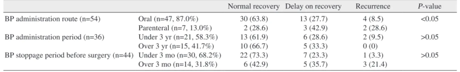
Patients who received intravenous BP medication had sig- nificantly worse prognosis after surgical management than those who took their medication orally (P<0.05). The BP medication duration in the 36 patients for whom such infor- mation was available was divided into less than and more than 3 years. There was no significant difference in prognosis between these two groups (P>0.05). In addition, the BP med- ication duration in the 36 patients for whom such informa- tion was available was divided into less than and more than 3 months. There was also no significant difference between these two groups (P>0.05).(Table 5)
The CTX value was determined after a morning fast 7 days
Table 5. Bisphosphonate (BP) administration and prognosis after surgery
Normal recovery Delay on recovery Recurrence P-value BP administration route (n=54)
BP administration period (n=36) BP stoppage period before surgery (n=44)
Oral (n=47, 87.0%) Parenteral (n=7, 13.0%) Under 3 yr (n=21, 58.3%) Over 3 yr (n=15, 41.7%) Under 3 mo (n=30, 68.2%) Over 3 mo (n=14, 31.8%)
30 (63.8) 2 (28.6) 13 (61.9) 10 (66.7) 22 (73.3) 6 (42.9)
13 (27.7) 3 (42.9) 6 (28.6) 5 (33.3) 7 (23.3) 5 (35.7)
4 (8.5) 2 (28.6) 2 (9.5) 0 (0) 1 (3.3) 3 (21.4)
<0.05
>0.05
>0.05
Values are presented as number (%).
Tae-Hwan Kim et al: Evaluation of the predisposing factors and involved outcome of surgical treatment in bisphosphonate-related osteonecrosis of the jaw cases including bone biopsies. J Korean Assoc Oral Maxillofac Surg 2016
Table 6. C-terminal cross-linking telopeptide (CTX) value and prognosis after surgery
CTX (pg/mL) (n=28) Normal
recovery Delay on
recovery Recurrence P-value
<100 (n=8, 28.6%) 100-150 (n=5, 17.9%)
>150 (n=15, 53.6%)
5 (62.5) 4 (80.0) 12 (80.0)
3 (37.5) 0 (0) 3 (20.0)
0 (0) 1 (20.0) 0 (0)
>0.05
Values are presented as number (%).
Tae-Hwan Kim et al: Evaluation of the predisposing factors and involved outcome of surgical treatment in bisphosphonate-related osteonecrosis of the jaw cases including bone biopsies. J Korean Assoc Oral Maxillofac Surg 2016
Table 7. Presence or absence of microorganisms and prognosis after surgery
Microorganism group (n=54)
Normal recovery
Delay on
recovery Recurrence P-value Presence (n=24, 44.4%)
Abscence (n=30, 55.6%) 12 (50.0)
20 (66.7) 9 (37.5)
7 (23.3) 3 (12.5)
3 (10.0) >0.05 Values are presented as number (%).
Tae-Hwan Kim et al: Evaluation of the predisposing factors and involved outcome of surgical treatment in bisphosphonate-related osteonecrosis of the jaw cases including bone biopsies. J Korean Assoc Oral Maxillofac Surg 2016
recovery after periodontal tissue destruction due to reduced immunity; this may lead to osteonecrosis27. Patients with BRONJ have a higher prevalence of diabetes mellitus than healthy persons. Therefore, diabetes mellitus is an important comorbidity with BRONJ. The pathogenesis of BRONJ in diabetes patients has been hypothesized. The cells of such pa- tients have lower glutathione levels, which induces oxidative stress. Accumulation of oxidative stress in tissues can lead to osteonecrosis28.
In this study, BRONJ patients with diabetes mellitus ex- hibited significantly greater rates of osteonecrosis recurrence and aggravation than those without diabetes mellitus, in agreement with previous reports9,10. However, diabetes type or HbA1c range, which can confirm blood glucose manage- ment, were not measured. The presence of diabetes mellitus was confirmed by interview during the investigation.
Steroid medication improves the prognosis of BRONJ sur- gical therapy, but there is a strong correlation between steroid treatment and osteonecrosis, particularly that caused by BPs.
However, previous studies focused on patients who took BP medication to treat or prevent malignancies such as breast cancer, prostate cancer, multiple myeloma, or bone metas- tasis. Treatment of these diseases generally includes oral administration of corticosteroids with intravenous BP medi- cation and chemotherapy as needed2. Malignancies influence the bone system irrespective of BP medication, and may undergo phagocytosis by osteoclasts. BPs can be divided into
two groups according to the presence of nitrogen. BP lack- ing nitrogen is metabolized by osteoclasts and transformed into adenosine triphosphate (ATP). This process hampers adenosine diphosphate (ADP)/ATP aminotransferase activ- ity, and triggers extinction of osteoclasts. Hence, the activity of osteoclasts, which resorb bones, is disturbed21. Nitrogen- containing BP (N-BP) resists bone resorption. In this case, osteoclasts resorb bone and N-BP, and exhibit cholesterol synthesis. During this process, farnesyl disphosphate is found in the mevalonate stage, and N-BP disturbs the synthesis of this enzyme, which hampers osteoclast activity. N-BP has a greater suppressive effect on bone resorption than BP1,23,24. In addition, if osteoclasts function normally, N-BP reduces the sites of bone metabolism. The pathogenesis of BRONJ is controversial because of the ambiguous effects of N-BP.
How much BP is metabolized and how much accumulates at membrane sites to influence wound treatment remains un- clear2.
We examined each patient’s systemic medical history to look for factors that affect the prognosis of surgical manage- ment of BRONJ. Diabetes mellitus increases the incidence of BRONJ due to its systemic effect. Osteonecrosis is caused by microvascular ischemia in bones, dysfunction of endothelial cells and decreased bone metabolism in patients with diabe- tes mellitus10,25,26. Patients with diabetes show delayed full
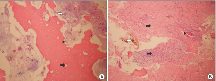
Fig. 4. A. Features about necrotic bone from biopsy specimen. The presence of cell components and blood vessels cannot be found and the formation of the new bones around the osteocystic lacunae are destroyed (black arrow). The boundaries adjacent to the necrotic bone sites are not smooth and show rugged feature in wavy aspects which is considered from loss of Howship’s lacunae (arrowhead), and there is microorganism colony around the necrotic bone (white arrow). B. Condition around soft tissue nearby the necrotic bone. It shows typical forms of granulation tissue which include distribution of inflammatory cells and fibrosis (black arrows). Although this specimen was col- lected a little far from the necrotic bone, we can find microorganism colony (white arrow), and there are also bone fragments considered broken down from necrotic bone (arrowhead).
Tae-Hwan Kim et al: Evaluation of the predisposing factors and involved outcome of surgical treatment in bisphosphonate-related osteonecrosis of the jaw cases including bone biopsies. J Korean Assoc Oral Maxillofac Surg 2016
A B
treatment has been considered as standard by many clinicians and led to the recommendation in the 2007 AAOMS posi- tion paper to discontinue BPs three months prior to surgery.
However, this approach was based only on experimental data, and BPs have a long half-life. The time of BP discon- tinuation before surgery was not significantly associated with prognosis after surgery in this study. These findings suggest that discontinuation of BP medication before BRONJ surgi- cal management is a non-optimal treatment protocol. Further studies are required to identify the optimum time at which BP medication should be discontinued in BRONJ patients2.
The carboxy-terminal collagen crosslinks, also known as CTX, is a telopeptide generated during bone resorption.
The C-terminal telopeptide is a metabolic product of bone resorption, mostly metabolized by collagen type 1. Serum CTX level is highly correlated with bone turnover rate and so is used to detect various diseases, as well as to predict the prognosis of osteoporosis, the risk of bone metastasis and di- agnose rheumatoid arthritis. In the serum test, the distribution of CTX is used as a biomarker of BRONJ severity16,32,33. The CTX value is affected by various factors, such as the age and sex of the patient, smoking, alcohol consumption, and un- derlying conditions such as diabetes mellitus, corticosteroid medication and fasting before the test.
Since Marx et al.16 reported CTX level as a biomarker of BRONJ severity, many studies have analyzed the association between CTX value and BRONJ. Several have reported that the CTX level not associated with BRONJ, in agreement with our findings34-36. Kunchur et al.35 reported that CTX value alone cannot determine the risk of osteonecrosis, because the jaw has 10-fold stronger metabolism than other parts of the skeleton and so changes in jaw metabolism do exert much influence on the CTX value. Berger et al.37 reported that the serum CTX level does not play a key role in diagnosing osteonecrosis due to differences in peripheral blood distribu- tion. Although this finding is based on necrosis in the thigh bones, not the jaw, it supports a reduced relevance for CTX in BRONJ.
Several studies have addressed the correlation between BRONJ and microorganisms. Ganguli et al.38 reported that hydroxyapatite covered with N-BP harbored a 60-fold greater number of bacteria than hydroxyapatite without N-BP. Kos et al.39 elucidated that this congregation of bacterial commu- nity is due to the ammonia in N-BP. Microorganisms were not detected in all biopsy specimens in this study, and may have been removed during decalcification for processing of the first biopsy sample. The presence of microorganisms was cause osteonecrosis irrespective of their effect on osteoclast
activity8. Chemotherapy decreases immunity and is involved in the development of osteonecrosis by provoking osteope- nia11,12.
Most individuals in this study were taking BP medication to prevent and treat osteoporosis. Thus, the results indicate the effect of steroid medication alone on the prognosis of BRONJ surgical management. Prognosis after surgery was not influenced by the presence of malignancies. Few patients with malignancies were included in this study, however, and further research is warranted.
Based on the scoring systems we applied, patients with more systemic disease factors and higher MUCONNS scores had worse prognosis after BRONJ surgical therapy. This finding suggests that prognosis can be predicted preopera- tively using systemic factors. Because the variables used were limited, we were unable to suggest a statistical process- ing standard such as a receiver-operating characteristic (ROC) curve. Therefore, further research should evaluate other fac- tors related to BRONJ.
Patients on a high dose of potent BP medication had a high incidence of BRONJ and aggravated BRONJ symptoms.
The AAOMS position paper in 2007 classified patients who had taken BP medication for more than 3 years as at high risk for BRONJ. However, other studies report no such cor- relation29,30. Kos et al.31 suggested a hypothesis regarding BRONJ stage classification: osteonecrosis may occur without bone exposure. If so, determining when osteonecrosis begins would be problematic. If bone exposure occurred in the ab- sence of clinical symptoms, it would be difficult to include such patients as subjects in studies of BRONJ. In this context, this hypothesis is strongly persuasive.
Of the patients, 58% had been taking BP medication for less than 3 years. They were assumed to be moderately safe in terms of BRONJ risk, but most were diagnosed with BRONJ stage II or III at the first medical examination. Thus, the majority of individuals were in progressive stages of os- teonecrosis, in agreement with the hypothesis of Kos et al.31. No significant correlation between BP medication duration and prognosis after surgery was identified. Therefore, BP medication duration was not predictive of the development of BRONJ or prognosis after surgery.
The prognosis according to time of BP discontinuation before surgery was determined in this study. Ruggiero et al.4 reported a positive treatment effect in patients who had dis- continued BP medication at least two months before surgery.
After that, the manual to cease BP medication before BRONJ
BP medication on osteoclasts and blood supply last. In such cases, the remnants of bacteria can act as a new medium and lead to recurrence of BRONJ, further complicating the deter- mination of prognosis after surgery.
In cases of purulent osteomyelitis not related to BPs, the inner part of the bone is filled with inflammatory cells. This condition indicates a natural immune reaction to resist bac- terial penetration and is not related to the osteoclast-killing effect of BPs45. The form of the osteonecrosis can be seen in the bone biopsies.(Fig. 4) Neither cell components for blood vessels were found, and osteocytes and new bone formed adjacent to the sites have disappeared. The type of osteone- crosis mentioned in this paper is highly related to BRONJ pathogenesis due to osteoclast extinction and decreased blood flow. The features of BRONJ described in this paper coincide with those reported by Marx and Tursun45. When BRONJ occurs, not only osteonecrosis, but also membrane exposure occurs. Bones provide blood flow to the periosteum and mucous membranes. Based on the bone biopsy results, BP medication decreases the blood flow in mucous membranes and the periosteum, leading directly to ischemic necrosis and ultimately bone exposure. BP medication could account for membrane exposure.
V. Conclusion
We reviewed 54 patients who were diagnosed with BRONJ and received surgical therapy. We evaluated whether differ- ent prognoses were associated with systemic medical history, the administration method of BP medication, BP medication duration and discontinuation duration, serum CTX value, and the presence of microorganism colonies during bone biopsy.
The histological features from necrotic bone samples were confirmed at osteonecrosis sites and surrounding tissue by bi-
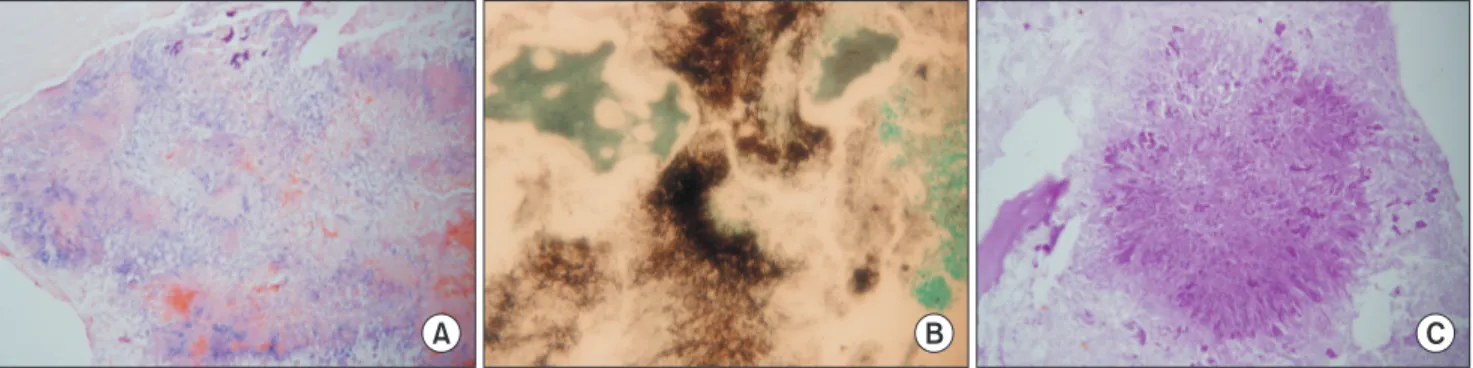
opsy of soft tissues.(Fig. 4. B) Therefore, even if the presence of microorganisms is not confirmed in a bone biopsy, micro- organisms adjacent to the osteonecrosis sites and surrounding tissue may cause clinical symptoms. To identify the micro- organisms, Gram, GMS and dPAS staining were performed and a filamentous form observed. This finding suggests Acti- nomyces species, and not a fungus.(Fig. 5)
The presence of Actinomyces species is correlated with BRONJ40,41. The anaerobic Gram-positive bacterium Actino- myces israelli causes actinomycosis in the mouth and is also found in the respiratory system and digestive system. It can- not pass through the oral mucosa, but physical pressure dur- ing mastication and iatrogenic stimulations such as exodontia or injuries from denture use may facilitate its penetration of the jaw, possibly leading to actinomycosis. Various Actino- myces species have been correlated with BRONJ. Naik and Russo42 reported that the Actinomyces species differ accord- ing to BRONJ stages and staining methods used to detect them. Types of bacteria other than Actinomyces related to BP in BRONJ cases have been investigated. Interactions between Actinomyces species and other bacteria, particularly strepto- cocci, have been reported43. Therefore, the presence of Acti- nomyces species can be used as an index of BRONJ clinical symptoms with infection caused by other bacteria44.
The presence of bacteria in BRONJ cases is accompanied by the following clinical symptoms. A. israelli is a resident of the mouth. Due to iatrogenic damage, A. israelli penetrates the submucosa and accumulates near the BP-affected jaw.
This leads to infection using osteonecrosis as a medium, which results in clinical symptoms of pain and infection8,45. So osteonecrosis of the jaw can be regenerated although once appropriate treatment is made if subsequent effects of
A B C
Fig. 5. A. Microorganism colony commonly appears not a capsular but a filamentous shape from each different stain (A: Gram staining,
×200, B: GMS staining, ×200, C: dPAS staining, ×200).
Tae-Hwan Kim et al: Evaluation of the predisposing factors and involved outcome of surgical treatment in bisphosphonate-related osteonecrosis of the jaw cases including bone biopsies. J Korean Assoc Oral Maxillofac Surg 2016
bone biopsies. New bone formation near the osteocytic lacu- nae sites was lost and destruction of the Howship’s lacunae occurred, which are typical forms of osteonecrosis. This type of necrotic bone is highly related to BRONJ pathogenesis be- cause of osteoclast extinction and decreased blood flow.
Based on these findings, prognosis after surgery can be pre- dicted by checking a patient’s medical history before BRONJ surgical treatment. Patients with diabetes, steroid medication usage or intravenous BP medication may be more likely to show delays in recovery after surgery or BRONJ recurrence.
Based on necrotic bone biopsies, typical changes in bone metabolism caused by BP medication were observed, and we confirmed that those changes caused osteonecrosis.
Conflict of Interest
No potential conflict of interest relevant to this article was reported.
ORCID
Tae-Hwan Kim, http://orcid.org/0000-0001-6226-2733 Won-Gyo Seo, http://orcid.org/0000-0002-6097-1322 Chul-Hong Koo, http://orcid.org/0000-0001-9904-0798 Jae-Hoon Lee, http://orcid.org/0000-0002-9959-4632
References
1. McClung MR. Bisphosphonates. Endocrinol Metab Clin North Am 2003;32:253-71.
2. Stanton DC, Balasanian E. Outcome of surgical management of bisphosphonate-related osteonecrosis of the jaws: review of 33 sur- gical cases. J Oral Maxillofac Surg 2009;67:943-50.
3. Marx RE. Pamidronate (Aredia) and zoledronate (Zometa) induced avascular necrosis of the jaws: a growing epidemic. J Oral Maxil- lofac Surg 2003;61:1115-7.
4. Ruggiero SL, Mehrotra B, Rosenberg TJ, Engroff SL. Osteonecro- sis of the jaws associated with the use of bisphosphonates: a review of 63 cases. J Oral Maxillofac Surg 2004;62:527-34.
5. Hinson AM, Smith CW, Siegel ER, Stack BC Jr. Is bisphospho- nate-related osteonecrosis of the jaw an infection? A histologi- cal and microbiological ten-year summary. Int J Dent 2014. doi:
10.1155/2014/452737.
6. Ohe JY, Kwon YD, Lee HW. Bisphosphonates modulate the ex- pression of OPG and M-CSF in hMSC-derived osteoblasts. Clin Oral Investig 2012;16:1153-9.
7. Kim RH, Lee RS, Williams D, Bae S, Woo J, Lieberman M, et al.
Bisphosphonates induce senescence in normal human oral kerati- nocytes. J Dent Res 2011;90:810-6.
8. Kos M, Brusco D, Kuebler J, Engelke W. Clinical comparison of patients with osteonecrosis of the jaws, with and without a his- tory of bisphosphonates administration. Int J Oral Maxillofac Surg 2010;39:1097-102.
9. Lee SH, Chang SS, Lee M, Chan RC, Lee CC. Risk of osteone- crosis in patients taking bisphosphonates for prevention of osteo-
also investigated. We found the following:
1. Most of the patients were women older than 60 years.
BRONJ occurred more than twice as much in the mandible as in the maxilla and most patients were BRONJ stage II or III in which symptoms could be detected. Osteonecrosis oc- curred most often after tooth extraction and osteoporosis was the main underlying indication for BP medication. Sauceriza- tion with sequestra removal was the most common surgical treatment of patients.
2. There were 32 cases (59.3%) of satisfactory recovery af- ter BRONJ surgery among a total of 54 cases. Delayed recov- ery for more than three months occurred in 16 cases (29.6%) and recurrence of BRONJ happened in 6 cases (11.1%) re- quiring re-operation.
3. Diabetes mellitus, steroid medication and malignancies at other sites were evaluated as factors from the systemic medical history. We confirmed that diabetes mellitus and ste- roid medication were correlated to the prognosis of surgery.
To sum up these factors and consider the weight of each fac- tor in the scoring system, a higher score was associated with worse prognosis.
4. Patients who took BP medication for less than 3 years made up 58% of the total. According to the AAOMS posi- tion paper in 2007, these patients should be segregated as a moderately safe group. However, most were at BRONJ stage II and III during the clinical examination, indicating that os- teonecrosis was already under way. Accordingly, the risk of BRONJ occurrence should not be assessed exclusively on BP medication duration.
5. BP administration method influenced prognosis after surgery. Patients with oral administration had better recovery, while BP medication duration and discontinuation duration before surgery were not statistically significant factors in prognosis.
6. CTX value was also not an independently absolute index to forecast prognosis.
7. Microorganism colonies were found during bone biopsy in 24 out of 54 cases (44.4%). The Actinomyces group was frequently observed. The presence of microorganism colo- nies in biopsy samples was not significantly associated with prognosis after surgery. It may be that the microorganisms were destroyed in the process of preparing the samples or that the microorganism groups were manifested on surrounding soft tissues rather than bone tissues. Clinical symptoms are expressed differently based on interactions between Actino- myces species and other bacteria.
8. Cell components and blood vessels were not found in
Endocrinol Metab 2007;92:817-8.
28. Ichiseki T, Ueda Y, Katsuda S, Kitamura K, Kaneuji A, Matsumoto T. Oxidative stress by glutathione depletion induces osteonecrosis in rats. Rheumatology (Oxford) 2006;45:287-90.
29. Ortega C, Montemurro F, Faggiuolo R, Vormola R, Nanni D, Goia F, et al. Osteonecrosis of the jaw in prostate cancer patients with bone metastases treated with zoledronate: a retrospective analysis. Acta Oncol 2007;46:664-8.
30. Vieillard MH, Maes JM, Penel G, Facon T, Magro L, Bonneterre J, et al. Thirteen cases of jaw osteonecrosis in patients on bisphos- phonate therapy. Joint Bone Spine 2008;75:34-40.
31. Kos M, Kuebler JF, Luczak K, Engelke W. Bisphosphonate-related osteonecrosis of the jaws: a review of 34 cases and evaluation of risk. J Craniomaxillofac Surg 2010;38:255-9.
32. Brown JE, Cook RJ, Major P, Lipton A, Saad F, Smith M, et al.
Bone turnover markers as predictors of skeletal complications in prostate cancer, lung cancer, and other solid tumors. J Natl Cancer Inst 2005;97:59-69.
33. Garnero P, Delmas PD. Noninvasive techniques for assessing skel- etal changes in inflammatory arthritis: bone biomarkers. Curr Opin Rheumatol 2004;16:428-34.
34. Fleisher KE, Welch G, Kottal S, Craig RG, Saxena D, Glickman RS. Predicting risk for bisphosphonate-related osteonecrosis of the jaws: CTX versus radiographic markers. Oral Surg Oral Med Oral Pathol Oral Radiol Endod 2010;110:509-16.
35. Kunchur R, Need A, Hughes T, Goss A. Clinical investigation of C-terminal cross-linking telopeptide test in prevention and man- agement of bisphosphonate-associated osteonecrosis of the jaws. J Oral Maxillofac Surg 2009;67:1167-73.
36. American Society for Bone and Mineral Research Task Force on Osteonecrosis of the Jaw, Khosla S, Burr D, Cauley J, Dempster DW, Ebeling PR, et al. Oral bisphosphonate-induced osteonecrosis:
risk factors, prediction of risk using serum CTX testing, preven- tion, and treatment. J Oral Maxillofac Surg 2008;66:1320-1; author reply 1321-2.
37. Berger CE, Kröner A, Kristen KH, Minai-Pour M, Leitha T, Engel A. Spontaneous osteonecrosis of the knee: biochemical mark- ers of bone turnover and pathohistology. Osteoarthritis Cartilage 2005;13:716-21.
38. Ganguli A, Steward C, Butler SL, Philips GJ, Meikle ST, Lloyd AW, et al. Bacterial adhesion to bisphosphonate coated hydroxy- apatite. J Mater Sci Mater Med 2005;16:283-7.
39. Kos M, Junka A, Smutnicka D, Bartoszewicz M, Kurzynowski T, Gluza K. Pamidronate enhances bacterial adhesion to bone hy- droxyapatite. Another puzzle in the pathology of bisphosphonate- related osteonecrosis of the jaw? J Oral Maxillofac Surg 2013;71:
1010-6.
40. Anavi-Lev K, Anavi Y, Chaushu G, Alon DM, Gal G, Kaplan I.
Bisphosphonate related osteonecrosis of the jaws: clinico-patholog- ical investigation and histomorphometric analysis. Oral Surg Oral Med Oral Pathol Oral Radiol 2013;115:660-6.
41. Hansen T, Kunkel M, Springer E, Walter C, Weber A, Siegel E, et al. Actinomycosis of the jaws--histopathological study of 45 pa- tients shows significant involvement in bisphosphonate-associated osteonecrosis and infected osteoradionecrosis. Virchows Arch 2007;451:1009-17.
42. Naik NH, Russo TA. Bisphosphonate-related osteonecrosis of the jaw: the role of actinomyces. Clin Infect Dis 2009;49:1729-32.
43. Kumar SK, Gorur A, Schaudinn C, Shuler CF, Costerton JW, Sedghizadeh PP. The role of microbial biofilms in osteonecrosis of the jaw associated with bisphosphonate therapy. Curr Osteoporos Rep 2010;8:40-8.
44. Sedghizadeh PP, Yooseph S, Fadrosh DW, Zeigler-Allen L, Thiaga- rajan M, Salek H, et al. Metagenomic investigation of microbes and viruses in patients with jaw osteonecrosis associated with bisphosphonate therapy. Oral Surg Oral Med Oral Pathol Oral Ra- porosis: a systematic review and meta-analysis. Osteoporos Int
2014;25:1131-9.
10. Khamaisi M, Regev E, Yarom N, Avni B, Leitersdorf E, Raz I, et al. Possible association between diabetes and bisphosphonate- related jaw osteonecrosis. J Clin Endocrinol Metab 2007;92:1172- 11. Sung EC, Chan SM, Sakurai K, Chung E. Osteonecrosis of the 5.
maxilla as a complication to chemotherapy: a case report. Spec Care Dentist 2002;22:142-6.
12. Tarassoff P, Csermak K. Avascular necrosis of the jaws: risk factors in metastatic cancer patients. J Oral Maxillofac Surg 2003;61:1238- 13. Migliorati CA, Schubert MM, Peterson DE, Seneda LM. Bisphos-9.
phonate-associated osteonecrosis of mandibular and maxillary bone: an emerging oral complication of supportive cancer therapy.
Cancer 2005;104:83-93.
14. Hewitt C, Farah CS. Bisphosphonate-related osteonecrosis of the jaws: a comprehensive review. J Oral Pathol Med 2007;36:319-28.
15. Leite AF, Figueiredo PT, Melo NS, Acevedo AC, Cavalcanti MG, Paula LM, et al. Bisphosphonate-associated osteonecrosis of the jaws. Report of a case and literature review. Oral Surg Oral Med Oral Pathol Oral Radiol Endod 2006;102:14-21.
16. Marx RE, Cillo JE Jr, Ulloa JJ. Oral bisphosphonate-induced osteo- necrosis: risk factors, prediction of risk using serum CTX testing, prevention, and treatment. J Oral Maxillofac Surg 2007;65:2397- 17. Freiberger JJ, Padilla-Burgos R, Chhoeu AH, Kraft KH, Boneta O, 410.
Moon RE, et al. Hyperbaric oxygen treatment and bisphosphonate- induced osteonecrosis of the jaw: a case series. J Oral Maxillofac Surg 2007;65:1321-7.
18. Vescovi P, Merigo E, Meleti M, Manfredi M. Bisphosphonate- associated osteonecrosis (BON) of the jaws: a possible treatment? J Oral Maxillofac Surg 2006;64:1460-2.
19. Lopes RN, Rabelo GD, Rocha AC, Carvalho PA, Alves FA. Surgi- cal therapy for bisphosphonate-related osteonecrosis of the jaw:
six-year experience of a single institution. J Oral Maxillofac Surg 2015;73:1288-95.
20. Stockmann P, Burger M, von Wilmowsky C, Ebker T, Lutz R, Bau- ersachs A, et al. The outcome after surgical therapy of bisphospho- nate-associated osteonecrosis of the jaw--results of a clinical case series with an average follow-up of 20 months. Clin Oral Investig 2014;18:1299-304.
21. Carlson ER, Basile JD. The role of surgical resection in the man- agement of bisphosphonate-related osteonecrosis of the jaws. J Oral Maxillofac Surg 2009;67(5 Suppl):85-95.
22. Reich W, Bilkenroth U, Schubert J, Wickenhauser C, Eckert AW.
Surgical treatment of bisphosphonate-associated osteonecrosis:
Prognostic score and long-term results. J Craniomaxillofac Surg 2015;43:1809-22.
23. Kim KW, Kim BJ, Lee CH. Clinical study of diagnosis and treat- ment of bisphosphonate-related osteonecrosis of the jaws. J Korean Assoc Oral Maxillofac Surg 2011;37:54-61.
24. Lehenkari PP, Kellinsalmi M, Näpänkangas JP, Ylitalo KV, Mönk- könen J, Rogers MJ, et al. Further insight into mechanism of action of clodronate: inhibition of mitochondrial ADP/ATP translocase by a nonhydrolyzable, adenine-containing metabolite. Mol Pharmacol 2002;61:1255-62.
25. Molcho S, Peer A, Berg T, Futerman B, Khamaisi M. Diabetes mi- crovascular disease and the risk for bisphosphonate-related osteo- necrosis of the jaw: a single center study. J Clin Endocrinol Metab 2013;98:E1807-12.
26. Watters AL, Hansen HJ, Williams T, Chou JF, Riedel E, Halpern J, et al. Intravenous bisphosphonate-related osteonecrosis of the jaw: long-term follow-up of 109 patients. Oral Surg Oral Med Oral Pathol Oral Radiol 2013;115:192-200.
27. Favus MJ. Diabetes and the risk of osteonecrosis of the jaw. J Clin
comparison and its implications for the mechanism of each disease.
Int J Oral Maxillofac Surg 2012;41:283-9.
diol 2012;114:764-70.
45. Marx RE, Tursun R. Suppurative osteomyelitis, bisphosphonate in- duced osteonecrosis, osteoradionecrosis: a blinded histopathologic