ORIGINAL ARTICLE
직장암에서 복강경과 개복 수술법의 장기 임상 결과의 비교: 단일 기관 후향적 연구
김재현, 안병권1, 박선자, 박무인, 김성은, 백승언1, 이승현1, 박시성2
고신대학교복음병원 소화기내과, 대장항문외과1, 정신과2
Long-term Outcomes of Laparoscopic versus Open Surgery for Rectal Cancer: A Single- center Retrospective Analysis
Jae Hyun Kim, Byung Kwon Ahn1, Seun Ja Park, Moo In Park, Sung Eun Kim, Sung Uhn Baek1, Seung Hyun Lee1, and Si Sung Park2 Departments of Gastroenterology, Colorectal Surgery1, and Neuropsychiatry2, Kosin University Gospel Hospital, Busan, Korea
Background/Aims: Laparoscopic surgery has been proven to be an effective alternative to open surgery in patients with colon cancer. However, data on laparoscopic surgery in patients with rectal cancer are insufficient. The aim of this study was to compare the long-term outcomes of laparoscopic and open surgery in patients with rectal cancer.
Methods: A total of 307 patients with rectal cancer who were treated by open and laparoscopic curative resection at Kosin University Gospel Hospital (Busan, Korea) between January 2002 and December 2011 were reviewed retrospectively.
Results: Regarding treatment, 176 patients underwent an open procedure and 131 patients underwent a laparoscopic procedure.
The local recurrence rate after laparoscopic resection was 2.3%, compared with 5.7% after open resection (p=0.088). Distant metastases occurred in 6.9% of the laparoscopic surgery group, compared with 24.4% in the open surgery group (p<0.001).
In univariate analysis, age (≥75 years vs. ≤60 years), preoperative staging, surgical approach (open vs. laparoscopic), elevated initial CEA level, elevated follow-up CEA level, number of positive lymph nodes, and postoperative chemotherapy affected overall survival and disease free survival. However, in multivariate analysis, the surgical approach apparently did not affect long-term oncologic outcome.
Conclusions: In this study, long-term outcomes after laparoscopic surgery for rectal cancer were not inferior to those after open surgery. Therefore, laparoscopic surgery would be an alternative operative tool to open resection for rectal cancer, although further investigation is needed. (Korean J Gastroenterol 2015;65:273-282)
Key Words: Rectal neoplasms; Conversion to open surgery; Laparoscopy
Received December 3, 2014. Revised March 7, 2015. Accepted March 22, 2015
CC This is an open access article distributed under the terms of the Creative Commons Attribution Non-Commercial License (http://creativecommons.org/licenses/
by-nc/4.0) which permits unrestricted non-commercial use, distribution, and reproduction in any medium, provided the original work is properly cited.
Copyright © 2015. Korean Society of Gastroenterology.
교신저자: 안병권, 602-702, 부산시 서구 감천로 262, 고신대학교 의과대학 외과학교실
Correspondence to: Byung Kwon Ahn, Department of Surgery, Kosin University College of Medicine, 262 Gamcheon-ro, Seo-gu, Busan 602-702, Korea. Tel: +82-51- 990-6462, Fax: +82-51-246-6093, E-mail: [email protected]
Financial support: None. Conflict of interest: None.
INTRODUCTION
Colorectal cancer is the third most commonly diagnosed cancer in males and the second in females worldwide.1 In the United States, an estimated 40,000 new cases of rectal can- cer will occur and an estimated 50,310 people will die from
rectal and colon cancer combined in 2014.2 In Eastern Asia, rectal cancer occurs in more than 16 per 100,000 individuals per year and accounts for more than 168,000 deaths per year.3 Over the last two decades, the introduction of total mesorectal excision and preoperative chemoradiation has led to improved local control and survival for patients with
rectal cancer.4,5 Several randomized controlled trials have confirmed that the long-term oncologic outcome of laparo- scopic resection is equivalent to that of open resection for co- lon cancer.6-9 The laparoscopic approach for colon cancer surgery has demonstrated earlier recovery of bowel function, less postoperative pain, and decreased hospital stay com- pared with open surgery.5,9-11 However, data regarding lapa- roscopic surgery in patients with rectal cancer are insuffi- cient. Several recent studies have reported on the short-term or long-term outcomes of laparoscopic surgery for rectal can- cer; these studies showed that laparoscopic surgery is fea- sible, with few complications and long-term outcomes similar to those for open surgery.12-15 Due to difficulties in pelvic ex- posure, rectal dissection, and sphincter preservation, lapa- roscopic surgery for rectal cancer is known to be technically more difficult than open surgery.
The aim of this study was to compare the long-term out- comes of laparoscopic surgery with those of open surgery in patients with rectal cancer, and to contribute to establishing the role of laparoscopic surgery in treatment of rectal cancer.
SUBJECTS AND METHODS
1. Patients
We began performing laparoscopic surgery for rectal can- cer in 2002. The analysis included 307 patients who under- went resection for rectal cancer between January 2002 and December 2011 at Kosin University Gospel Hospital (Busan, Korea). Rectal cancer was defined as a cancer that forms in the tissues of the rectum (located 15 cm or less from the anal verge on rigid sigmoidoscopy) according to the National Can- cer Institute definition.16 The location of the tumor was cate- gorized as lower (less than 5 cm from anal verge), middle (6-10 cm from anal verge), or upper rectum (11-15 cm from anal verge). For preoperative staging, patients underwent a full pre-operative workup including a colonoscopy, as well as flexible sigmoidoscopy and abdominal CT with additional MRI or ano-rectal ultrasound. PET-CT was also performed af- ter September 2004. Patients with stage IV rectal cancer were excluded. The medical records of all patients were re- viewed retrospectively, including patient characteristics, sur- gical procedures, pathologic findings, and long-term follow- up data. This study was approved by the institutional review board of Kosin University College of Medicine (KUGH IRB No.
14-104).
2. Treatment
Among patients with T3N0 or greater TNM preoperative staging, neoadjuvant concurrent chemoradiotherapy (CCRT) was selected by decision of a multidisciplinary team confer- ence. Radiotherapy consisted of 56.0 Gy total in 28 fractions (200 cGy daily, Monday-Friday), delivered with an energy of 10 MV photons via a three-field box technique to the primary tumor and to the mesorectal, presacral, and internal iliac lymph nodes. CCRT consisted of a continuous infusion of 5-fluorouracil (425 mg/m2) plus leucovorin (20 mg/m2) on days 1-5 and 29-33 of radiotherapy, and one or two rounds of chemotherapy were added after CCRT. At six to ten weeks after CCRT, patients were re-staged with CT and proceeded to surgery.
Both open and laparoscopic surgery for rectal cancer were performed by three experienced colorectal surgeons (A.B.K., B.S.U., and L.S.H.). Selection of the surgical approach was de- cided by the surgeon and patient with full information regard- ing the procedures, including the possibility of morbidity. The same oncologic guidelines were followed in performance of all procedures in both groups: adequate resection margins,
‘en bloc’ vascular resection and lymphadenectomy and mini- mal intraoperative manipulation of the tumor. Patients with upper rectal tumors underwent partial mesorectal excision with a 5-cm gross tumor margin from the inferior pole of the tumor. Patients with middle and lower rectal tumors under- went total mesorectal excision. Our first laparoscopic surgery was performed in 2002, and the frequencies of these proce- dures increased each year. Laparoscopic resection was per- formed using the 5-trocar technique with high ligation of the inferior mesenteric artery, medial to lateral mobilization of the left colon and splenic flexure, rectal transection with a laparoscopic stapler, and double-stapled anastomosis. Pro- tective ileostomy was rarely performed. Patients who under- went surgery using an initial laparoscopic approach, which was then converted to open surgery, belonged to the open surgery group. Conversion to open surgery was performed in most of these patients, as an abdominal incision larger than that necessary for specimen retrieval was created.
After surgery, patients with pathologic T3N0 or greater TNM staging received additional chemotherapy consisting of oral folinic acid, oral capecitabine, intravenous 5-fluorouracil
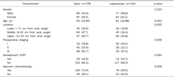
Table 1. Patient Characteristics
Characteristic Open (n=176) Laparoscopy (n=131) p-value
Gender 0.153
Male 89 (50.6) 77 (58.8)
Female 87 (49.4) 54 (41.2)
Age (yr) 59 (22-85) 61 (33-88) 0.252
Location 0.002
Lower (≤5 cm from anal verge) 45 (25.6) 26 (19.8)
Middle (6-10 cm from anal verge) 84 (47.7) 45 (34.4)
Upper (11-15 cm from anal verge) 47 (26.7) 60 (45.8)
Preoperative staging 0.056
I 33 (18.8) 40 (30.5)
II 45 (25.6) 29 (22.1)
III 98 (55.7) 62 (47.3)
Neoadjuvant CCRT 0.293
Yes 26 (14.8) 14 (10.7)
No 150 (85.2) 117 (89.3)
Adjuvant chemotherapy 0.008
Yes 130 (73.9) 78 (59.5)
No 46 (26.1) 53 (40.5)
Values are presented as n (%) or mean (range).
CCRT, concurrent chemoradiotherapy.
plus leucovorin, or FOLFOX regimen (combination of oxali- platin, 5-fluorouracil, and leucovorin). However, postopera- tive radiotherapy was rarely performed.
3. Follow-up
After the operation, patients were seen at 3-month inter- vals for the first 2 years, then at 6-month intervals for the next 3 years, and after 5 years, annually at an outpatient clinic.
Follow-up examination included CEA measurement per three months, chest X-ray and abdominal CT per six months during the first 2 years, and annually thereafter. CEA is routinely de- tected as a tumor biomarker and an auxiliary indicator for the preoperative diagnosis of colorectal cancer, as well as an ear- ly predictor of recurrence. Elevated initial CEA level refers to higher than normal range at the time of diagnosis, and ele- vated follow-up CEA level refers to elevation of CEA level dur- ing the follow-up period. Colonoscopy and PET-CT were also performed annually. Recurrence was diagnosed by endo- scopic biopsy, surgical resection, and/or radiological imag- ing study. Local recurrence was defined as any recurrence within the pelvic cavity, and distant recurrence was defined as any recurrence outside the pelvic cavity.
4. Statistical analysis
Student’s t-test and chi-square test for continuous and cat- egorical variables, as appropriate, were performed to de-
termine significant differences between open and laparo- scopic resection. Kaplan-Meier method was used for estima- tion of the overall survival (OS) and disease-free survival (DFS). The OS was measured from the date of diagnosis of rectal cancer to the date of death or of the final follow-up. DFS was measured from the date of diagnosis of rectal cancer to the date of disease progression or of the final follow-up. The log-rank test was used for comparison of time-to-event dis- tributions; the Cox proportional-hazards regression model was used for univariate and multivariate models. p-values lower than 0.05 were considered to indicate statistical signi- ficance. Statistical analysis was performed using IBM SPSS Statistics version 20.0 (IBM Co., Armonk, NY, USA).
RESULTS
1. Patient characteristics
Between January 2002 and December 2011, a total of 307 patients underwent surgery for rectal cancer; 176 pa- tients underwent open surgery (open surgery group), and 131 patients underwent laparoscopic surgery (laparoscopic surgery group). Baseline characteristics of both groups are summarized in Table 1. There were no significant differences in sex, age, or tumor stage between the two groups, but tumor locations differed significantly.
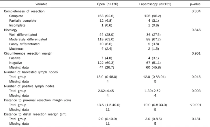
Table 2. Pathological Data
Variable Open (n=176) Laparoscopy (n=131) p-value
Completeness of resection 0.304
Complete 163 (92.6) 126 (96.2)
Partially complete 12 (6.8) 4 (3.1)
Incomplete 1 (0.6) 1 (0.8)
Histology 0.846
Well differentiated 44 (28.0) 36 (27.5)
Moderately differentiated 118 (63.0) 88 (67.2)
Poorly differentiated 10 (6.6) 5 (3.8)
Mucinous 4 (2.4) 2 (1.5)
Circumference resection margin 0.951
Positive 7 (4.0) 4 (3.1)
Negative 122 (69.3) 67 (51.1)
Missing data 47 (26.7) 60 (45.8)
Number of harvested lymph nodes
Total group 13.0 (0-48.0) 12.0 (0-83.04) 0.946
Missing data 4 5
Number of positive lymph nodes
Total group 2.62±4.45 1.39±2.52 0.003
Missing data 4 4
Distance to proximal resection margin (cm)
Total group 13.5 (1.5-40.0) 10.0 (0.8-33.0) <0.001
Missing data 11 5
Distance to distal resection margin (cm)
Total group 2.0 (0-10.0) 3.0 (0-8.5) 0.181
Missing data 11 5
Values are presented as n (%), median (range), n only, or mean±SD.
2. Pathological data
Macroscopically incomplete resected specimens were re- corded in 4 of 131 patients (3.1%) after laparoscopic surgery and 12 of 176 patients (6.8%) after open surgery. No sig- nificant difference in tumor histology was observed between the two groups. The proportion of patients with a positive cir- cumferential resection margin was 4.0% in the laparoscopic surgery group and 3.1% in the open surgery group (p=0.951).
The median number of lymph nodes harvested after surgery was not significantly different in the two groups (p=0.946);
however, the mean number of positive lymph nodes after open surgery was significantly higher than that after laparo- scopic surgery (p=0.003). The median proximal resection margin was 10 cm after laparoscopic surgery and 13.5 cm af- ter open surgery; the distal resection margin was 3.0 cm after laparoscopic surgery and 2.0 cm after open surgery (Table 2).
3. Postoperative recurrence rates
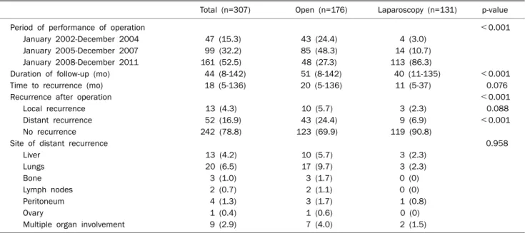
Open surgery was performed more often than laparo- scopic surgery from 2002 to 2007, whereas between 2008
and 2011, laparoscopic surgery was performed more often than open surgery. The overall median time of follow-up peri- od was 44 months (8-142 months), and 51 months (8-142 months) and 40 months (11-135 months) for the open sur- gery group and laparoscopic surgery group, respectively. The overall median time to recurrence was 18 months (5-136 months), and 11 months (5-37 months) and 21 months (5-136 months) for the open surgery group and laparoscopic surgery group, respectively (Table 3).
The recurrence rate for the open surgery group was 30.1%, and that of the laparoscopic surgery group was 9.2%. In the open surgery group, 10 patients (5.7%) had a local recur- rence and 43 patients (24.4%) had distant recurrence. In the laparoscopic surgery group, 3 patients (2.3%) had a local re- currence and 9 patients (6.9%) had distant recurrence. A statistically significant difference was observed between the two groups (p<0.001). The sites of distant recurrence in both groups are summarized in Table 3.
4. Overall survival and disease free survival
The OS of the open surgery group and laparoscopic surgery
Table 3. Postoperative Recurrence according to Surgical Procedure
Total (n=307) Open (n=176) Laparoscopy (n=131) p-value
Period of performance of operation <0.001
January 2002-December 2004 47 (15.3) 43 (24.4) 4 (3.0)
January 2005-December 2007 99 (32.2) 85 (48.3) 14 (10.7)
January 2008-December 2011 161 (52.5) 48 (27.3) 113 (86.3)
Duration of follow-up (mo) 44 (8-142) 51 (8-142) 40 (11-135) <0.001
Time to recurrence (mo) 18 (5-136) 20 (5-136) 11 (5-37) 0.076
Recurrence after operation <0.001
Local recurrence 13 (4.3) 10 (5.7) 3 (2.3) 0.088
Distant recurrence 52 (16.9) 43 (24.4) 9 (6.9) <0.001
No recurrence 242 (78.8) 123 (69.9) 119 (90.8)
Site of distant recurrence 0.958
Liver 13 (4.2) 10 (5.7) 3 (2.3)
Lungs 20 (6.5) 17 (9.7) 3 (2.3)
Bone 3 (1.0) 3 (1.7) 0 (0)
Lymph nodes 2 (0.7) 2 (1.1) 0 (0)
Peritoneum 4 (1.3) 3 (1.7) 1 (0.8)
Ovary 1 (0.4) 1 (0.6) 0 (0)
Multiple organ involvement 9 (2.9) 7 (4.0) 2 (1.5)
Values are presented as n (%) or median (range).
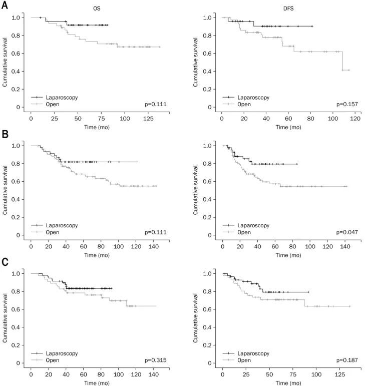
group at 5 years was 72.5% and 84.8%, respectively. Statisti- cally significant OS and DFS according to preoperative stag- ing between the two groups were observed only in patients with stage 3; however, statistical significance was not ob- served in patients with stage 1 and stage 2 (Fig. 1). In addi- tion, excluding DFS in patients with middle rectal tumor, the OS and DFS according to tumor location between the two groups were not statistically significant (Fig. 2).
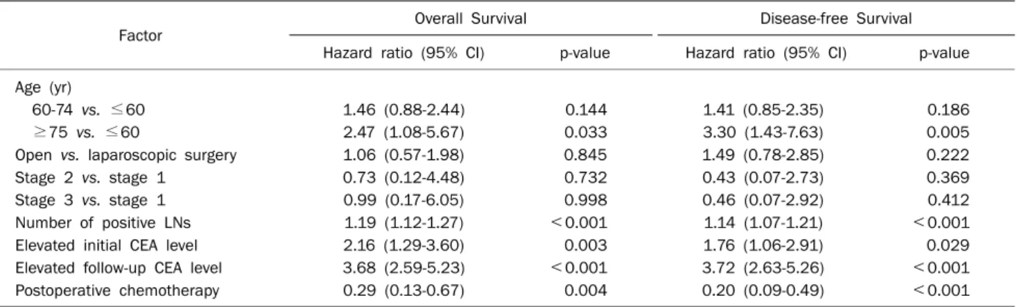
Cox regression analysis was performed for identification of prognostic factors of OS and DFS. Results of univariate analy- sis of factors of OS and DFS are shown in Table 4. In multi- variate analysis, the prognostic factors affecting OS were age (≥75 years vs. ≤60 years, hazard ratio [HR] 2.47, 95% CI 1.08-5.67, p=0.033), elevated initial CEA level (HR 2.16, 95% CI 1.29-3.60, p=0.003), elevated follow-up CEA level (HR 3.68, 95% CI 2.59-5.23, p<0.001), number of positive lymph nodes (HR 1.19, 95% CI 1.12-1.27, p<0.001), and postoperative chemotherapy (HR 0.29, 95% CI 0.13-0.67, p=0.004). The prognostic factors affecting DFS were age (≥75 years vs. ≤60 years, HR 3.30, 95% CI 1.43-7.63, p=0.005), elevated initial CEA level (HR 1.76, 95% CI 1.06-2.91, p=0.029), elevated follow-up CEA level (HR 3.72, 95% CI 2.63-5.26, p<0.001), number of positive lymph no- des (HR 1.14, 95% CI 1.07-1.21, p<0.001), and post- operative chemotherapy (HR 0.20, 95% CI 0.09-0.49, p
<0.001) (Table 5).
DISCUSSION
Results of this study shows that long-term outcomes after laparoscopic resection are better than those after conven- tional open resection for rectal cancer. The COREAN trial, which compared open and laparoscopic surgery for middle or lower rectal cancer after neoadjuvant chemotherapy, showed that laparoscopic resection for locally advanced rec- tal cancer after preoperative chemoradiotherapy provides similar outcomes with respect to DFS as open resection.16 As reported in the COLOR II trial, in selected patients treated by skilled surgeons, laparoscopic surgery provided safety, re- section margins, and completeness of resection similar to those of open surgery, and in-hospital recovery time was de- creased after laparoscopic surgery.17 In this study, sig- nificantly higher OS was observed for the laparoscopic sur- gery group compared with the open surgery group (p=0.012), and the DFS was also significantly higher in the laparoscopic surgery group compared with the open surgery group (p=0.005).
Patients with rectal cancer who underwent laparoscopic sur- gery also had lower recurrence rates than those who under- went open surgery (p<0.001). In our hospital, laparoscopic surgery was performed mainly after January 2008 (Table 3), and the mean follow-up duration of the laparoscopic surgery group was shorter than that of the open surgery group (41 months vs. 56 months, p<0.001). This may be a function of
Fig. 1. Comparison of overall survival (OS) and disease-free survival (DFS) according to preoperative staging between open surgery group and laparoscopic surgery group. (A) Stage 1. (B) Stage 2. (C) Stage 3.
the lower recurrence rates and better OS or DFS of the laparo- scopic surgery group compared with those of the open sur- gery group.
In this study, we evaluated long-term outcomes according to preoperative tumor stage. da Luz Moreira et al.12 reported on the recurrence rates of a laparoscopic surgery group and
an open surgery group; they found that rates for stage I were 3.7% (1/27) vs. 6.3% (2/32); for stage II, 29.4% (5/17) vs.
19.0% (4/21); and for stage III, 17.9% (5/28) vs. 33.3% (6/
18). Ng et al.18 reported a recurrence rate of 20.0% (8/40) for a laparoscopic surgery group and 25.0% (9/36) for an open surgery group. In the current study, the OS and DFS ac-
Fig. 2. Comparison of overall survival (OS) and disease-free survival (DFS) according to tumor location between open surgery group and laparoscopic surgery group. (A) Upper (11-15 cm from anal verge). (B) Middle (6-10 cm from anal verge). (C) Lower (≤5 cm from anal verge).
cording to preoperative staging between the two groups was not statistically significant in patients with stage 1 and stage 2; however, the OS and DFS of the laparoscopic surgery group were better than those of the open surgery group in patients with stage 3 (Fig. 1). These results are attributed to a larger number of positive lymph nodes in the open surgery group
compared with the laparoscopic surgery group (2.62±4.45 vs. 1.39±2.52, p=0.003; Table 2). We also evaluated re- currence rates according to tumor location in the rectum. A distal rectal tumor may easily metastasize initially to the lungs because the inferior rectal vein drains into the inferior vena cava bypassing the portal venous system.19 Tarantino
Table 4. Univariate Analysis of Overall Survival and Disease-free Survival
Factor Overall Survival Disease-free Survival
Hazard ratio (95% CI) p-value Hazard ratio (95% CI) p-value Age (yr)
60-74 vs. ≤60 1.46 (0.88-2.44) 0.144 1.51 (0.94-2.44) 0.091
≥75 vs. ≤60 2.47 (1.08-5.67) 0.033 3.79 (1.85-7.77) <0.001
Male vs. female 1.21 (0.78-1.89) 0.404 1.31 (0.84-2.04) 0.232
Open vs. laparoscopic surgery 1.94 (1.14-3.28) 0.014 2.09 (1.24-3.52) 0.006
Tumor location
Middle vs. lower 1.62 (0.89-2.92) 0.114 1.60 (0.89-2.90) 0.120
Upper vs. lower 1.08 (0.57-2.08) 0.807 1.04 (0.54-1.99) 0.910
Stage 2 vs. stage 1 3.29 (1.22-8.93) 0.019 3.45 (1.27-9.36) 0.015
Stage 3 vs. stage 1 6.19 (2.48-15.45) <0.001 5.59 (2.24-13.97) <0.001
Histology (well/moderate vs. poorly/mucinous) 0.94 (0.38-2.32) 0.887 0.85 (0.34-2.11) 0.727 Completeness of resection (R1/R2 vs. R0) 2.00 (0.96-4.16) 0.063 1.98 (0.95-4.12) 0.067
Positive CRM 0.86 (0.67-1.10) 0.234 0.83 (0.64-1.06) 0.138
Number of harvested LNs 0.99 (0.97-1.02) 0.787 1.00 (0.97-1.03) 0.983
Number of positive LNs 1.17 (1.12-1.23) <0.001 1.15 (1.11-1.20) <0.001
Distance to proximal RM 0.99 (0.96-1.02) 0.558 0.99 (0.96-1.02) 0.622
Distance to distal RM 1.01 (0.89-1.14) 0.861 0.99 (0.88-1.12) 0.882
Elevated initial CEA level 3.12 (1.97-4.95) <0.001 2.91 (1.84-4.60) <0.001
Elevated follow-up CEA level 3.46 (2.59-4.61) <0.001 3.69 (2.76-4.93) <0.001
Preoperative CCRT 0.82 (0.44-1.51) 0.519 0.93 (0.50-1.72) 0.810
Postoperative chemotherapy 0.18 (0.08-0.39) <0.001 0.19 (0.09-0.41) <0.001
CRM, circumferential resection margin; LNs, lymph nodes; RM, resection margin; CCRT, concurrent chemoradiotherapy.
Table 5. Multivariate Analysis of Overall Survival and Disease-free Survival
Factor Overall Survival Disease-free Survival
Hazard ratio (95% CI) p-value Hazard ratio (95% CI) p-value Age (yr)
60-74 vs. ≤60 1.46 (0.88-2.44) 0.144 1.41 (0.85-2.35) 0.186
≥75 vs. ≤60 2.47 (1.08-5.67) 0.033 3.30 (1.43-7.63) 0.005
Open vs. laparoscopic surgery 1.06 (0.57-1.98) 0.845 1.49 (0.78-2.85) 0.222
Stage 2 vs. stage 1 0.73 (0.12-4.48) 0.732 0.43 (0.07-2.73) 0.369
Stage 3 vs. stage 1 0.99 (0.17-6.05) 0.998 0.46 (0.07-2.92) 0.412
Number of positive LNs 1.19 (1.12-1.27) <0.001 1.14 (1.07-1.21) <0.001
Elevated initial CEA level 2.16 (1.29-3.60) 0.003 1.76 (1.06-2.91) 0.029
Elevated follow-up CEA level 3.68 (2.59-5.23) <0.001 3.72 (2.63-5.26) <0.001
Postoperative chemotherapy 0.29 (0.13-0.67) 0.004 0.20 (0.09-0.49) <0.001
LNs, lymph nodes.
et al.20 reported that the distance of tumor from anal verge (<5 cm) is one of the predictors for poor OS (HR 1.93, 95%
CI 1.11-3.37, p=0.039). On the other hand, Das et al.21 found that a greater distance from anal verge (>5 cm) indepen- dently predicted a lower downstaging rate in patients who re- ceived preoperative chemoradiation for rectal cancer. In this study, excluding DFS in patients with middle rectal tumor, the OS and DFS according to tumor location between two groups was not statistically significant (Fig. 2). To date, long-term out- comes according to tumor location in the rectum remain con-
troversial; therefore further studies are needed.
OS rates ranging from 62.8% to 91.0% following laparo- scopic rectal resection have been reported.18,22-25 In this study, the 5-year OS of the open surgery group and of the lapa- roscopic surgery group were 72.5% and 84.8%, respectively.
The independent predictors of OS were old age (≥75 years), elevated initial CEA level, elevated follow-up CEA level, num- ber of positive lymph nodes, and postoperative chemother- apy. These factors were also included among independent predictors of DFS (Table 5). According to some authors, the
prognosis for colorectal cancer in the elderly is not sig- nificantly different from that of younger patients.26-29 Howev- er, the relation between age and outcomes from colorectal cancer surgery is complex and may be confounded by differ- ences in stage at presentation, tumor location, pre-existing comorbidities, and type of treatment received.28 Results of univariate Cox regression analysis showed that long-term on- cologic outcome was better in the laparoscopic surgery group compared with the open surgery group (Table 4); however, in multivariate Cox regression analysis, the surgical approach (laparoscopic vs. open) apparently did not affect long-term oncologic outcome (Table 5). Based on these results, pa- tients with old age, elevated initial CEA level, elevated fol- low-up CEA level, larger number of positive lymph nodes, or without preoperative chemotherapy require more careful fol- low-up observation after surgery than other patients.
Data from this study indicate that long-term outcomes af- ter laparoscopic surgery are not inferior to those after open surgery for rectal cancer. Although this study was a retro- spective, single-center study, our results suggest that laparo- scopic surgery would be a valid alternative operative tool to open surgery for rectal cancer. Further randomized trials evaluating the outcomes of laparoscopic surgery for rectal cancer will be needed to enable clinical acceptance of laparo- scopic surgery for treatment of rectal cancer.
REFERENCES
1. Jemal A, Bray F, Center MM, Ferlay J, Ward E, Forman D. Global cancer statistics. CA Cancer J Clin 2011;61:69-90.
2. Siegel R, Ma J, Zou Z, Jemal A. Cancer statistics, 2014. CA Cancer J Clin 2014;64:9-29.
3. Ferlay J, Shin HR, Bray F, Forman D, Mathers C, Parkin DM.
Estimates of worldwide burden of cancer in 2008: GLOBOCAN 2008. Int J Cancer 2010;127:2893-2917.
4. Kapiteijn E, Kranenbarg EK, Steup WH, et al. Total mesorectal excision (TME) with or without preoperative radiotherapy in the treatment of primary rectal cancer. Prospective randomised tri- al with standard operative and histopathological techniques.
Dutch ColoRectal Cancer Group. Eur J Surg 1999;165:410-420.
5. Sauer R, Becker H, Hohenberger W, et al; German Rectal Cancer Study Group. Preoperative versus postoperative chemoradio- therapy for rectal cancer. N Engl J Med 2004;351:1731-1740.
6. Clinical Outcomes of Surgical Therapy Study Group. A compar- ison of laparoscopically assisted and open colectomy for colon cancer. N Engl J Med 2004;350:2050-2059.
7. Buunen M, Veldkamp R, Hop WC, et al; Colon Cancer Laparo- scopic or Open Resection Study Group. Survival after laparo- scopic surgery versus open surgery for colon cancer: long-term
outcome of a randomised clinical trial. Lancet Oncol 2009;10:
44-52.
8. Jayne DG, Guillou PJ, Thorpe H, et al; UK MRC CLASICC Trial Group. Randomized trial of laparoscopic-assisted resection of colorectal carcinoma: 3-year results of the UK MRC CLASICC Trial Group. J Clin Oncol 2007;25:3061-3068.
9. Lacy AM, García-Valdecasas JC, Delgado S, et al. Laparoscopy- assisted colectomy versus open colectomy for treatment of non- metastatic colon cancer: a randomised trial. Lancet 2002;359:
2224-2229.
10. Delaney CP, Chang E, Senagore AJ, Broder M. Clinical outcomes and resource utilization associated with laparoscopic and open colectomy using a large national database. Ann Surg 2008;247:
819-824.
11. Kemp JA, Finlayson SR. Outcomes of laparoscopic and open co- lectomy: a national population-based comparison. Surg Innov 2008;15:277-283.
12. da Luz Moreira A, Mor I, Geisler DP, Remzi FH, Kiran RP. Laparos- copic resection for rectal cancer: a case-matched study. Surg Endosc 2011;25:278-283.
13. Lujan J, Valero G, Biondo S, Espin E, Parrilla P, Ortiz H. Laparos- copic versus open surgery for rectal cancer: results of a pro- spective multicentre analysis of 4,970 patients. Surg Endosc 2013;27:295-302.
14. Ng SS, Lee JF, Yiu RY, et al. Laparoscopic-assisted versus open total mesorectal excision with anal sphincter preservation for mid and low rectal cancer: a prospective, randomized trial. Surg Endosc 2014;28:297-306.
15. Seshadri RA, Srinivasan A, Tapkire R, Swaminathan R. Laparos- copic versus open surgery for rectal cancer after neoadjuvant chemoradiation: a matched case-control study of short-term outcomes. Surg Endosc 2012;26:154-161.
16. Jeong SY, Park JW, Nam BH, et al. Open versus laparoscopic sur- gery for mid-rectal or low-rectal cancer after neoadjuvant che- moradiotherapy (COREAN trial): survival outcomes of an open- label, non-inferiority, randomised controlled trial. Lancet Oncol 2014;15:767-774.
17. van der Pas MH, Haglind E, Cuesta MA, et al; COlorectal cancer Laparoscopic or Open Resection II (COLOR II) Study Group. Lapar- oscopic versus open surgery for rectal cancer (COLOR II): short- term outcomes of a randomised, phase 3 trial. Lancet Oncol 2013;14:210-218.
18. Ng SS, Leung KL, Lee JF, et al. Laparoscopic-assisted versus open abdominoperineal resection for low rectal cancer: a pro- spective randomized trial. Ann Surg Oncol 2008;15:2418-2425.
19. Chiang JM, Hsieh PS, Chen JS, Tang R, You JF, Yeh CY. Rectal cancer level significantly affects rates and patterns of distant metastases among rectal cancer patients post curative-intent surgery with- out neoadjuvant therapy. World J Surg Oncol 2014;12:197.
20. Tarantino I, Warschkow R, Worni M, et al. Elevated preoperative CEA is associated with worse survival in stage I-III rectal cancer patients. Br J Cancer 2012;107:266-274.
21. Das P, Skibber JM, Rodriguez-Bigas MA, et al. Predictors of tumor response and downstaging in patients who receive preoperative chemoradiation for rectal cancer. Cancer 2007;109:1750-1755.
22. Baik SH, Gincherman M, Mutch MG, Birnbaum EH, Fleshman
JW. Laparoscopic vs open resection for patients with rectal can- cer: comparison of perioperative outcomes and long-term sur- vival. Dis Colon Rectum 2011;54:6-14.
23. Braga M, Frasson M, Vignali A, Zuliani W, Capretti G, Di Carlo V.
Laparoscopic resection in rectal cancer patients: outcome and cost-benefit analysis. Dis Colon Rectum 2007;50:464-471.
24. Laurent C, Leblanc F, Wütrich P, Scheffler M, Rullier E. Laparos- copic versus open surgery for rectal cancer: long-term oncologic results. Ann Surg 2009;250:54-61.
25. Lujan J, Valero G, Hernandez Q, Sanchez A, Frutos MD, Parrilla P. Randomized clinical trial comparing laparoscopic and open surgery in patients with rectal cancer. Br J Surg 2009;96:982- 989.
26. Irvin TT. Prognosis of colorectal cancer in the elderly. Br J Surg 1988;75:419-421.
27. Mulcahy HE, Patchett SE, Daly L, O'Donoghue DP. Prognosis of elderly patients with large bowel cancer. Br J Surg 1994;81:736- 738.
28. Colorectal Cancer Collaborative Group. Surgery for colorectal cancer in elderly patients: a systematic review. Lancet 2000;
356:968-974.
29. Vironen JH, Sainio P, Husa AI, Kellokumpu IH. Complications and survival after surgery for rectal cancer in patients younger than and aged 75 years or older. Dis Colon Rectum 2004;47:1225- 1231.