⋅교신저자: 임윤경. 대전광역시 동구 용운동 대전대학교 한의과대 학 경락경혈학교실. Tel. 042-280-2610, Fax.
042-280-2641, E-mail : [email protected]
․ 투고 : 2009/02/28 심사 : 2009/03/17 채택 : 2009/03/20
음곡에 시술한 구맥 약침이 LPS로 유도된 흰쥐의 신장염에 미치는 영향
김경민1․이현2․강희정3․이용흠4․임윤경1
대전대학교 한의과대학4 1경락경혈학교실, 2침구학교실, 3대요메디(주)
연세대학교 보건과학대학 의공학과 한의공학연구실,
Anti-inflammatory Effect of Dianthi Semen Herbal-acupuncture at KI10 on nephritis in rats
Kyung-Min Kim1, Hyun Lee2, Hee-Jung Kang3, Yong-Heum Lee4, Yun-Kyoung Yim1 Dept. of 1Acupoint & Meridian, 2Acupuncture & Moxibution, Oriental Medicine College, Daejeon University,
3Daeyomedi Co., Ltd. 4Dept. of Biomedical Engineering, College of Health Science, Yonsei University Abstract
Objective : This study aimed to evaluate the effects of Dianthi Semen Herba herbal-acupuncture (DS-HA) at KI10 (Umgok) on nephritis induced by LPS in rat.
Methods : Rats were injected with LPS and treated with DS-HA at KI10 3 times for a week(DS-HA), N.P. group was treated 26 gauge needle at KI10, saline group was treated with normal saline at KI10. To estimate the effects of DS-HA at KI10 on nephritis in rats, WBC, neutrophils in blood, BUN, creatinine TNF-α, CINC-1 in serum, urinal volume and creatinean and total protein in urine, reanl TNF-α, renal MPO were measured and histological analysis of renal tissue was performed.
Results : DS-HA at KI10 significantly decreased WBC and neutrophil in blood and BUN and creatinin in serum, MPO in kidney, and significantly increased urinary volume in LPS-stimulated rats. DS-HA at KI10 reduced accumulation of neutrophil in renal tissue of LPS-stimulated rats.
Conclusions : Taken together, DS-HA at KI10 has a protective or therapeutic effect for nephritis in LPS-stimulated rat. Therefore, it is suggested that DS-HA at KI10 may be an useful therapeutics in clinical field after further researches.
Key words : Nephritis, Herbal-acupuncture, KI10, Dianthi Semen.
Ⅰ. 서 론
신장은 생명유지에 필수적인 기관으로 체 내의 모든 세표의 환경을 일정하게 유지시 키는 역할을 한다. 이 신장은 타 장기에 비 해 많은 혈류를 받아 이를 여과 배설시키는 역할을 하므로 혈액 속에 포함된 신독성 물
질 및 약물에 손상받기 쉬운 여건을 가지고 있다1).
LPS(Lipopolysaccharide)는 그람음성박테 리아의 외부 세포막을 구성하는 성분으로 대식세포의 활성화 인자이며 자극된 대식세 포는 tumor necrosis factor-alpha(TNF-α), Interleukin-1(IL-1), IL-6, IL-10 등과 같은 cytokine을 생성하여2,3,4) 세포매개성 면역반 응 및 염증반응에 관여한다5,6).
瞿麥(Dianthi herba)은 石竹科 (Caryophyllaceae)에 속한 다년생 초목인 술 패랭이꽃과 패랭이꽃의 지상부를 건조한 것 으로, 性은 寒 無毒하고, 味는 苦하며 歸經 은 心, 小腸經이며 利尿通淋, 破血通經하는 효능이 있다7).
음곡(KI10)은 足少陰腎經의 合水穴로서 滋補肝腎의 效能이있어 腎虛로 인한 각종 증상을 主治한다8).
급성 신염은 신원에의 급성염증에 의하여 육안적 혹은 현미경적 혈뇨와 핍뇨, 부종, 고혈압이나 사구체여과율 감소 등 급격한 신기능 약화에 의한 증상을 보인다1).
급성신부전 및 신장독성에 관한 기존의 실험 연구로는 육미지황탕9), 단삼10), 녹용11) 등의 약물을 이용한 연구가 시행되어 유효 한 효과를 보인 예가 있으나 음곡에 자침한 구맥 약침 실험은 접하지 못하였다.
이에 저자는 補腎하는 음곡穴에 滋腎淸熱 하는 구맥약침을 시술하여 혈액과 소변, 신 장내 염증활성, 조직학적 분석을 통하여 유 의한 결과를 얻었기에 보고하는 바이다.
Ⅱ. 실 험 1. 재 료
1) 동 물
동물은 6 주령의 수컷 SD rat을 (주)대한 바이오링크(경기도, 한국)에서 공급받아 1주 일간 실험실 환경에 적응시킨 후 실험에 사 용하였다. 실험에 사용된 동물은 실험 당일 까지 고형사료(삼양사료, 한국)와 물을 충분
히 공급하고, 실험실은 실온(22±2℃)과 습도 20
~
60%을 유지하였다.2) 약재
본 실험에 사용된 구맥(
Dianthi Semen
, DS)는 대전대학교 한방병원에서 구입하여 실온에서 보관되었으며, 사용하기 전 정선하 여 초음파 세척을 실시하였다.3) 시약 및 기기
⑴ 시약
시 약 명 제조사 제조국
Phosphate buffer saline(PBS) Collagenase typeⅣHBSS MTTLPS
gum-sucrose formalin absolute EtOH Harris Hematoxlin Eosin B
Sigma
U.S.A RPMI 1640 media
Antibiotics
FBS Hyclone
BCA kit BD
TNF-alpha ELISA Kit Creatinine assay kit CINC-1 ELISA Kit MPO ELISA Kit
Oxford Bio Assay Systems
ALPCO Cellscience Ethyl ether
⑵ 기기
기 기 명 제조사 제조국
Bio-freezer Sanyo
Japan Rotary evaporator Eyela
Digital camera
Microscope Nicon
ELISA reader TARAN Canada
Sonicate Fisher USA
Ice maker Vision science
Korea
Metabolic cage B&P
Cytological centrifuge
Hanil Micro centrifuge
Cryotome Shandon U.K
2. 방 법
1) 약침액의 제조
분쇄기를 이용해서 구맥 55g을 분쇄하여 분말로 만들어, 삼각 flask에 넣고 증류수 500 ㎖를 가하여 3시간 동안 shaking water bath에서 유출한 후, 유출액을 여과하였다.
여과액을 여과지로 3회 여과한 후, rotary evaporator에 감압농축 하였다. 농축액에 95% ethyl alcohol 30 ㎖를 가하여, 실온에 서 교반한 후 방치하여, 침전물이 생성되게 한 후 여과하였다. 이 여과액을 rotary evaporator로 감압농축하한 후, 농축액을 다 시 여과하였다. 이 여과액에 85% ethyl alcohol 30 ㎖를 가하여 잠시 교반 후 방치 하여, 침전물이 생성되게 한 후 여과하였다.
여과액에 75% ethyl alcohol 30 ㎖를 가하 고 교반한 후 방치하였다가 다시 여과하는 조작을 2회 반복하였다. 여과액중의 ethyl alcohol 성분을 rotary evaporator로 감압 제 거하고, 남은 농축액이 20 ㎖가 되게 하였 다. 구맥 55g에서 얻은 구맥약침액 건조 분 말은 1.86g(수율 3.38%)이었다. 1N NaOH를 이용하여 농축액을 pH 6.8이 되도록 조절하 고, 4℃에서 12시간 방치한 후, 침전물을 제 거하기 위해 syringe filtering을 실시하였다.
여과된 농축액에 PBS를 첨가하여 5%로 희 석하여 약침액으로 사용하였다(Scheme 1).
2) 세포독성
⑴ 간세포 분리
본 실험모델과 동일한 6주령 수컷 SD rat
DS (55 g)
- Extracted with D.W. 500 ㎖ for 3 hr Filtrated and evaporated three times
Residue discarded Extracts(D.W.)
- Suspended in D.W.
Residue discarded Filtrate
- Precipitated in 95% EtOH and standing at cooling temperature - Filtration
Precipitated Filtrate
- Vacuum concentration and cooling it off to room temperature - Precipitated in 85% EtOH and
standing at cooling temperature - Filtration
Precipitated Filtrate
- Vacuum concentration and cooling it off to room temperature - Precipitated in 75% EtOH and
standing at cooling temperature (repeated twice)
- Filtration
Precipitated Filtrate
- Evaporated 2.5 ㎖ and add PBS 47.5 ㎖ DS Herbal-acupuncture solution (DS-HA)
Scheme 1. M anufacturing procedure of Dianthi Semen Herbal acupuncture Solution.
의 간조직을 사용하였다. Rat을 Ethyl ether 를 이용하여 마취시킨 후, 대동맥 혈관에 HBSS(Ca2+, Mg2+ free)를 투여하며 복부쪽 혈관을 절단시켜 동물의 혈액을 모두 배출 시켰다. 조직을 잘게 잘라서 RPMI 1640 media(with 10% FBS)와 collagenase type
Ⅳ(300u/㎖)를 넣고 실온에서 90분간 incubation 한다. 이 때 20분에 한 번씩 흔 들어 줌으로써 간세포가 잘 분리되도록 한 다. 얻어진 간세포는 RPMI 1640 media에 세척한 후, 세포를 계수한다.
⑵ MTT assay
분리된 간 실질세포의 viability를 trypan blue를 이용하여 확인한 뒤, 96 well plate에 세포를 1×10⁵ cells/well으로 분주하고, FBS
10%와 antibiotics를 첨가한 RPMI 1640 media에 구맥 전탕액을 농도별로 처리하고, 72시간동안 세포 배양을 실시한다. 72시간 후에, MTT solution(5 ㎎/㎖, Cat No.
135038, Sigma, USA)을 각 well에 20 ㎕씩 분주하고 5시간 동안 37℃에서 incubation을 실시한다. 5시간 후, 각 well에 있는 medium을 100 ㎕씩 버리고, solubilizing solution을 100 ㎕씩 분주한 뒤 pipetting을 강하게 하여 well에 dark blue crystals가 침전하는 정도를 ELISA reader를 이용하여 570 ㎚에서 optical densities로 확인한다.
3) 신장염 동물 모델
6주령 수컷 SD rat에 LPS(2㎎/kg)를 복 강에 투여하여 신장염을 유도하였다. 각 실 험군에는 실험동물을 8마리씩 배정하였다.
LPS 투여 1시간 뒤, 각 실험군에서 5마리의 혈액 sample을 채취하였으며, LPS 투여 3 시간 후에는 혈액을 채취했던 동물 5 마리 를 sacrifice하여 신장을 적출하였다. 각 실 험군의 나머지 3마리로부터 LPS 투여 후 12시간 동안 소변 sample을 채취하였다 (Scheme 2).
Scheme 2. Experimental Procedure
4) 실험군 분류 및 처치
실험동물은 정상군(normal), LPS군(LPS), N.P.군(needle prick, N.P.), saline대조군
(Saline), 구맥약침군(DS-HA)의 5군으로 나 누었다. 정상군을 제외한 LPS군, N.P.군, saline대조군, DS-HA군은 각각 2㎎/kg의 LPS를 복강에 투여하였다. N.P.군과 saline 대조군, DS-HA군은 LPS 투여 1 주일 전에 좌우 교대로 음곡(KI10) 상응부위에 NP(needle prick)자극, saline 주입, 구맥 약 침처치를 주3회 실시하였다. 각 실험군에 대한 처치는 다음과 같다.
(1) 정상군(Normal) : 아무 처치하지 않은 정상 SD rat.
(2) LPS군(LPS) : 실험 당일 LPS를 2㎎/kg 으로 복강 투여한 군.
(3) N.P.군(N.P.) : 실험 전 1 주일간 주 3회 음곡(KI10) 상응부위에 26 gauge 주사 기를 0.5 cm 정도 깊이로 자입한 후 즉 시 제거하고, 실험 당일 LPS 2㎎/kg을 복강에 투여한 군.
(4) Saline대조군(Saline) : 실험전 1 주일간 saline 0.2 ㎖를 주 3회 음곡(KI10) 상응 부위에 주입하고, 실험 당일 LPS 2㎎
/kg을 복강에 투여한 군.
(5) 구맥약침군(DS-HA) : 실험전 1 주일간 5%의 구맥 약침액 0.2 ㎖를 주 3회 음 곡(KI10) 상응부위에 주입하고, 실험 당 일 LPS 2㎎/kg을 복강에 투여한 군.
5) 신장염 평가
(1) 혈액 분석LPS 처리 1시간 뒤, ethyl ether를 이용하 여 rat를 마취하고 심장 채혈 법으로 각 동 물에서 4 ㎖의 혈액을 채취하였다. (주) 이 원 임상검사센터(대전. 한국)에 의뢰하여 혈
중 WBC, Neutrophils의 비율을 측정하였으 며, 혈청 BUN의 농도를 확인하였다. 혈청 TNF-alpha, CINC-1, creatinine의 농도는 ELISA kit를 이용하여 확인하였다.
(2) 소변 분석
Metabolic cage에서 12시간동안 소변을 채취하여, 배뇨량과 뇨중 total protein과 creatinine 농도를 kit를 이용하여 측정하였 다.
(3) 신장 내 염증활성 평가
LPS 처리 3시간 후, ethyl ether를 이용하 여 rat를 마취하고 대동맥 혈관에 HBSS (Ca2+, Mg2+ free)를 투여하여 방혈한 후 신 장을 적출하였다. 적출된 신장조직은 0.05%
sodium azide가 첨가된 0.1M phosphate buffer (pH7.4)를 넣고 sonicate(Fisher, USA)하여 잘게 간 후, 2000×g에서 10분간 원심분리하였다. 분리된 supernatant는 -8 0℃에서 보관되었다가 ELISA kit을 이용하 여 TNF-alpha와 MPO의 농도를 측정하였 다.
(4) 조직학적 분석
LPS 처리 3시간 후, 실험동물의 신장을 적취하여, 10%의 formalin에 고정한다. 고정 된 조직은 왁스에 format 시켜 5 ㎛의 두께 로 박절한 후 조직 절편을 슬라이드에 부착 시키고 실내에서 30분 이상 방치하였다. 슬 라이드에 부착된 조직은 Harris hematoxylin에서 1분 정도 염색을 실시하고 흐르는 물에 씻어준다. 물기를 제거하고 1%
acid alcohol에 2, 3번 정도 넣었다 빼 준 뒤, 흐르는 물에 슬라이드를 씻어준 후, 0.3% ammonia water에 중화시키기 위해
5~10번 정도 넣었다 빼 주었다. 다시 흐르 는 물에 슬라이드를 씻어주고, Eosin에서 3 분 정도 염색처리한 뒤, 95% alcohol에 10번 정도 넣었다 빼 주고, 다시 깨끗한 95%
alcohol에서 10번 정도 넣었다 빼 주었다.
남은 alcohol성분은 흡수지를 이용하여 완전 하게 alcohol성분을 제거하고, 100% alcohol 에 10번 정도 넣었다 빼 준 후, 다시 깨끗한 100% alcohol에서 10번 정도 넣었다 빼 주 었다. 남은 alcohol 성분을 제거하고, Xylene 에서 10번 정도 넣었다 빼 주고, 다시 깨끗 한 Xylene에서 10번 정도 넣었다 빼 준 뒤, 봉입한다. 봉입을 끝낸 조직은 광학현미경 (ECLIPSE E600, Nikon, Japan)을 이용하여 관찰한 후, digital camera(DXM 1200F, Nickon, Japan)를 이용하여 사진을 촬영하 였다.
6) 통계분석
실험 결과는 각 실험군에서 얻은 결과값 의 평균과 표준편차로 나타내었다. 통계프로 그램은 SPSS(14.0 KOR)을 이용하여 분석 하였다. Fig. 1의 데이터는 ANOVA-test를 이용하여 여러 농도간 결과값의 평균을 비 교하였으며, Fig. 2와 Fig. 3에서는 t-test를 이용하여 LPS 처리군과 비처리군의 평균을 비교하였다. 이후 Fig. 4부터는 정상군과 LPS군의 평균을 t-test로 비교하고, 다시 LPS군, N.P.군, saline군, DS-HA군의 평균 값을 ANOVA test를 이용하여 비교하였으 며, 사후검정은 Scheffe's test를 이용하였 다. 신뢰도는 95%이상(p<0.05)일 때 유의성 이 있는 것으로 판정하였다.
Ⅲ. 성 적 1. 구맥 약침액의 세포독성
구맥 약침액을 1%, 2.5%, 5%, 10%로 희 석하여 정상 rat의 간세포에 대한 세포독성 을 확인하였다. 구맥 약침액의 농도가 높을 수록 세포의 viability가 증가함을 알 수 있 었다(Fig. 1).
Fig. 1. Cytotoxicity of DS-HA on rat liver cells.
Liver cells from normal SD rat were cultured in RPMI 1640 with 10% FBS medium for 72h with or without various concentrations of DS-HA (Dianthi Semen herbal acupuncture solution). The cell viability were measured by MTT assay.
Values represent the means ± SD of 3 independent experiments.
Control : culture medium without DS-HA
10%, 5%, 2.5%, 1% DS-HA : culture medium with 10%, 5%, 2.5%, 1% DS-HA respectively
*** : p<0.001, compared to control by ANOVA test
††† : p<0.001, compared to 10% DS-HA by ANOVA test
‡ : p<0.05, compared to 5% DS-HA by ANOVA test
2. LPS투여 따른 TNF-α의 농도 변화
1) Serum TNF-α
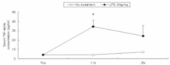
Rat에 LPS를 복강투여하여 신장염을 유 도하고, 1시간 후와 3시간 후에 serum TNF-α의 농도를 측정하였다. LPS 투여 1 시간 후에 serum TNF-α의 농도가 크게 증 가하여 LPS를 투여하지 않은 군에 비하여 유의하게 높았으며, LPS 투여 3시간 후에는 1시간 후에 비하여 serum TNF-α가 감소하 여 No-treatment군과 유의한 차이를 나타내 지 않았다(Fig. 2).
Fig. 2. Effect of LPS on serum TNF-α level in SD rats.
The male SD rats were injected intra-peritoneally with LPS(2㎎/㎏). Blood samples were taken from rat heart at 1 hr or 3 hr after the LPS injection, and serum TNF-α level was analysed by ELISA. Data were expressed as mean ± SD (n=5).
No treatment : normal SD rat.
LPS 2㎎/㎏: SD rat with LPS (2㎎/㎏) challenge
* : p<0.05, compared to no-treatment group by t-test
2) Renal TNF-α
Rat에 LPS를 복강투여하여 신장염을 유 도하고, 1시간 후와 3시간 후에 각각 신장을 적출하여 renal TNF-α의 농도를 측정하였 다. LPS 복강투여 1시간 후에 renal TNF-α 의 농도는 LPS를 투여하지 않은 군에 비하 여 차이를 보이지 않았으나, LPS 투여 3시 간 후 renal TNF-α의 농도는 LPS를 투여 하지 않은 군에 비하여 유의하게 증가하였 다(Fig. 3).
Fig. 3. Effect of LPS on renal TNF-α level in SD rats.
The male SD rats were injected intra-peritoneally with LPS(2㎎/㎏). Rat kidney was removed at 1 hr or 3 hr after the LPS injection, and renal TNF-α level was analysed by ELISA. Data were expressed as mean ± SD (n=5).
No treatment : normal SD rat without LPS challenge LPS 2 ㎎/kg: SD rat with LPS (2㎎/㎏) challenge
** : p<0.001, compared to no-treatment group by t-test
3. 음곡에 시술한 구맥 약침이 LPS에 의한 rat의 신장염에 미치는 영향
1) 혈액학적 분석
Rat에 LPS로 신장염을 유도하고, 1시간 후에 rat의 혈액을 채취하여 WBC의 수와 WBC 중 neutrophil의 비율을 측정하였다.
(1) WBC
Rat에 LPS로 신장염을 유도하고, 1시간 후에 rat의 혈중 WBC를 측정하였다. LPS 군의 혈중 WBC는 정상군에 비하여 유의하 게 증가하였다. N.P.군과 saline대조군, DS-HA군의 혈중 WBC는 LPS군에 비하여 유의하게 감소하였으며, DS-HA군의 혈중 WBC는 saline대조군에 비하여 유의하게 낮 았다(Fig. 4)
Fig. 4. Effect of DS-HA on W BC count in blood of LPS-stimulated rats.
The male SD rats were treated as described in the materials and methods and injected intra-peritoneally with LPS (2㎎/㎏). Blood samples were taken from rat hearts 1 hr after the LPS injection and WBC count was analysed.
Data were expressed as mean ± SD (n=5).
Normal : normal SD rat LPS : LPS (2㎎/㎏) challenge
N.P. : LPS (2㎎/㎏) challenge and a needle prick at KI10 Saline : LPS (2㎎/㎏) challenge and saline(200㎕/rat) injcetion at KI10
DS-HA : LPS (2㎎/㎏) challenge and DS-HA(5%, 200㎕
/rat) at KI10
** : p<0.01, compared to normal group by t-test
††† : p<0.001, †† : p<0.01 compared to LPS group by ANOVA test
# : p<0.01 compared to saline group by ANOVA test
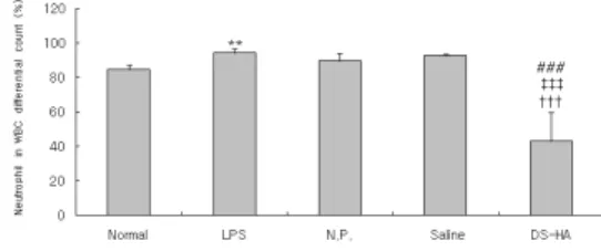
(2) Neutrophil
Rat에 LPS로 신장염을 유도하고, 1시간 후에 rat의 혈액을 채취하여 WBC 중 neu- trophil 비율을 측정하였다. LPS군의 neu- trophil 비율은 정상군에 비하여 유의하게 증가하였다. DS-HA군에서는 LPS군, N.P.
군 및 saline군에 비하여 neutrophil 비율이 유의하게 감소하였다(Fig. 5).
Fig. 5. Effect of DS-HA on neutrophil count in blood of LPS-stimulated rats.
The male SD rats were treated as described in the materials and methods and injected intra-peritoneally with LPS (2㎎/㎏). Blood samples were taken from rat hearts 1 hr after the LPS injection and the percentage of neutrophil out of WBC count was analysed. Data were expressed as mean ± SD (n=5).
Normal : normal SD rat LPS : LPS (2㎎/㎏) challenge
N.P.: LPS (2㎎/㎏) challenge and a needle prick at KI10 Saline : LPS (2㎎/㎏) challenge and saline(200㎕/rat) injcetion at KI10
DS-HA : LPS (2㎎/㎏) challenge and DS-HA(5%, 200㎕
/rat) at KI10
** : p<0.01, compared to normal group by t-test
††† : p<0.001 compared to LPS group by ANOVA test
‡‡‡ : p<0.001 compared to N.P. group by ANOVA test
### : p<0.001 compared to saline group by ANOVA test
2) Blood chemistry
Rat에 LPS로 신장염을 유도하고, 1시간 후에 rat의 혈액을 채취하여 혈청을 분리한 후, 혈청 BUN, creatinine, TNF-alpha 그리 고 CINC-1의 농도를 측정하였다.
(1) BUN (blood urea nitrogen)
Rat에 LPS로 신장염을 유도하고 1시간 후에 rat의 혈청 BUN 농도를 확인하였다.
LPS군에서는 정상군에 비하여 혈청 BUN 농도가 유의하게 증가하였다. N.P.군 및 saline대조군, DS-HA군에서는 LPS군에 비 하여 혈청 BUN 농도가 유의하게 감소하였 다(Fig. 6).
Fig. 6. Effects of DS-HA on serum BUN level in LPS-stimulated rats.
The male SD rats were treated as described in the materials and methods and injected intra-peritoneally with LPS (2㎎/
㎏). Blood samples were taken from rat hearts 1 hr after the LPS injection and serum BUN level was analysed. Data were expressed as mean ± SD (n=5).
Normal : normal SD rat LPS : LPS (2㎎/㎏) challenge
N.P.: LPS (2㎎/㎏) challenge and a needle prick at KI10 Saline : LPS (2㎎/㎏) challenge and saline(200㎕/rat) injcetion at KI10
DS-HA : LPS (2㎎/㎏) challenge and DS-HA(5%, 200㎕/rat) at KI10
* : p<0.05 compared to normal group by t-test
††† : p<0.001, †† : p<0.01 compared to LPS group by ANOVA test
‡‡ : p<0.01 compared to N.P. group by ANOVA test
## : p<0.01 compared to saline group by ANOVA test
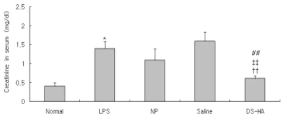
(2) Creatine
Rat에 LPS로 신장염을 유도하고 1시간 후에 rat의 혈청 creatine 농도를 확인하였 다. LPS군에서는 정상군에 비하여 혈청 creatine이 유의하게 증가하였다. DS-HA군 에서는 LPS군에 비하여 혈청 creatine 농도 가 유의하게 감소하였다(Fig. 7).
Fig. 7. Effects of DS-HA on serum creatine level in LPS-stimulated rats.
The male SD rats were treated as described in the materials and methods and injected intra-peritoneally with LPS (2㎎/
㎏). Blood samples were taken from rat hearts 1 hr after the LPS injection and serum creatine level was analysed. Data were expressed as mean ± SD (n=5).
Normal : normal SD rat
HJ : oral administration with HJ(0.5㎎/㎖, 1㎖) LPS : LPS (2㎎/㎏) challenge
N.P.: LPS (2㎎/㎏) challenge and a needle prick at KI10 Saline : LPS (2㎎/㎏) challenge and saline(200㎕/rat) injcetion at KI10
DS-HA : LPS (2㎎/㎏) challenge and DS-HA(5%, 200㎕/rat) at KI10
* : p<0.05 compared to normal group by t-test
†† : p<0.01 compared to LPS group by ANOVA test
‡‡ : p<0.01 compared to N.P. group by ANOVA test
## : p<0.01 compared to saline group by ANOVA test
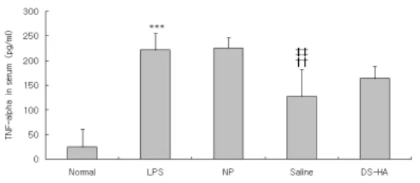
(3) TNF-α
Rat에 LPS로 신장염을 유도하고 1시간 후에 rat의 혈청 TNF-α 농도를 확인하였 다. LPS군에서는 정상군에 비하여 혈청 TNF-α가 현저하게 증가하였다. Saline대조 군에서는 LPS군에 비하여 혈청 TNF-α 농 도가 유의하게 감소하였다. DS-HA군은 LPS군에 비하여 혈청 TNF-α 농도가 감소 하였으나 유의성은 없었다(Fig. 8).
(4) CINC-1 (Cytokine-induced neutrophil chemoattractant-1)
Rat에 LPS로 신장염을 유도하고 1시간 후에 rat의 혈청 CINC-1 농도를 확인하였 다. LPS군에서는 정상군에 비하여 혈청 CINC-1이 유의하게 증가하였다. NP군, sal- ine대조군 및 DS-HA군에서는 LPS군에 비
하여 혈청 CINC-1 농도가 감소하였으나 유 의성은 없었다(Fig. 9).
Fig. 8. Effects of DS-HA on serum TNF-alpha level in LPS-stimulated rats.
The male SD rats were treated as described in the materials and methods and injected intra-peritoneally with LPS (2㎎/㎏). Blood samples were taken from rat hearts 1 hr after the LPS injection and serum TNF-alpha level was analysed. Data were expressed as mean ± SD (n=5).
Normal : normal SD rat LPS : LPS (2㎎/㎏) challenge
N.P.: LPS (2㎎/㎏) challenge and a needle prick at KI10 Saline : LPS (2㎎/㎏) challenge and saline(200㎕/rat) injcetion at KI10
DS-HA : LPS (2㎎/㎏) challenge and DS-HA(5%, 200㎕/rat) at KI10
*** : p<0.001 compared to normal group by t-test
††: p<0.01 compared to LPS group by ANOVA test
‡‡: p<0.01 compared to N.P. group by ANOVA test
Fig. 9. Effects of DS-HA on serum CINC-1 level in LPS-stimulated rats.
The male SD rats were treated as described in the materials and methods and injected intra-peritoneally with LPS (2㎎/
㎏). Blood samples were taken from rat hearts 1 hr after the LPS injection and serum CINC-1 level was analysed. Data were expressed as mean ± SD (n=5).
Normal : normal SD rat LPS : LPS (2㎎/㎏) challenge
N.P.: LPS (2㎎/㎏) challenge and a needle prick at KI10 Saline : LPS (2㎎/㎏) challenge and saline(200㎕/rat) injcetion at KI10
DS-HA : LPS (2㎎/㎏) challenge and DS-HA(5%, 200㎕/rat) at KI10
*** : p<0.001 compared to normal group by t-test
3) 소변 분석
Rat에 LPS로 신장염을 유도하고 12시간 동안 소변을 채취하여 이뇨활성과 뇨중 creatinine과 total protein의 양을 측정하였 다.
⑴ 이뇨 활성
Rat에 LPS로 신장염을 유도한 후 metabolic cage에 한 마리씩 수용하고 12시 간동안 소변을 채취하여 배뇨량을 측정하였 다. LPS군에서는 정상군에 비하여 배뇨량이 유의하게 감소하였다. DS-HA에서는 LPS대 조군, N.P.군 및 saline대조군에 비하여 배뇨 량이 유의하게 증가하였다(Fig. 10).
Fig. 10. Effect of PS on urinary volume in LPS-stimulated rats.
The male SD rats were treated as described in the materials and methods and injected intra-peritoneally with LPS (2㎎/
㎏). The rats were kept in metabolic cages and the urine was collected for 12 hours. Data were expressed as mean ± SD (n=3).
Normal : normal SD rat LPS : LPS (2㎎/㎏) challenge
N.P.: LPS (2㎎/㎏) challenge and a needle prick at KI10 Saline : LPS (2㎎/㎏) challenge and saline(200㎕/rat) injcetion at KI10
DS-HA : LPS (2㎎/㎏) challenge and DS-HA(5%, 200㎕/rat) at KI10
* : p<0.05 compared to normal group by t-test
† : p<0.05 compared to LPS group by ANOVA test
‡ : p<0.05 compared to N.P. group by ANOVA test
## : p<0.01 compared to saline group by ANOVA test
⑵ Creatinine
Rat에 LPS로 신장염을 유도하고 12시간
동안 소변을 채취하여 뇨중 creatinine 양을 측정하였다. LPS군의 뇨중 creatinine 농도 는 정상군에 비하여 유의하게 증가하였다.
DS-HA군에서는 LPS군에 비하여 뇨중 creatinine 농도가 감소하였으나 유의성은 없었다(Fig. 11).
Fig. 11. Effects of DS-HA on urinary creatinine level in LPS-stimulated rats.
The male SD rats were treated as described in the materials and methods and injected intra-peritoneally with LPS (2㎎/
㎏). The rats were kept in the metabolic cages and the urine was collected for 12 hours. And the urinary creatinine level was analysed. Data were expressed as mean ± SD (n=3).
Normal : normal SD rat LPS : LPS (2㎎/㎏) challenge
N.P.: LPS (2㎎/㎏) challenge and a needle prick at KI10 Saline : LPS (2㎎/㎏) challenge and saline(200㎕/rat) injcetion at KI10
DS-HA : LPS (2㎎/㎏) challenge and DS-HA(5%, 200㎕/rat) at KI10
* : p<0.05 compared to normal group by t-test
⑶ Total protein
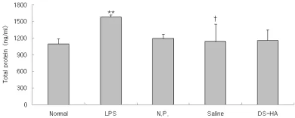
Rat에 LPS로 신장염을 유도하고 12시간 동안 소변을 채취하여 뇨중 total protein 양 을 측정하였다. LPS군에서는 정상군에 비하 여 뇨중 total protein이 유의하게 증가하였 다. DS-HA군의 뇨중 total protein은 LPS 군에 비하여 감소하였으나 유의성은 없었다 (Fig. 12).
Fig. 12. Effects of DS-HA on total protein level in urine of LPS-stimulated rats.
The male SD rats were treated as described in the materials and methods and injected intra-peritoneally with LPS (2㎎/
㎏). The rats were kept in the metabolic cages and the urine was collected for 12 hours. And, total protein level in urine was analysed. Data were expressed as mean ± SD (n=3).
Normal : normal SD rat LPS : LPS (2㎎/㎏) challenge
N.P.: LPS (2㎎/㎏) challenge and a needle prick at KI10 Saline : LPS (2㎎/㎏) challenge and saline(200㎕/rat) injcetion at KI10
DS-HA : LPS (2㎎/㎏) challenge and DS-HA(5%, 200㎕/rat) at KI10
** : p<0.01 compared to normal group by t-test
† : p<0.05 compared to LPS group by ANOVA test
4) 신장 내 염증활성
Rat에 LPS로 신장염을 유도하고 3시간 후에 신장을 적출하여 신장조직을 분획한 후 TNF-alpha와 MPO의 농도를 측정하였 다.
(1) TNF-α
Rat에 LPS로 신장염을 유도하고 3시간 후에 신장을 적출하여 renal TNF-alpha의 농도를 측정하였다. LPS군에서는 정상군에 비하여 renal TNF-alpha가 유의하게 증가 하였다. DS-HA군의 renal TNF-alpha는 LPS군에 비하여 감소하였으나 유의성은 없 었다(Fig. 13).
Fig. 13. Effects of DS-HA on renal TNF-alpha level in LPS-stimulated rats.
The male SD rats were treated as described in the materials and methods and injected intra-peritoneally with LPS (2㎎/㎏). Three hours after the LPS stimulation, rat kidney was removed and renal TNF-alpha level was analysed by ELISA. Data were expressed as mean ± SD (n=5).
Normal : normal SD rat LPS : LPS (2㎎/㎏) challenge
N.P.: LPS (2㎎/㎏) challenge and a needle prick at KI10
Saline : LPS (2㎎/㎏) challenge and saline(200㎕/rat) injcetion at KI10DS-HA : LPS (2㎎/㎏) challenge and DS-HA(5%, 200㎕/rat) at KI10
* : p<0.05 compared to normal group by t-test
(2) MPO (myeloperoxidase)
Rat에 LPS로 신장염을 유도하고 3시간 후에 신장을 적출하여 renal MPO 농도를 측정하였다. LPS군에서는 정상군에 비하여 renal MPO가 현저하게 증가하였다.
DS-HA군의 renal MPO는 LPS군에 비하여 유의하게 감소하였다(Fig. 14).
Fig. 14. Effects of DS-HA on renal MPO level in LPS-stimulated rats.
The male SD rats were treated as described in the materials and methods and injected intra-peritoneally with LPS (2㎎/㎏). Three hours after the LPS stimulation, rat kidney was removed and renal MPO level was analysed by ELISA. Data were expressed as mean
± SD (n=5).
Normal : normal SD rat LPS : LPS (2㎎/㎏) challenge
N.P.: LPS (2㎎/㎏) challenge and a needle prick at KI10 Saline : LPS (2㎎/㎏) challenge and saline(200㎕/rat) injcetion at KI10DS-HA : LPS (2㎎/㎏) challenge and DS-HA(5%, 200㎕/rat) at KI10*** : p<0.001 compared to normal group by ANOVA test
† : p<0.05 compared to LPS group by ANOVA test
5) 조직학적 분석
Rat에 LPS로 신장염을 유도하고 3시간 후에 신장을 적출하여 Hematoxilin and Eosin(H&E) 염색법으로 조직을 염색한 후 광학현미경으로 관찰하였다. LPS군의 신장 조직은 정상군에 비하여 사구체에 netrophil 의 집중과 조직의 조밀도 저하를 나타내었 다. DS-HA군의 신장 조직에서는 LPS군에 비하여 neutrophil의 집중이 감소된 것으로 나타내었다(Fig. 15).
Fig. 15. Histological analysis of renal tissue in LPS-stimulated rats. (H&E stain)
The male SD rats were treated as described in the materials and methoDS and injected intra-peritoneally with LPS (2㎎/
㎏). Three hours after the LPS stimulation, rat kidney was removed. The renal tissue was sliced and embedded in OCT compound, and 8㎛ sections were stained with hematoxilin and eosin.(↑ : glomerulus, ×400)
Normal : normal SD rat LPS : LPS (2㎎/㎏) challenge
N.P.: LPS (2㎎/㎏) challenge and a needle prick at KI10 Saline : LPS (2㎎/㎏) challenge and saline(200㎕/rat) injcetion at KI10
DS-HA : LPS (2㎎/㎏) challenge and DS-HA(5%, 200㎕/rat) at KI10
Ⅳ. 고 찰
신장은 1쌍의 암적색을 띤 완두형의 장기 로서 지방피맥에 싸여 복막과 후복벽 사이 에 있는 복막후 장기의 하나로 체액의 양 및 전해질 조성 유지, 대사로 생긴 노폐물
배설, 독소 및 독소의 대사산물의 해독 또는 체외배설, 세포외액의 양과 혈압의 내분비성 조절, 적혈구 총량 조절, 광물질 대사의 내 분비성 조절, peptide 호르몬의 분해 및 이 화작용, 경쇄 또는 beta-2 microglobulin 등 작은 단백의 이화작용, 대사과정에서 중간변 환 수행 등의 역할을 한다1). 따라서 신사구 체 및 세뇨관의 내피세포는 핼액 중의 각종 물질에 넓게 노출되어 있으며, 신세뇨관세포 는 많은 효소계의 왕성한 대사작용으로 인 해 독성물질에 대한 감수성이 높은 특징을 가지고 있으므로 혈액중에 포함된 신독성 물질 및 약물에 손상받기 쉬운 여건에 있다
12).
염증이란 혈관이 있는 조직 내에서 여러 가지 염증 유발성 자극에 의하여 발생한 손 상에 대한 생체의 반응이며 염증의 화학 매 개체란 자신이 염증의 원인이 되는 것이 아 니고, 혈장, 세포 및 손상받은 조직으로부터 유래하여 여러 가지의 반응을 중개하는 물 질로 이중의 하나가 cytokine이다. 그중 염 증에 관여하는 2가지의 중요한 cytokine에 는 IL-1과 TNF-α가 있다. 염증에 관여하는 세포는 백혈구로 염증의 경과와 종류에 의 해 염증 반응에서 백혈구의 종류가 변한다.
일반적인 염증에서는 초기에는 Neutrophil가 주역을 행하고 후기에는 림프구, 형질세포와 대식구가 이에 대신한다. 염증의 초기에 주 요 역할을 담당하는 Neutrophil는 골수에서 유래하고 세포질 내에 호중성의 과립을 가 지며 염증이 발생하면 혈관의 내피 사이를 빠져나가 혈관 밖으로 유주하고 화학 주성 에 의해 항원과 이물에 이동하고 부착한 후
이것들을 탐식하는 역할을 한다. 박테리아의 표면에서 만들어진 LPS는 대식구를 자극하 여 IL-1과 TNF-α를 분비하여 혈관의 내피 세포와 섬유모세포, 상피세포에 영향을 미쳐 Neutrophil 화학주성을 가진 IL-8과 단핵구 화학주성을 가진 MCP를 분비하도록 한다
13).
약침요법은 신침요법의 일종으로 혈위주 사요법이라고도 하며, 경락학설과 약물이론 에 근거하여 환자의 질병에 상응하는 경혈 과 약물을 선택하고 약침액을 경혈내에 자 입하여 경혈과 약물이 질병에 대해 종합적 인 작용을 발휘하여 질병을 치료하는 방법 으로 약물의 흡수가 빠르고14), 경구투여가 불가능한 경우에 처치할 수 있으며 환처에 처치할 수 있는 등의 장점이 있다8,15).
陰谷(KI10)은 足少陰腎經의 經穴로 膝下 內 輔骨後大筋下 小筋上에 위치한다. 足少陰 의 合水穴이며 自性穴로서, 祛濕通溲, 滋腎 淸熱, 疎泄厥氣, 利導下焦 등의 效能이 있어 膝關節炎, 股關節痛, 膝不能屈伸, 尿道炎, 陰 道炎, 陰萎, 子宮出血, 帶下, 痔核, 小便難, 腹脹滿, 臍腹痛, 崩漏 등의 치료혈이 된다8).
瞿麥(Dianthi herba)은 石竹科 (Caryophyllaceae)에 속한 다년생 초목인 술 패랭이꽃과 패랭이꽃 의 지상부를 건조한 것으로, 性은 寒 無毒하고, 味는 苦하며 歸 經은 心, 小腸經 이며 利尿通淋, 破血通經 하는 효능이 있어 熱淋, 血淋, 石淋, 小便不 通, 淋瀝澁痛, 月經閉止 등을 치료한다. 신장 의 질환은 대부분 요형성의 장애를 일으키 며 소변불리의 증상을 가지고 있으므로 瞿 麥은 상기 증상에 응용할 수 있을 것으로
사료되었다7).
LPS는 여러 종류의 세포를 강하게 자극 하여 cytokine을 분비시키며 증가된 혈청내 proinflammatory cytokine은 LPS에 의한 신 체손상을 매개한다. 신장의 경우 LPS는 신 장의 혈행역학장애를 유발하며 IL-1, IL-6, TNF-α 등의 cytokine을 분비시키고 IL-8, MCP-1 등의 chemokine을 분비하여 면역세 포의 화학주성을 유발한다. 또 LPS는 메산 지움 세포에 많은 양의 nitric oxide 분비를 유발시키며 메산지움 세포와 tubular epithelial cell에 Fos 발현을 증가시킴으로서 직간접적으로 신장손상을 초래하며16,17,18), 최 근의 실험에서 신장독성으로 인한 사구체신
염17,18,19), 급성 신부전20,21), 간질성신염22), 신
독성신염23) 등의 병태모델에 많이 사용되고 있다.
이에 저자는 음곡에 시술한 구맥 약침의 신장염에 대한 보호효과를 알아보고자 구맥 약침액을 시료로 정상 rat의 간세포에 대한 세포독성을 확인하고 LPS 투여 후 혈청과 신장내 TNF-α의 농도를 측정하였고, 음곡 에 시술한 구맥 약침 투여 후 혈액 및 혈청 분석, 소변분석, 신장내 염증활성, 조직학석 분석을 시행하였다.
정상 rat의 간세포에 DS-HA를 여러 가 지 농도로 투여한 후 세포생존율을 확인한 결과 DS-HA의 농도가 높을수록 세포의 viability가 유의하게 증가됨을 확인하였다 (Fig 1).
LPS 투여에 따른 TNF-α의 농도 변화를 알아보고자 각각 혈청과 신장 조직에서 정 상군과 LPS투여군의 TNF-α 농도를 측정하
였다. 혈청 검사에서는 LPS 투여 후 1시간 과 3시간 후 모두 TNF-α가 증가하였고 1 시간 후에는 유의한 증가를 보였으며 3시간 후에는 유의한 차이를 보이지 않으며 신장 조직검사에서는 투여하고 3시간 이후에 유 의한 증가를 보였다(Fig 2, 3). 따라서 LPS 투여에 따른 효과를 측정하기 위해서 혈청 검사는 투여하고 1시간 후에 측정하고 소변 검사는 12시간 동안 소변을 채취하여 실험 을 진행하였다.
Neutrophil는 골수에서 유래하고 세포질 내에 호중성의 과립을 가지는 백혈구로 염 증이 발생하면 혈관의 내피 사이를 빠져나 가 혈관 밖으로 유주하고 화학주성에 의해 항원과 이물에 이동하고 부착한 후 탐식한 다13). 본 실험에서 LPS 투여군에 비해 DS-HA군에서 백혈구와 백혈구 중 Neutrophil의 비율이 유의하게 감소하였고 N.P.군에서 혈중 WBC가 감소하여 음곡에 시술한 구맥약침과 음곡혈 자침이 신장염 모델에서 염증반응을 억제한 것으로 사료된 다(Fig 4, 5).
proimflammatory cytokine인 TNF-α는24) 그람-음성 박테리아와 다른 감염성 미생물 에 대한 급성염증반응의 주된 매개자로, 내 피세포와 대식세포를 자극하여 백혈구의 주 화성과 보충을 유도하는 케모카인을 분비토 록하며, 혈액 단핵구와 T 림프구, Neutrophil, 혈관내피세포, 피부섬유아세포 등의 세포에서 IL-8 유전자 발현을 유도하
여19,25) IL-8의 분비26) 및 활성화와
Neutrophil의 침투로 인한 조직손상을 통해 서 lysozomal 효소와 Neutrophil에 의해 과
산화음이온을 방출시킨다27). TNF-α로 유도 된 chemokine인 IL-8은 염증반응에서 Neutrophil를 유도하며13) CINC-1은 IL-8 family의 일원이다. 최근의 연구에서 쥐의 염증반응 모델에서 CINC-1 수치를 측량하 여 IL-8 발생을 조사한 예가 있다28). 본 실 험에서는 염증반응에서 Neutrophil의 활성화 정도를 알아보기 위하여 CINC-1 수치를 측 정하였다.
Blood urea nitrogen(BUN)은 혈중에 존재 하는 요소(urea)내 질소를 나타내는 것으로 요소는 간의 요소회로(urea cycle)에서 단백 질대사의 최종산물로서 생성되어 혈류를 거 쳐 신사구체에서 여과된 다음 세뇨관에서 일부는 재흡수 되고 나머지는 요중으로 배 설되는데, 신기능이 저하하여 사구체여과율 이 감소하면 혈중 농도가 상승하므로 신기 능의 지표가 된다29).
혈액내의 creatinine phosphate에서 생성 된 creatine이 탈수되어 생긴 대사의 최종산 물로 creatinine량은 인체 근육량에 비례한 다. 오로지 신장을 통해서만 체외로 배설되 고 체내에서 재순환을 하지 않으므로 creatinine의 요중 배설량은 체내의 creatinine 생성량과 동일하며 사구체의 여 과에 따라 변하므로 creatinine 수치는 신기 능의 지표가 된다1,3).
본 실험에서 rat의 혈청에서 BUN, creatinine, TNF-α, CINC-1의 농도를 측정 한 결과 BUN과 creatinine은 음곡에 시술한 구맥 약침에 의해 대조군에 비해 유의하게 감소하였고, TNF-α와 CINC-1은 대조군에 비해 감소하였으나 유의성은 없었다. (Fig
6~9).
신장의 여러 역할 중 뇨를 형성하여 체액 의 양과 전해질 조성을 정상으로 유지하는 것은 가신장의 가장 핵심적인 역할이며 정 상적인 사구체에서 혈액 속의 적혈구나 단 백물질은 사구체를 통과할 수 없다. 그러나 사구체에 질환이 있어 파괴되거나 투과성이 증가되면 단백물질이 나오게 된다1). 그러므 로 본 실험에서는 rat에서 소변을 채취하여 소변양과 뇨중 creatinine, total protein을 측 정하였다. 소변량은 음곡에 시술한 구맥 약 침에 의해 유의하게 증가하여 정상 수준으 로 회복되었으나, 뇨중 creatinine과 total protein은 대조군에 비하여 감소하기는 하였 으나 유의성은 없었으며 이는 구맥의 약리 학적 효과로 보인다(Fig 10~12).
MPO는 Neutrophil로부터 주로 방출되는 효소이므로 MPO 분석은 염증의 지표로서 조직속의 Neutrophil의 수를 측량하는데 사 용된다30).
본 실험에서는 신장 실질내 염증활성도 를 측정하기 위해 TNF-α와 MPO의 농도를 측정하였는데 TNF-α의 농도는 음곡에 시 술한 구맥 약침에 의해 감소하였으나 통계 적 유의성은 없었다. MPO의 농도는 음곡에 시술한 구맥 약침에 의해 유의하게 감소하 였다(fig 13~14).
신장을 적출하여 손상을 관찰한 결과 LPS대조군은 사구체 내 neutrophil이 집중 되고 조직의 조밀도 저하를 나타내었으나 DS-HA군에서는 비교적 neutrophil의 집중 이 감소되고 조직손상이 덜한 것으로 나타 났다(Fig 15). 따라서 DS-HA군에서는 LPS
와 같은 독성물질이 유발한 신장염의 조직 손상을 막는데 효과가 있을 것으로 사료된 다.
이상의 결과, 음곡에 시술한 구맥 약침은 LPS로 유도된 rat의 신장염에 대하여 혈액 내 백혈구와 Neutrophil의 증가를 억제하며, 혈청내에서 BUN, creatinine 등의 노폐물 축적을 감소시키고, inflammatory cytokine 을 억제하였으며, 여과율의 감소로 인한 핍 뇨를 개선시켰으며, 신장 실질내의 염증반응 또한 억제하는 효과를 나타내었다. 이러한 효과는, 음곡에 saline을 투여한 군에서 혈 청내 BUN, TNF-α를 유의하게 감소시킨 것이나, NP군에서 혈중 WBC, 혈청 BUN을 유의하게 감소시킨 것 등으로 보아, 구맥의 약리적 작용뿐만 아니라 음곡의 경혈 특이 성도 작용한 것으로 보인다. 따라서 음곡에 대한 구맥약침은 사구체신염과 급성 신부전, 간질성 신염과 같은 신장의 염증반응의 치 료에 효과가 있을 것으로 생각되는 바 향후 이에 대한 지속적 연구와 임상연구가 필요 할 것으로 사료된다.
Ⅴ. 결 론
음곡(KI10)에 시술한 구맥 약침이 LPS로 유도된 신장염 모델 rat에 미치는 영향을 알 아보고자 구맥 약침액을 시료로 정상 rat의 간세포에 대한 세포독성을 확인하고 LPS 투여 후 혈청과 신장내 TNF-α의 농도를 측정하였고, 음곡에 시술한 구맥 약침 투여 후 혈액 및 혈청 분석, 소변분석, 신장내 염 증활성, 조직학석 분석을 시행한 결과, 다음
과 같은 결론을 얻었다.
혈액에서 WBC와 neutrophil과 혈청에서 BUN, creatinine을 유의하게 감소시켰으며 소변량을 유의하게 증가시켰고, 신장실질내 의 MPO를 유의하게 감소시켰다. 따라서 이 상의 결과로 볼때, 음곡에 시술한 구맥 약침 은 신장의 염증반응을 억제하는 효과가 있 는 것으로 사료된다.
參考文獻
1. 杜鎬京. 동의신계학. 서울 : 동양과학연구 회. 1993 : 226, 326, 256-7, 334-6, 361-2, 454-9, 514-6.
2. Medzhitov R, Preston-Hurlburt P, Janeway CA Jr. A human homologue of the Drosophila Toll protein signal activation of adaptive immunity. Nature.
1997 ; 388(6640) : 394-7.
3. Aderem A, Ulevitch RJ. Toll-like receptors in the induction of the innate immune response. Nature. 2000 ; 406(6797) : 782-7.
4. Hoshino K, Takeuchi O, Kawai T, Sanjo H, Ogawa T, Takeda Y et.
Toll-like receptor 4(TLR4)-deficient mice are hyporesponsive to lipopolysaccharide : evidence for TLR4 as the Lps gene product. J immunol.
1999 ; 162(7) : 3749-52.
5. Lee BG, Kim SH, Zee OP, Lee KR, Lee HY, Han JW et. Suppression of inducible nitric oxide synthase
expression in RAW 264.7 macrophages by two-carboline alkaloids extracted from Melia azedarach. Eur J Pharmacol.
2000 ; 406(3) : 301-9.
6 Seo WG, Pae HO, Oh GS, Chai KY, Yun YG, Kwon TO et. Inhibitory effect of ethyl acetate fraction from Cudrania tricuspidata on the expression of nitric oxide synthase gene in RAW 264.7 macrophages stimulated with interferon-and lipopolysaccharide. Gen Pharmacol. 2000 ; 35(1) : 21-8.
7. 康秉秀, 高雲彩, 金圭烈, 金先熙, 盧昇鉉, 朴涌基 외. 本草學. 서울 : 永林社. 1991 : 323-4.
8. 전국한의과대학침구경혈학교실. 鍼灸學 (上). 서울 : 집문당. 2001 : 531-63, 1457-6.
9. 최정배, 이경환, 김용성. 육미지황탕가황 기인삼 추출물의 Cisplatin 유도 신장독성 보호효과. 대한한방내과학회지. 2003 ; 24(3) : 579-87.
10. 황영근, 정지천. 수은으로 유발된 토끼의 신장 기능 손상에 대한 단삼(丹參)의 효 과. 대한한방내과학회지. 2000 ; 21(2) : 227-34.
11. 윤철호, 정지천, 신의섭. Bromobenzene 으로 유도된 신장기능저하에 미치는 녹 용 약침의 항산화 효과. 대한한방내과학 회지. 1999 ; 20(2) : 287-302.
12. Ernest hodgson, Patricia E. Levi. A text of mordern toxicology. America : APPLETON & LANGE. 1987 : 207-9.
13. 송계용, 지제근, 함의근. 핵심병리학. 서 울 : 고려의학. 1998 : 63-82.
14. 대한약침학회. 약침제재와 임상응용. 서 울 : 대한약침학회. 1997 : 1-5.
15. 崔旼燮. 수침요법에 관한 고찰. 대한한의 학회지. 1990 ; 11(1) : 315-6, 324-6.
16. Glauser MP, Zanetti G, BaumgartnerJ-D, Cohen J. Septic shock : Pathogenesis. Lancet. 1991 ; 338 : 732-6.
17. Ohmori Y, Hamilton TA. A macrophage LPS-inducible early gene encodes the murine homologue of IP-10. Biochem Biophys Res Commun 1990 ; 168 : 1261-7.
18. Tannenbaum CS, Koemer TJ, Jansen MM, Hamilton TA. Characteriation of Lipopolysaccharide-induced macrophage gene expression. J immunol 1988 ; 140 : 3640-5.
19. Akihisa Harada, Nobuaki Sekido, Tohru Akahoshi, Takashi Wada, Naofumi Mukaida, Kouji Matsushima.
Essential involvement of interleukin-8 (IL-8) in acute inflammation. Journal of Leukocyte Biology. 1994 ; 56 : 559-64.
20. Nestor Schor. Acute renal failure and the sepsis syndrome. Kidney International. 2002 ; 61 : 764–76.
21. Patrick N. Cunningham, Hristem M.
Dyanov, Pierce Park, Jun Wang, Kenneth A. Newell, Richard J. Quigg.
Directly on TNF Receptor-1 in Kidney.
The Journal of Immunology. 2002 ; 168 : 5817-23.
22. Patricia Wahl, Roland Schoop, Grozdana Bilic, Jörg Neuweiler, Michel Le Hir, Steven K. Yoshinaga et. Renal Tubular Epithelial Expression of the Costimulatory Molecule B7RP-1 (Inducible Costimulator Ligand). Journal of american society of Nephrology.
2002 ; 13 : 1517-26.
23. A. M. KARKAR, Y. KOSHINO, S. J.
CASHMAN, A. C. DASH, J.
BONNEFOY, A. MEAGERt et. Passive immunization against tumour necrosis factor-alpha (TNF-a) and IL-1 protects from LPS enhancing glomerular injury in nephrotoxic nephritis in rats. Clin.
exp. Immunol. 1992 ; 90 : 312-8.
24. 강재성 외 역. 세포분자이역학(4판). 서 울 : 범문사. 2002 : 23-4, 240-3, 567-8.
25. Matsushima, K., Morishita, K., Yoshimura, T., Lavu, S., Kobayashi, Y., Lew, W. E. et. Molecular cloning of a human monocyte-derived neutrophil chemotactic factor (MDNCF) and the induction of MDNCF mRNA by interleukin 1 and tumor necrosis factor.
J. Exp. Med. 1988 ; 167 : 1883–93.
26. Strieter, R.M., Kunkel, S. L., Showell, H. J., Remick, D.G., Phan, S. H., Ward,
P. A. et. Endothelial cell gene expression of a neutrophil chemotactic factor by TNF-alpha, LPS, and IL-1 beta. Science. 1989 ; 243 : 1467-9.
27. Sekido, N., Mukaida, N., Harada, A., Nakanishi, I., Watanabe, Y., and Matsushima, K.. Prevention of lung reperfusion injury in rabbits by a monoclonal antibody against interleukin-8. Nature. 1993 ; 365 : 654-7.
28. Nakagawa, H., Komori, N., Shibata, F., Ikesue, A., Konishi, K., Fujioka, M. et.
Identification of cytokine-induced neutrophil chemoattractants (CINC), rat GRO/CINC-2 alpha and CINC-2 beta, produced by granulation tissue in culture: purification, complete amino acid sequences and characterization.
Biochem. J.. 1994 ; 301 : 545–50.
29. 이귀녕, 이종순. 임상병리파일(제3판). 서 울 : 의학문화사. 2000 ; 334-5.
30. Krawisz, J.E., Sharon, P., and Stenson, W.F.. Quantitive assay for acute intestinal inflammation based on myeloperoxidase activity. Assessment of inflammation in rat and hamster models. Gastroenterology. 1984 ; 87 : 1344-55.