pISSN 1229-1137
Original Article
The Effect of Junci Medulla Herbal-acupuncture at KI 10 on LPS induced Nephritis in Rats
Jee Soon Ihm, Tae Hwan Cho and Yun Kyoung Yim*
Dept. of Meridian and Acupoint, College of Oriental Medicine, Daejeon University
Objectives : This study aimed to evaluate the effects of Junci Medulla Herbal-acupuncture(JM-HA) at KI
10( Umgok ) on nephritis induced by lipopolysaccharide(LPS) in rats.
Methods : Rats with nephritis induced by LPS, were treated with JM-HA at KI
103 times a week. The rats in the NP group and the saline group were treated with a needle prick and a saline injection respectively. To evaluate the effects of JM-HA at KI
10on nephritis in rats, WBC, neutrophils in blood, BUN, creatinine, TNF-α in serum, creatinine, total protein in urine and renal MPO were measured.
Results : JM-HA at KI
10significantly inhibited the increase of WBC and neutrophils in blood, BUN, creatinine, TNF-α in serum, and MPO in kidney of LPS-stimulated rats.
Conclusion : JM-HA at KI
10has therapeutic effects on nephritis in LPS-stimulated rats. Therefore, it is suggested that JM-HA at KI
10may be a useful therapy in clinical field after further researches.
Key Words : nephritis, herbal-acupuncture, KI
10, Junci Medulla , LPS
1)
음곡에 시술한 등심초약침이 LPS로 유도된 흰쥐의 신장염에 미치는 영향
임지순․조태환․임윤경*
대전대학교 한의과대학 경락경혈학교실
Acceptance : 2012. 11. 12. Adjustment : 2012. 11. 20. Adoption : 2012. 11. 20.
Corresponding author : Yun Kyoung Yim, Department. of Meridian and Acupoint, College of Oriental Medicine, Daejeon University, Yongun-dong, Dong-gu, Daejeon, 300-176, Republic of Korea Tel : +82-42-280-2610 E-mail : [email protected]
This is an Open-Access article distributed under the terms of the Creative Commons Attribution Non-Commercial License(http://creativecommons.org/licenses/by-nc/3.0) which permits unrestricted non-commercial use, distribution, and reproduction in any medium, provided the original work is properly cited.
Copylight ⓒ The Journal of Korean Acupuncture & Moxibustion Medicine Society
Abstract
Ⅰ. 서 론
등심초(Junci Medulla, JM)는 골풀과(Juncaceae) 에 속한 다년생 초본인 골풀의 莖髓를 8∼9월에 채취 하여 曬乾한 다음 莖髓를 取出한 것으로, 性은 微寒하 고, 味는 甘淡하며, 歸經은 心ㆍ肺ㆍ 小腸經이며, 淸心 火 利小便하는 효능이 있다
1).
음곡(KI
10)은 足少陰腎經의 合水穴로서 袪濕通溲, 滋腎淸熱, 疏泄厥氣, 利導下焦의 효능이 있어 尿道炎, 陰道炎, 小便難 등의 증상을 主治한다
2).
신장은 타 장기에 비해 많은 혈류를 받아 이를 여 과 배설시키는 역할을 하므로 혈액 속에 포함된 신독 성 물질 및 약물에 의해 손상받기 쉬운 여건이고
3), 감 염으로 인한 혈중의 면역복합체나 항체가 사구체기저 막에 침착되면 사구체신염이 발생한다. 사구체기저막 에 침착된 면역복합체나 세균에 대한 항체는 면역시 스템을 활성화시켜 사구체를 손상시킴으로써 혈뇨, 단백뇨 등을 유발하고, 여과기능의 손상까지 진행되 면, 핍뇨와 요독증이 발생한다
4).
LPS(lipopolysaccharide)는 그람음성박테리아의 외 부 세포막을 구성하는 성분으로, 내독소 쇼크(endotoxin shock)라 불리는 병리적 연쇄반응의 주요한 시작인자 이다
5). LPS는 단핵구(monocyte)를 자극하여 tumor necrosis factor-alpha(TNF-α)와 같은 cytokine을 분 비하도록 함으로써 패혈증을 유발한다
6). 패혈증이 진 행되는 동안, 활성화된 호중구(neutrophil)와 같은 체 액성 면역물질들이 체순환계로 분비되고
7), 대량으로 활성화된 호중구는 호중구 엘라스타제(neutrophil elastase) 및 초과산화물로 유도된 활성산소(superoxide derived free radical)와 같은 염증물질을 분비함으로써 조직을 손상시킨다
8,9).
음곡에 시술한 약침이 신장염에 미치는 영향에 관한 기존의 실험 연구로는 금전초약침
10)ㆍ동과인약침
11)ㆍ 차전자약침
12)ㆍ구맥약침
13)등을 이용하여 유효한 효 과를 보인 연구가 보고된 바 있으나, 등심초약침(Junci Medulla herbal acupuncture, JM-HA)을 이용한 실 험은 아직 보고된 바 없다.
이에 저자는 음곡 등심초약침이 독소에 의한 신장 염에 미치는 영향을 알아보고자, 등심초약침액(Junci Medulla herbal acupuncture solution, JM-HAS)을 만들어 흰쥐의 음곡 상응부위에 약침시술하고, LPS로 신장염을 유도하여 혈액분석, 소변분석, 신장 내 MPO(myeloperoxidase) 활성평가 등을 통하여 유의한
결과를 얻었기에 보고하는 바이다.
Ⅱ. 실 험
1. 재료 1) 동물
동물은 6주령의 웅성 SD rat을 (주) 대한바이오링 크(경기도, 한국)에서 공급받아 1주일간 실험실 환경 에 적응시킨 후 실험에 사용하였다. 실험에 사용된 동 물은 실험 당일까지 고형사료(삼양사료, 한국)와 물을 충분히 공급하였고, 실험실은 실온(22±2 ℃)과 습도 50∼60 %를 유지하였으며 대전대학교 동물실험 윤리 위원회의 규정을 따랐다.
2) 약재
본 실험에 사용된 등심초(Junci Medulla, JM)는 대전대학교 한방병원에서 구입하여 실온에서 보관되 었으며, 사용하기 전 초음파 세척을 실시하였다.
3) 시약 및 기기
(1) 시약
Reagent Manufacturer Country Phosphate buffer saline
(PBS)
Sigma
USA HBSS
Collagenase type Ⅳ MTT
Lipopolysaccharide(LPS) Gum-sucrose
Absolute EtOH RPMI 1640 media
Hyclone Antibiotics
FBS
BCA kit BD
TNF-alpha ELISA kit Oxford Creatinine assay kit Bio Assay
Systems MPO ELISA Kit Cell Science Ethyl ether
Table 1. Reagents
(2) 기기
Instrument Manufacturer Country
Bio-freezer Sanyo
Japan Rotary evaporator Eyela
ELISA reader TARAN Canada
Sonicate Fisher USA
Ice maker Vision science
Korea Metabolic cage B&P
Cytological centrifuge
Hanil Micro centrifuge
Table 2. Instruments
2. 방 법
1) 약침액의 제조
분쇄기를 이용해서 등심초 55 g을 분쇄하여 분말로 만들어, 삼각 flask에 넣고 증류수 500 ㎖를 가하여 3 시간 동안 shaking water bath에서 유출한 후, 유출 액을 여과하였다. 유출액을 여과지로 3회 여과한 후, rotary evaporator에 감압농축 하였다. 농축액에 95 % ethyl alcohol 30 ㎖를 가하여, 실온에서 교반한 후 방 치하여, 침전물이 생성되게 한 후 여과하였다. 이 여 과액을 rotary evaporator로 감압농축한 후, 농축액을 다시 여과하였다. 이 여과액에 85 % ethyl alcohol 30 ㎖를 가하여 잠시 교반 후 방치하여, 침전물이 생 성되게 한 후 여과하였다. 여과액에 75 % ethyl alcohol 30 ㎖를 가하고 교반한 후 방치하였다가 다시 여과하 는 조작을 2회 반복하였다. 여과액 중의 ethyl alcohol 성분을 rotary evaporator로 감압 제거하고, 남은 농 축액이 20 ㎖가 되게 하였다. 등심초 55 g에서 얻은 약침액을 감압농축하여 수분을 모두 제거하였을 때 건조분말은 0.141 g이었다(수율 0.26 %). 1N NaOH 를 이용하여 농축액을 pH 6.8이 되도록 조절하고, 4
℃에서 12시간 방치한 후, 침전물을 제거하기 위해 syringe filtering을 실시하였다. 여과된 농축액에 PBS 를 첨가하여 10 %로 희석하여 약침액으로 사용하였 다(Scheme 1).
2) 약침액의 세포독성측정
(1) 세포 분리
본 실험모델과 동일한 6주령 수컷 SD rat의 간조직 을 사용하였다. Rat을 ethyl ether를 이용하여 마취시
킨 후, 대동맥 혈관에 HBSS(Ca
2+, Mg
2+free)를 투여 하며 복부쪽 혈관을 절단시켜 동물의 혈액을 모두 배 출시켰다. 조직을 잘게 잘라서 RPMI 1640 media (with 10 % FBS)와 collagenase type Ⅳ(300 u/㎖)를 넣고 실온에서 90분간 incubation하였다. 이때 20분에 한 번씩 흔들어 줌으로써 간세포가 잘 분리되도록 하 였다. 얻어진 간세포는 RPMI 1640 media에 세척한 후, 세포를 계수하였다.
(2) MTT assay
분리된 간 실질세포의 viability를 trypan blue를 이 용하여 확인한 뒤, 96 well plate에 세포를 1×10
5cells/well으로 분주하고, FBS 10 %와 antibiotics를 첨가한 RPMI 1640 media에 등심초 전탕액을 농도별 로 처리하고, 72시간 동안 세포 배양을 실시하였다.
72시간 후에, MTT solution(5 ㎎/㎖, Cat No. 135038, Sigma, USA)을 각 well에 20 ㎕씩 분주하고 5시간 동안 37 ℃에서 incubation을 실시한다. 5시간 후, 각 well에 있는 medium을 100 ㎕씩 버리고, solubilizing solution을 100 ㎕씩 분주한 뒤 pipetting을 강하게 하여 well에 dark blue crystals가 침전하는 정도를 ELISA reader를 이용하여 570 ㎚에서 optical densities로 확 인하였다.
3) 신장염 동물 모델
6주령 수컷 SD rat에 LPS (2 ㎎/kg)를 복강에 투 여하여 신장염을 유도하였다. 각 실험군에는 실험동 물을 8마리씩 배정하였다. LPS 투여 1시간 뒤, 각 실 험군에서 5마리의 혈액 sample을 채취하였으며, LPS 투여 3시간 후에는 혈액을 채취했던 동물 5 마리를 sacrifice하여 신장을 적출하였다. 각 실험군의 나머지 3마리로부터 LPS 투여 후 12시간 동안 소변 sample 을 채취하였다(Scheme 2).
4) 실험군 분류 및 처치
실험동물은 정상군(normal), LPS군(LPS), NP군
(needle prick, NP), saline군(Saline), 등심초약침군
(JM-HA)의 5군으로 나누었으며, 각 실험군에는 실험
동물을 8마리씩 배정하였다. 정상군을 제외한 LPS군,
NP군, saline군, 등심초약침군은 각각 2 ㎎/kg의 LPS
를 복강에 투여하였다. NP군과 saline군, 등심초약침
군은 LPS 투여 1 주일 전에 좌우 교대로 음곡(KI
10)
상응부위에 NP(needle prick)자극, saline 주입, 등심
JM(55 g)
-
Extracted with DW 500 ㎖ for 3 hr Filtrated and evaporated three timesResidue discarded Extracts(DW)
-
Suspended in DWResidue discarded Filtrate
-
Precipitated in 95 % EtOH and standing at cooling temperature - FiltrationPrecipitated Filtrate
- Vacuum concentration and cooling it off to room temperature
- Precipitated in 85 % EtOH and standing at cooling temperature - Filtration
Precipitated Filtrate
- Vacuum concentration and cooling it off to room temperature
- Precipitated in 75 % EtOH and standing at cooling temperature (repeated twice)
- Filtration
Precipitated Filtrate
- Evaporated 5 ㎖ and add PBS 45 ㎖ JM Herbal-acupuncture solution
(JM-HAS)
Scheme 1. Manufacturing procedure of Junci Medulla herbal acupuncture solution
초 약침시술을 주3회 실시하였다. 각 실험군에 대한 처치는 다음과 같다.
① 정상군(Normal) : 아무 처치하지 않은 정상 SD rat
② LPS군(LPS) : 실험 당일 LPS를 2 ㎎/kg으로 복강 투여한 군
③ NP군(NP) : 실험 전 1주일간 주 3회 음곡(KI
10) 상응부위에 26 gauge 주사기를 0.5 cm 정도 깊
이로 자입한 후 즉시 제거하고, 실험 당일 LPS 2 ㎎/kg을 복강에 투여한 군
④ Saline군(Saline) : 실험 전 1 주일간 saline 0.2
㎖를 주 3회 음곡(KI
10) 상응부위에 주입하고, 실험 당일 LPS 2 ㎎/kg을 복강에 투여한 군
⑤ 등심초약침군(JM-HA) : 실험 전 1 주일간 10%
의 등심초약침액 0.2 ㎖를 주 3회 음곡(KI
10) 상
Scheme 2. Experimental procedure
응부위에 주입하고, 실험 당일 LPS 2 ㎎/kg을 복강에 투여한 군
5) 신장염 평가
(1) 혈액 분석
기존의 연구보고
10-13)에 따르면 LPS로 신장염을 유 도하고 1시간 후에 serum TNF-α의 농도가 유의하게 증가한다고 하였다. 이에 본 실험에서도 LPS 처리 1 시간 뒤, ethyl ether를 이용하여 rat를 마취하고 심장 채혈법으로 각 동물에서 4 ㎖의 혈액을 채취하였다.
(주) 이원 임상검사센터(대전, 한국)에 의뢰하여 혈중 WBC의 수와 WBC 중 Neutrophil의 비율을 측정하 였으며, 혈청 BUN의 농도를 확인하였다. 혈청 TNF- α , creatinine의 농도는 ELISA kit를 이용하여 확인하 였다.
(2) 소변 분석
Metabolic cage에서 12시간동안 소변을 채취하여 요중 total protein과 creatinine 농도를 kit를 이용하여 측정하였다.
(3) 신장 내 염증활성 평가
기존의 연구보고
10-13)에 따르면 LPS로 신장염을 유 도하고 3시간 후에 renal TNF-α의 농도가 유의하게 증가한다고 하였다. 이에 본 실험에서도 LPS 처리 3시 간 후, ethyl ether를 이용하여 rat를 마취하고 대동맥 혈관에 HBSS(Ca
2+, Mg
2+free)를 투여하여 방혈한 후 신 장을 적출하였다. 적출된 신장조직은 0.05 % sodium azide가 첨가된 0.1 M phosphate buffer(pH 7.4)를 넣고 sonicate (Fisher, USA)하여 잘게 간 후, 2,000×g에서 10 분간 원심분리하였다. 분리된 supernatant는 -80 ℃에서 보관하였다가 ELISA kit을 이용하여 MPO의 농도를 측 정하였다.
6) 통계분석
실험 결과는 각 실험군에서 얻은 결과값의 평균과 표준편차로 나타내었다. 통계프로그램은 SPSS(14.0 KO)를 이용하여 분석하였다. 등심초약침액의 세포독 성 데이터는 ANOVA test를 이용하여 여러 농도간 결과값의 평균을 비교하였다(Fig. 1), 이후의 분석에 서는 정상군과 LPS군의 평균을 t-test로 비교하고, 다 시 LPS군, NP군, saline군, 등심초약침군의 평균값을 ANOVA test를 이용하여 비교하였으며, ANOVA test의 사후검정은 Scheffe’s test를 이용하였다(Fig.
2~9). 신뢰도는 95 % 이상(p<0.05)일 때 유의성이 있는 것으로 판정하였다.
Ⅲ. 성 적
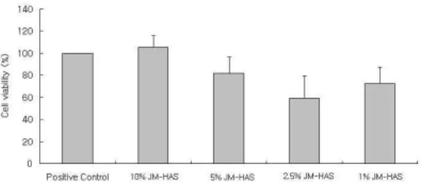
1. 등심초약침액의 세포독성
등심초약침액을 1 %, 2.5 %, 5 %, 10 %로 희석하여 정상 흰쥐의 간세포에 대한 세포독성을 확인하였다.
등심초약침액의 농도가 높을수록 세포의 viability가 증가되는 경향을 보였으나 유의성은 없었다(Fig. 1).
2. 음곡에 시술한 등심초약침이 LPS로 유도된 흰쥐의 신장염에 미치는 영향 1) 혈액학적 분석
흰쥐에 LPS로 신장염을 유도하고, 1시간 후에 흰 쥐의 혈액을 채취하여 WBC의 수와 WBC 중 호중구 의 비율을 측정하였다.
(1) WBC
흰쥐에 LPS로 신장염을 유도하고, 1시간 후에 흰
Fig. 1. Cytotoxicity of JM-HAS on rat liver cells
Liver cells from normal SD rat were cultured in RPMI 1640 with 10 % FBS medium for 72 h with or without various concentrations of JM-HAS(Junci Medulla herbal acupuncture solution). The cell viability were measured by MTT assay. Values represent the means ± SEM of 3 independent experiments.Control : culture medium without JM-HAS.
10 %, 5 %, 2.5 %, 1 % JM-HAS : culture medium with 10 %, 5 %, 2.5 %, 1 % JM-HAS respectively.
Fig. 2. Effect of JM-HA on WBC count in blood of LPS-stimulated rats
The male SD rats were treated as described in the materials and methods and injected intra-peritoneally with LPS(2 ㎎/㎏). Blood samples were taken from rat hearts 1 hr after the LPS injection and WBC count was analysed.
Data were expressed as mean ± SD(n=5).
Normal : normal SD rat.
LPS : LPS(2 ㎎/㎏) challenge.
NP : LPS(2 ㎎/㎏) challenge and a needle prick at KI10. Saline : LPS(2 ㎎/㎏) challenge and saline(200 ㎕/rat)
injection at KI10.
JM-HA : LPS(2 ㎎/㎏) challenge and JM-HA(10 %, 200
㎕/rat) at KI10.
** : p<0.01 compared to normal group by t-test.
††: p<0.01.
†: p<0.05 compared to LPS group by ANOVA test.
쥐의 혈중 WBC 수를 측정하였다. LPS군의 혈중 WBC는 정상군에 비하여 유의하게 증가하였다. NP군 과 saline군, 등심초약침군의 혈중 WBC는 LPS군에 비하여 유의하게 감소하였다(Fig. 2).
(2) 호중구(neutrophil)
흰쥐에 LPS로 신장염을 유도하고, 1시간 후에 흰 쥐의 혈액을 채취하여 WBC 중 neutrophil 비율을 측 정하였다. LPS군과 saline군의 호중구 비율은 정상군
Fig. 3. Effect of JM-HA on neutrophil count in blood of LPS-stimulated rats
The male SD rats were treated as described in the materials and methods and injected intra-peritoneally with LPS(2 ㎎/㎏). Blood samples were taken from rat hearts 1 hr after the LPS injection and the percentage of neutrophil out of WBC count was analysed. Data were expressed as mean ± SD(n=5).
Normal : normal SD rat.
LPS : LPS(2 ㎎/㎏) challenge.
NP : LPS(2 ㎎/㎏) challenge and a needle prick at KI10. Saline : LPS(2 ㎎/㎏) challenge and saline(200 ㎕/rat)
injection at KI10.
JM-HA : LPS(2 ㎎/㎏) challenge and JM-HA(10%, 200 ㎕ /rat) at KI10.
** : p<0.01 compared to normal group by t-test.
††: p<0.01 compared to LPS group by ANOVA test.
‡‡: p<0.01 compared to NP group by ANOVA test.
## :p<0.01 compared to saline group by ANOVA test.
에 비하여 유의하게 증가하였다. 등심초약침군에서는 LPS군, NP군 및 saline군에 비하여 호중구 비율이 유 의하게 감소하였다(Fig. 3).
2) Blood chemistry
흰쥐에 LPS로 신장염을 유도하고, 1시간 후에 흰 쥐의 혈액을 채취하여 혈청을 분리한 후, 혈청 TNF- α , creatinine, BUN의 농도를 측정하였다.
(1) TNF-α
흰쥐에 LPS로 신장염을 유도하고 1시간 후에 흰쥐 의 혈청 TNF-α 농도를 확인하였다. LPS군에서는 정 상군에 비하여 혈청 TNF-α가 현저하게 증가하였다.
Saline군 및 등심초약침군은 LPS군에 비하여 혈청 TNF-α 농도가 유의하게 감소하였다(Fig. 4).
(2) Creatinine
흰쥐에 LPS로 신장염을 유도하고 1시간 후에 흰쥐
의 혈청 creatinine 농도를 확인하였다. LPS군에서는
정상군에 비하여 혈청 creatinine이 유의하게 증가하
였다. 등심초약침군에서는 LPS군에 비하여 혈청
creatinine 농도가 유의하게 감소하였다(Fig. 5).
Fig. 4. Effects of JM-HA on serum TNF-α level in LPS-stimulated rats
The male SD rats were treated as described in the materials and methods and injected intra-peritoneally with LPS(2
㎎/㎏). Blood samples were taken from rat hearts 1 hr after the LPS injection and serum TNF-α level was analysed. Data were expressed as mean ± SD(n=5).
Normal : normal SD rat LPS : LPS(2 ㎎/㎏) challenge
NP : LPS(2 ㎎/㎏) challenge and a needle prick at KI10. Saline : LPS(2 ㎎/㎏) challenge and saline(200 ㎕/rat)
injection at KI10.
JM-HA : LPS(2 ㎎/㎏) challenge and JM-HA(10%, 200
㎕/rat) at KI10.
*** : p<0.001 compared to normal group by t-test
†:p<0.05 compared to LPS group by ANOVA test
‡:p<0.05 compared to N.P. group by ANOVA test
Fig. 5. Effects of JM-HA on serum creatinine level in LPS-stimulated rats
The male SD rats were treated as described in the materials and methods and injected intra-peritoneally with LPS (2
㎎/㎏). Blood samples were taken from rat hearts 1 hr after the LPS injection and serum creatinine level was analysed. Data were expressed as mean ± SD(n=5).
Normal : normal SD rat.
LPS : LPS(2 ㎎/㎏) challenge.
NP : LPS(2 ㎎/㎏) challenge and a needle prick at KI10. Saline : LPS(2 ㎎/㎏) challenge and saline(200 ㎕/rat)
injection at KI10.
JM-HA : LPS(2 ㎎/㎏) challenge and JM-HA(10 %, 200
㎕/rat) at KI10.
* : p<0.05 compared to normal group by t-test.
† : p<0.05 compared to LPS group by ANOVA test.
## : p<0.01 compared to saline group by ANOVA test.
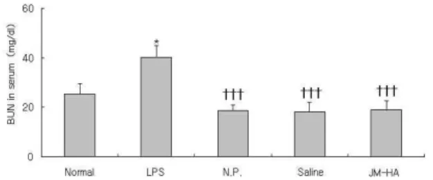
(3) BUN(blood urea nitrogen)
흰쥐에 LPS로 신장염을 유도하고 1시간 후에 흰쥐 의 혈청 BUN 농도를 확인하였다. LPS군에서는 정상 군에 비하여 혈청 BUN 농도가 유의하게 증가하였다.
NP군, saline군, 등심초약침군 모두 LPS군에 비하여 혈청 BUN 농도가 유의하게 감소하였다(Fig. 6).
Fig. 6. Effects of JM-HA on serum BUN level in LPS-stimulated rats
The male SD rats were treated as described in the materials and methods and injected intra-peritoneally with LPS(2 ㎎/㎏). Blood samples were taken from rat hearts 1 hr after the LPS injection and serum BUN level was analysed. Data were expressed as mean ± SD(n=5).
Normal : normal SD rat.
LPS : LPS(2 ㎎/㎏) challenge.
NP : LPS(2 ㎎/㎏) challenge and a needle prick at KI10. Saline : LPS(2 ㎎/㎏) challenge and saline(200 ㎕/rat)
injection at KI10.
JM-HA : LPS(2 ㎎/㎏) challenge and JM-HA(10 %, 200
㎕/rat) at KI10.
* : p<0.05 compared to normal group by t-test.
†††: p<0.001 compared to LPS group by ANOVA test.
3) 소변 분석
흰쥐에 LPS로 신장염을 유도하고 12시간 동안 소 변을 채취하여 요중 creatinine과 total protein의 양을 측정하였다.
(1) Creatinine
흰쥐에 LPS로 신장염을 유도하고 12시간 동안 소 변을 채취하여 요중 creatinine 양을 측정하였다. LPS
Fig. 7. Effects of JM-HA on urinary creatinine level in LPS-stimulated rats
The male SD rats were treated as described in the materials and methods and injected intra-peritoneally with LPS(2 ㎎ /㎏). The rats were kept in the metabolic cages and the urine was collected for 12 hours. And the urinary creatinine level was analysed. Data were expressed as mean ± SD(n=3).
Normal : normal SD rat.
LPS : LPS(2 ㎎/㎏) challenge.
NP : LPS(2 ㎎/㎏) challenge and a needle prick at KI10. Saline : LPS(2 ㎎/㎏) challenge and saline(200 ㎕/rat)
injection at KI10.
JM-HA : LPS(2 ㎎/㎏) challenge and JM-HA(10 %, 200
㎕/rat) at KI10.
* : p<0.05 compared to normal group by t-test.
군의 creatinine 농도는 정상군에 비하여 요중 creati- nine은 유의하게 증가하였다. 등심초약침군, NP군, saline 군에서 모두 LPS군에 비하여 요중 creatinine 농도가 감소하였으나 유의성은 나타나지 않았다(Fig. 7).
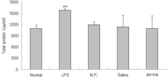
(2) Total protein
흰쥐에 LPS로 신장염을 유도하고 12시간 동안 소
Fig. 8. Effects of JM-HA on total protein level in urine of LPS-stimulated rats
The male SD rats were treated as described in the materials and methods and injected intra-peritoneally with LPS(2 ㎎ /㎏). The rats were kept in the metabolic cages and the urine was collected for 12 hours. And, total protein level in urine was analysed. Data were expressed as mean ± SD(n=3).
Normal : normal SD rat.
LPS : LPS(2 ㎎/㎏) challenge.
NP : LPS(2 ㎎/㎏) challenge and a needle prick at KI10. Saline : LPS(2 ㎎/㎏) challenge and saline(200 ㎕/rat)
injection at KI10.
JM-HA : LPS(2 ㎎/㎏) challenge and JM-HA(10 %, 200
㎕/rat) at KI10.
** : p<0.01 compared to normal group by t-test.
Fig. 9. Effects of JM-HA on renal MPO level in LPS-stimulated rats
The male SD rats were treated as described in the materials and methods and injected intra-peritoneally with LPS(2 ㎎/㎏). 3 hours after the LPS stimulation, rat kidney was removed and renal MPO level was analysed by ELISA. Data were expressed as mean ± SD(n=5).
Normal : normal SD rat.
LPS : LPS(2 ㎎/㎏) challenge.
NP : LPS(2 ㎎/㎏) challenge and a needle prick at KI10. Saline : LPS(2 ㎎/㎏) challenge and saline(200 ㎕/rat)
injection at KI10.
JM-HA : LPS(2 ㎎/㎏) challenge and JM-HA(10 %, 200
㎕/rat) at KI10.
*** : p<0.001 compared to normal group by t-test.
††: p<0.01 compared to LPS group by ANOVA test.
‡: p<0.05 compared to NP group by ANOVA test.
변을 채취하여 요중 total protein량을 측정하였다.
LPS군에서는 정상군에 비하여 요중 total protein이 유의하게 증가하였다. 등심초약침군의 요중 total protein 은 LPS군에 비하여 감소하였으나 유의성은 없었다 (Fig. 8).
4) 신장 내 MPO 활성