⋅교신저자: 임윤경, 대전 동구 용운동 대전대학교 한의과대학 경락 경혈학교실, Tel. 042-280-2610, Fax. 042-280-2641, E-mail : [email protected]
․ 투고 : 2009/02/28 심사 : 2009/03/18 채택 : 2009/06/20
음곡 차전자 약침이 LPS로 유도된 흰쥐의 신장염에 미치는 영향
박병문, 허부, 임윤경
대전대학교 한의과대학 경락경혈학교실
Anti-inflammatory Effect of Plantaginis Semen Herbal-acupuncture at KI10 on LPS-induced nephritis in rats
Byong-mun park, Hur Boo, Yun-Kyoung Yim
Dept. of Meridian & Acupoint, College of Oriental Medicine, Daejeon University Abstract
Objective : This study aimed to evaluate the effects of Plantaginis Semen herbal-acupuncture (PS-HA) at KI10 (Umgok) on nephritis induced by lipopolysaccharide (LPS) in rat.
Methods : The authors performed several experimental items including measurements of urinary volume, WBC in blood, BUN, creatine, TNF-α, CINC-1 in serum, creatinine, total protein in urine, TNF-α, MPO in kidney and histological analysis of renal tissue.
Results : PS-HA at KI10 significantly reduced WBC in blood, BUN, TNF-α in serum of LPS-stimulated rats. PS-HA at KI10 significantly increased urinary volume in LPS-stimulated rats. And PS-HA at KI10 significantly reduced MPO in kidney of LPS-stimulated rats.
Conclusion : Taken together, PS-HA at KI10 has a therapeutic effect on nephritis in LPS-stimulated rat.
Therefore, it is suggested that PS-HA at KI10 may be an useful therapeutics for nephritis in clinical field.
Key words : Nephritis, Herbal-acupuncture, KI10, Plantaginis Semen
Ⅰ. 서 론
신장은 후복막장기로 전해질과 수분을 조 절하여 순환혈액량을 조절하고, 암모니아 대 사산물인 요소를 포함한 독성물질을 배출함 으로써 체내환경을 유지하는 장기로 순환혈 액의 20~25%가 공급되며, 여과와 재흡수를 위하여 얇은 막구조를 가지고 있어 독성물 질에 의한 손상에 취약하다1).
신장의 사구체에 발생하는 사구체신염은 감염에 의한 면역복합체나, 항체가 사구체기
저막에 침착되어 발생한다. 사구체기저막에 침착된 면역복합체나 세균에 대한 항체는 면역시스템을 활성화시켜 사구체를 손상시 켜, 혈뇨, 단백뇨 등을 유발하고, 여과기능의 손상까지 진행되면, 핍뇨와 요독증이 발생한 다2).
LPS(Lipopolysaccharide)는 그람음성박테 리아의 세포막의 구성성분으로 대식세포의 활성화 인자이며 자극된 대식세포는 TNF- α, IL-1, IL-6, IL-10 등과 같은 cytokine을 생성하여3-5), 세포매개성 면역반응 및 염증 반응을 유발한다. 또 LPS는 메산지움 세포 에 많은 양의 nitric oxide 분비를 유발시키
며 메산지움 세포와 tubular epithelial cell에 Fas 발현을 증가시킴으로서 직간접적으로 신장손상을 초래한다16-18).
음곡(KI10)은 足少陰腎經의 合水穴이며, 足少陰腎經, 衝脈, 陰維脈의 交會穴로서, 補 腎培元, 調經利水의 효능이 있어 陽萎, 月經 不調, 崩漏, 小便難 등을 치료한다6). 차전자 (Plantaginis Semen)는 질경이과(차전 과:Planaraginaceae)에 속한 다년생 초본인 질경이 또는 털질경이의 성숙한 종자로 性 은 微寒하고, 味는 甘하며, 歸經은 腎, 肝, 肺, 小腸經이며, 淸熱利尿, 滲濕通淋, 明目, 袪痰의 효능이 있다7).
급성신부전 및 신장독성에 관한 기존의 연구로는 육미지황탕8), 단삼9), 녹용10) 등의 약물을 이용하여 유효한 효과를 보인 연구 가 보고된 바 있었으며, 차전자약침을 이용 한 연구로는, 권 등11)의 肝兪 차전자 약침이 간손상에 미치는 영향에 대한 연구, 조 등12) 의 腎兪 차전자 약침이 glycerol로 유발된 급성신부전에 미치는 영향에 대한 연구, 박 등13)의 腎兪 차전자 약침이 cisplatine을 이 용한 항암치료에서 유발되는 급성신부전에 미치는 영향에 대한 보고가 있었으나, 陰谷 에 시술한 차전자약침이 LPS로 유발된 신 장염에 미치는 영향을 보고한 예는 없었다.
이에 저자는 음곡 차전자 약침이 독소에 의한 신장염에 미치는 영향을 알아보고자, 차전자 약침액을 만들어, LPS로 유도된 신 장염 병태모델 흰쥐의 음곡 상응부위에 시 술하고, 이뇨활성, 혈액과 소변, 신장내 염증 활성평가, 조직학적 분석 등을 통하여 유의 한 결과를 얻었기에 보고하는 바이다.
Ⅱ. 실 험 1. 재 료
1) 동 물
동물은 6 주령의 웅성 SD rat을 (주)대한 바이오링크(경기도, 한국)에서 공급받아 1주 일간 실험실 환경에 적응시킨 후 실험에 사 용하였다. 실험에 사용된 동물은 실험 당일 까지 고형사료(삼양사료, 한국)와 물을 충분 히 공급하고, 실험실은 실온(22±2 ℃)과 습 도 20~60%를 유지하였다.
2) 약재
본 실험에 사용된 차전자 (Plantaginis Semen, PS)는 대전대학교 부속 한방병원에 서 구입한 것을 정선하여 실온에서 보관하 였으며, 사용하기 전 초음파세척을 실시하였 다.
3) 시약 및 기기
⑴ 시약
시 약 명 제조사 제조국
Phosphate buffer saline(PBS) Collagenase typeⅣHBSS Lipopolysaccharide(LPS)MTT
gum-sucrose formalin absolute EtOH Harris Hematoxlin
Eosin B
Sigma
USA RPMI 1640 media
Antibiotics
FBS Hyclone
TNF-alpha ELISA Kit Creatinine assay kit
CINC-1 ELISA Kit MPO ELISA Kit
Oxford Bio Assay Systems
ALPCO Cellscience
Ethyl ether Samchun Korea
⑵ 기기
기 기 명 제조사 제조국
Bio-freezer Sanyo
Japan Rotary evaporator Eyela
Digital camera
Microscope Nicon
ELISA reader TARAN Canada
Sonicate Fisher USA
Ice maker Vision science
Korea Metabolic cage B&P
Cytological centrifuge Hanil Micro centrifuge
Cryotome Shandon UK
2. 방 법
1) 약침액의 제조
분쇄기를 이용해서 차전자 55 g을 분쇄하 여 분말로 만들어, 삼각 flask에 넣고 증류 수 500 ㎖를 가하여 3시간 동안 shaking water bath에서 유출한 후, 유출액을 여과 하였다. 여과액을 여과지로 3회 여과한 후, rotary evaporator에 감압농축 하였다. 농축 액에 95% ethyl alcohol 30 ㎖를 가하여, 실 온에서 교반한 후 방치하여, 침전물이 생성 되게 한 후 여과하였다. 이 여과액을 rotary evaporator로 감압농축한 후, 농축액을 다시 여과하였다. 이 여과액에 85% ethyl alcohol 30 ㎖를 가하여 잠시 교반 후 방치하여, 침 전물이 생성되게 한 후 여과하였다. 여과액 에 75% ethyl alcohol 30 ㎖를 가하고 교반 한 후 방치하였다가 다시 여과하는 조작을 2회 반복하였다. 여과액중의 ethyl alcohol 성분을 rotary evaporator로 감압 제거하고, 남은 농축액이 20 ㎖가 되게 하였다. 차전자 55 g에서 얻은 차전자약침액 건조 분말은
1.203 g(수율 2.19%)이었다. 1N NaOH를 이 용하여 농축액을 pH 6.8이 되도록 조절하 고, 4℃에서 12시간 방치한 후, 침전물을 제 거하기 위해 syringe filtering을 실시하였다.
여과된 농축액에 PBS를 첨가하여 5%로 희 석하여 약침액으로 사용하였다(Scheme 1).
PS (Plantaginis Semen) (55 g)
- Extracted with D.W. 500 ㎖ for 3 hr Filtrated and evaporated three times
Residue discarded Extracts(D.W.) - Suspended in D.W.
Residue discarded Filtrate
- Precipitated in 95% EtOH and standing at cooling temperature
- Filtration
Precipitated Filtrate
- Vacuum concentration and cooling it off to room temperature - Precipitated in 85% EtOH and
standing at cooling temperature - Filtration
Precipitated Filtrate
- Vacuum concentration and cooling it off to room temperature
- Precipitated in 75% EtOH and standing at cooling temperature
(repeated twice) - Filtration
Precipitated Filtrate
- Evaporated 2.5 ㎖ and add PBS 47.5 ㎖ PS Herbal-acupuncture solution (PS-HAS)
Scheme 1. M anufacturing procedure of PS Herbal acupuncture Solution.
2) 세포독성
⑴ 간세포 분리
본 실험모델과 동일한 6주령 수컷 SD rat 의 간조직을 사용하였다. 백서를 ethyl ether 를 이용하여 마취시킨 후, 대동맥 혈관에 HBSS(Ca2+, Mg2+ free)를 투여하며 복부쪽
혈관을 절단시켜 동물의 혈액을 모두 배출 하였다. 조직을 잘게 잘라서 RPMI 1640 media(with 10% FBS)와 collagenase type
Ⅳ(300 u/㎖)를 넣고 실온에서 90분간 incubation 하였다. 이 때 20분에 한 번씩 흔들어 줌으로써 간세포가 잘 분리되도록 하였다. 얻어진 간세포는 RPMI 1640 media 에 세척한 후, 세포를 계수하였다.
⑵ MTT assay
분리된 간 실질세포의 viability를 trypan blue를 이용하여 확인한 뒤, 96 well plate에 세포를 1×10⁵ cells/well으로 분주하고, FBS 10%와 antibiotics를 첨가한 RPMI 1640 media에 차전자 전탕액을 1%, 2.5%, 5%
10%로 처리하고, 72시간동안 세포 배양을 실시하였다. 72시간 후에, MTT solution (5
㎎/㎖, Cat No. 135038, Sigma, USA)을 각 well에 20 ㎕씩 분주하고 5시간 동안 37℃
에서 incubation을 실시하였다. 5시간 후, 각 well에 있는 medium을 100 ㎕씩 버리고, solubilizing solution을 100 ㎕씩 분주한 뒤 pipetting을 강하게 하여 well에 dark blue crystals가 침전하는 정도를 ELISA reader 를 이용하여 570 ㎚에서 optical densities로 확인하였다.
3) 신장염 동물 모델
6주령 수컷 SD rat에 LPS(2 ㎎/kg)를 복 강 투여하여 신장염을 유도하였다14,15). 각 실험군에는 실험동물을 8마리씩 배정하였다.
LPS 투여 1시간 뒤, 각 실험군의 8마리 중 5마리에서 혈액 sample을 채취하였으며, LPS 투여 3시간 후에는 혈액을 채취했던 5
마리를 sacrifice하여 신장을 적출하였다. 각 실험군의 8마리 중 나머지 3마리는 LPS 투 여 후 metabolic cage에 넣어 12시간 동안 소변 sample을 채취하였다.
Scheme 2. Experimental Procedure
4) 실험군 분류 및 처치
실험동물은 정상군(normal), LPS군(LPS), NP군(needle prick, NP), saline대조군 (Saline), 차전자약침군(PS-HA)의 5군으로 나누었으며, 각 실험군에는 실험동물을 8마 리씩 배정하였다. 정상군을 제외한 LPS군, NP군, saline대조군, 차전자약침군은 각각 2 ㎎/kg의 LPS를 복강에 투여하였다. NP군 과 saline대조군, 차전자약침군은 LPS 투여 1 주일 전에 음곡(KI10) 상응부위에 좌우 교대로 NP(needle prick)자극, saline 주입, 차전자약침처치를 주3회 실시하였다. 각 실 험군에 대한 처치는 다음과 같다.
(1) 정상군(Normal) : 아무 처치하지 않은 정상 SD rat군.
(2) LPS군(LPS) : 실험 당일 LPS를 2 ㎎ /kg으로 복강 투여한 군.
(3) NP군(N.P.) : 실험 전 1 주일간 주 3회 음곡(KI10) 상응부위에 26 gauge 주사 기를 0.5 cm 정도 깊이로 자입한 후 즉 시 제거하고(needle prick, NP), 실험 당 일 LPS 2 ㎎/kg을 복강에 투여한 군.
(4) Saline대조군(LPS+saline) : 실험전 1 주
일간 saline 0.2 ㎖를 주 3회 음곡(KI10) 상응부위에 주입하고, 실험 당일 LPS 2
㎎/kg을 복강에 투여한 군.
(5) 차전차 약침군(PS-HA) : 실험전 1 주일 간 10%의 차전차 약침액 0.2 ㎖를 주 3 회 음곡(KI10) 상응부위에 주입하고, 실 험 당일 LPS 2㎎/kg을 복강에 투여한 군.
5) 신장염 평가
(1) 이뇨활성 평가이뇨활성을 확인하기 위해, 실험동물을 한 마리씩 metabolic cage에 수용하고 12시간 동안 소변을 채취하여 배뇨량을 측정하였다.
(2) 혈액 분석
LPS 처리 1시간 뒤, ethyl ether를 이용하 여 rat를 마취하고 심장 채혈 법으로 각 동 물에서 4 ㎖의 혈액을 채취하였다. (주) 이 원 임상검사센터(대전. 한국)에 의뢰하여 혈 중 WBC와, WBC 중 neutrophils의 비율을 측정하였으며, 혈청 BUN의 농도를 확인하 였다. 혈청 TNF-α, CINC-1 (Cytokine-induced neutrophil chemoattractant-1), creatinine의 농도는 ELISA kit를 이용하여 확인하였다.
(3) 소변 분석
Metabolic cage에서 12시간동안 채취한 소변을 (주)이원 임상검사센터(대전. 한국)에 의뢰하여 뇨중 total protein과 creatinine 농 도를 측정하였다.
(4) 신장 내 염증활성 평가
LPS 처리 3시간 후, ethyl ether를 이용하
여 백서를 마취하고 대동맥 혈관에 HBSS (Ca2+, Mg2+ free)를 투여하여 방혈한 후 신 장을 적출하였다. 적출된 신장조직은 0.05%
sodium azide가 첨가된 0.1M phosphate buffer (pH7.4)를 넣고 sonicate (Fisher, USA)하여 잘게 간 후, 2000×g에서 10분간 원심분리하였다. 분리된 supernatant는 -80
℃에서 보관되었다가 ELISA kit을 이용하여 TNF-α와 MPO(myeloperoxydase)의 농도를 측정하였다.
(5) 조직학적 분석
LPS 처리 3시간 후, 실험동물의 신장을 적취하여, 10%의 formalin에 고정한다. 고정 된 조직은 왁스에 format 시켜 5 ㎛의 두께 로 박절한 후 조직 절편을 슬라이드에 부착 시키고 실내에서 30분 이상 방치하였다. 슬 라이드에 부착된 조직은 Harris hematoxylin에서 1분 정도 염색을 실시하고 흐르는 물에 씻어주었다. 물기를 제거하고 1% acid alcohol에 2, 3번 정도 넣었다 빼 준 뒤, 흐르는 물에 슬라이드를 씻어준 후, 0.3% ammonia water에 중화시키기 위해 5~10번 정도 넣었다 빼 주었다. 다시 흐르 는 물에 슬라이드를 씻어주고, Eosin에서 3 분 정도 염색처리한 뒤, 95% alcohol에 10번 정도 넣었다 빼 주고, 다시 깨끗한 95%
alcohol에서 10번 정도 넣었다 빼 주었다.
남은 alcohol성분은 흡수지를 이용하여 완전 하게 alcohol성분을 제거하고, 100% alcohol 에 10번 정도 넣었다 빼 준 후, 다시 깨끗한 100% alcohol에서 10번 정도 넣었다 빼 주 었다. 남은 alcohol 성분을 제거하고, xylene 에서 10번 정도 넣었다 빼 주고, 다시 깨끗
한 xylene에서 10번 정도 넣었다 빼 준 뒤, 봉입하였다. 봉입을 끝낸 조직은 광학현미경 을 이용하여 관찰한 후, digital camera를 이용하여 사진을 촬영하였다.
6) 통계분석
실험 결과는 각 실험군에서 얻은 결과값 의 평균과 표준편차로 나타내었다. 통계프로 그램은 SPSS(14.0 KOR)을 이용하여 분석 하였다. Fig. 1의 데이터는 ANOVA-test를 이용하여 여러 농도간 결과값의 평균을 비 교하였으며, Fig. 2와 Fig. 3에서는 t-test를 이용하여 LPS 처리군과 비처리군의 평균을 비교하였다. 이후 Fig. 4부터는 정상군과 LPS군의 평균을 t-test로 비교하고, 다시 LPS군, N.P.군, saline군, PS-HA군의 평균 값을 ANOVA test를 이용하여 비교하였으 며, ANOVA test의 사후검정은 Scheffe's test를 이용하였다. 신뢰도는 95%이상 (p<0.05)일 때 유의성이 있는 것으로 판정하 였다.
Ⅲ. 성 적 1. 세포독성
차전차 약침액 원액을 1%, 2.5%, 5%, 10%로 희석하여 정상 백서의 간세포에 대
한 세포독성을 확인하였다. 차전차 약침액의 농도가 높을수록 세포의 viability가 증가하 였다(Fig. 1).
Fig. 1. Cytotoxicity of PS-HAS on rat liver cells.
Liver cells from normal SD rat were cultured in RPMI 1640 with 10% FBS medium for 72 h with or without various concentrations of PS-HAS (Plantaginis Semen herbal acupuncture solution). The cell viability were measured by MTT assay. Values represent the means ± SD of 3 independent experiments.
Control : culture medium without PS-HAS
10%, 5%, 2.5%, 1% PS-HAS : culture medium with 10%, 5%, 2.5%, 1% PS-HAS respectively
*** : p<0.001, compared to control by ANOVA test
††† : p<0.001, †† : p<0.01 compared to 10% PS-HAS by ANOVA test
2. LPS투여 따른 TNF-α의 농도 변화
1) Serum TNF-α
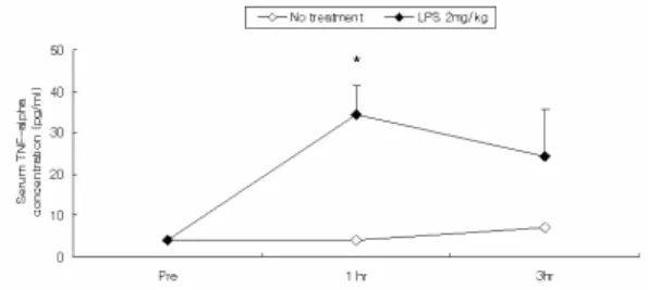
신장염을 유도하고, 1시간 후와 3시간 후 에 serum TNF-alpha의 농도를 관찰하였다.
LPS 복강투여 1시간 후에 serum TNF-alpha의 농도가 크게 증가하여 LPS를 투여하지 않은 군에 비하여 유의하게 높았 다. 3시간 후에는 1시간 후에 비하여 LPS 투여군의 serum TNF-alpha가 감소하여 no-treatment군과 유의한 차이를 나타내지 않았다(Fig. 2).
Fig. 2. Effect of LPS on serum TNF-alpha level in SD rats.
The male SD rats were injected intra-peritoneally with LPS(lipopolysaccharide; 2 ㎎/㎏). Blood samples were taken from rat heart 1 hr and 3 hr after the LPS injection, and serum TNF-alpha level was analysed by ELISA. Data were expressed as mean ± SD (n=3).
No treatment : normal SD rat.
LPS 2 ㎎/kg: SD rat with LPS (2㎎/㎏) challenge
* : p<0.05, compared to no-treatment group by t-test
2) Renal TNF-α
신장염을 유도하고, 1시간 후와 3시간 후 에 각각 신장을 적출하여 renal TNF-alpha 의 농도를 관찰하였다. LPS 복강투여 1시간 후에 renal TNF-alpha의 농도는 LPS를 투 여하지 않은 군에 비하여 차이를 보이지 않 았다. LPS 투여 3시간 후 renal TNF-alpha 의 농도는 LPS를 투여하지 않은 군에 비하 여 유의하게 증가하였다(Fig. 3).
Fig. 3. Effect of LPS on renal TNF-alpha level in SD rats.
The male SD rats were injected intra-peritoneally with LPS (2 ㎎/㎏). Rat kidney was removed and renal TNF-alpha level was analysed by ELISA. Data were expressed as mean
± SD (n=3).
No treatment : normal SD rat
LPS 2 ㎎/kg: SD rat with LPS (2㎎/㎏) challenge
** : p<0.001, compared to no-treatment group by t-test
3. 차전자 전탕액 경구투여가 LPS에 의 한 rat의 신장염에 미치는 영향
1) 이뇨 활성
신장염을 유도한 후 metabolic cage에 한 마리씩 수용하고 12시간동안 소변을 채취하 여 배뇨량을 측정하였다. LPS군에서는 정상 군에 비하여 배뇨량이 유의하게 감소하였다.
차전자약침군(PS-HA)에서는 LPS군, NP군 및 saline군에 비하여 배뇨량이 유의하게 증 가하였다.
Fig. 4. Effect of PS on urinary volume in LPS-stimulated rats.
The male SD rats were treated as described in the materials and methods and injected intra-peritoneally with LPS (2 ㎎/
㎏). The rats were kept in metabolic cages and the urine was collected for 12 hours and the amount was measured.
Data were expressed as mean ± SD (n=3).
Normal : normal SD rat LPS : LPS (2 ㎎/㎏) challenge
N.P.: LPS (2 ㎎/㎏) challenge and needle prick at KI10 Saline : LPS (2 ㎎/㎏) challenge and saline(200 ㎕) injcetion at KI10
PS-HA : LPS (2 ㎎/㎏) challenge and PS-HA(10%, 200 ㎕) at KI10
* : p<0.05 compared to normal group by t-test
†† : p<0.01 compared to LPS group by ANOVA test
‡‡ : p<0.01 compared to N.P. group by ANOVA test
## : p<0.01 compared to saline group by ANOVA test
2) 혈액 분석
신장염을 유도하고, 1시간 후에 혈액을 채 취하여 WBC의 수와 WBC 중 neutrophil의 비율을 측정하였다.
Table 1. Blood cell counts in LPS-stimulated rats.
Normal LPS N.P. Saline PS-HA (×10WBC3Cell/㎖) 3.07
±2.40 11.624
±0.63 2.934
±2.43 6.25
±3.33 1.97
±1.02 Neutrophil
(%) 84.6
±2.79 94.2
±2.39 89.6
±4.88 92.6
±1.14 89.4
±5.03
The male SD rats were treated as described in the materials and methods and injected intra-peritoneally with LPS (2 ㎎/
㎏). Blood samples were taken from rat hearts and the blood cells were counted. Data were expressed as mean ± SD (n=5).
Normal : normal SD rat LPS : LPS (2 ㎎/㎏) challenge
N.P.: LPS (2 ㎎/㎏) challenge and administration with needle prick at KI10
Saline : LPS (2 ㎎/㎏) challenge and saline(200 ㎕) injcetion at KI10
PS-HA : LPS (2 ㎎/㎏) challenge and PS-HA(10%, 200 ㎕) at KI10
(1) WBC
신장염을 유도하고, 1시간 후에 혈중 WBC 수를 측정하였다. LPS군의 혈중 WBC는 정상군에 비하여 유의하게 증가하 였다. NP군, saline군 및 차전자약침군에서 는 LPS군에 비하여 혈중 WBC가 유의하게 감소하였다(Fig. 5).
(2) Neutrophil
신장염을 유도하고, 1시간 후에 혈액을 채 취하여 WBC 중 neutrophil 비율을 측정하 였다. LPS군과 saline군의 neutrophil 비율은 정상군에 비하여 유의하게 증가하였다. 차전 자약침군에서는 LPS군에 비하여 neutrophil 비율이 약간 감소하였으나 유의성은 없었다 (Fig. 6).
Fig. 5. Effect of HJ on W BC count in blood of LPS-stimulated rats.
The male SD rats were treated as described in the materials and methods and injected intra-peritoneally with LPS (2 ㎎/
㎏). Blood samples were taken from rat hearts and WBC count was analysed. Data were expressed as mean ± SD (n=5).
Normal : normal SD rat LPS : LPS (2 ㎎/㎏) challenge
N.P.: LPS (2 ㎎/㎏) challenge and administration with needle prick at KI10
Saline : LPS (2 ㎎/㎏) challenge and saline(200 ㎕) injcetion at KI10
PS-HA : LPS (2 ㎎/㎏) challenge and PS-HA(10%, 200 ㎕) at KI10
** : p<0.001, compared to normal group by t-test
††† : p<0.001, †† : p<0.01 compared to LPS group by ANOVA test
# : p<0.05 compared to Saline group by ANOVA test
Fig. 6. Effect of HJ on neutrophil count in blood of LPS-stimulated rats.
The male SD rats were treated as described in the materials and methods and injected intra-peritoneally with LPS (2 ㎎/
㎏). Blood samples were taken from rat hearts and the percentage of neutrophil out of WBC count was analysed.
Data were expressed as mean ± SD (n=5).
Normal : normal SD rat LPS : LPS (2 ㎎/㎏) challenge
N.P.: LPS (2 ㎎/㎏) challenge and administration with needle prick at KI10
Saline : LPS (2 ㎎/㎏) challenge and saline(200 ㎕) injcetion at KI10
PS-HA : LPS (2 ㎎/㎏) challenge and PS-HA(10%, 200 ㎕) at KI10
** : p<0.01, compared to normal group by ANOVA test
3) Blood chemistry
신장염을 유도하고, 1시간 후에 혈액을 채 취하여 혈청을 분리한 후, 혈청 BUN, creatinine, TNF-alpha 그리고 CINC-1의 농도를 측정하였다.
Table 2. Blood chemistry analysis in LPS-stimulated rats.
LPS N.P. Saline PS-HA
BUN(㎎/㎗) 25.52±4.1 40.16
±4.8 18.74
±2.3 18.26
±3.8 21.48
±3.3 Creatinine
(㎎/㎗) 0.412
±0.1 1.40
±0.2 1.088
±0.3 1.593
±0.2 0.808
±0.4 TNF-alpha
(pg/㎖) 25.029
±14.1 222.143
±38.1 225.857
±3.3 127.314
±48.5 117
±29.1 CINC-1
(pg/㎖) 255.429
±19.0 612.714
±21.3 378.357
±104.4 411
±212.4 311.143
±43.5
The male SD rats were treated as described in the materials and methods and injected intra-peritoneally with LPS (2 ㎎/
㎏). Blood samples were taken from rat hearts and serum was isolated. The levels of IgG, BUN, creatinine, TNF-alpha and CINC-1 in serum were analysed. Data were expressed as mean ± SD (n=5).
Normal : normal SD rat LPS : LPS (2 ㎎/㎏) challenge
N.P.: LPS (2 ㎎/㎏) challenge and administration with needle prick at KI10
Saline : LPS (2 ㎎/㎏) challenge and saline(200 ㎕) injcetion at KI10
PS-HA : LPS (2 ㎎/㎏) challenge and PS-HA(10%, 200 ㎕) at KI10
(1) BUN (Blood urea nitrogen)
신장염을 유도하고 1시간 후에 혈청 BUN 농도를 확인하였다. LPS군에서는 정상군에 비하여 혈청 BUN 농도가 유의하게 증가하 였다. NP군, saline군 및 차전자약침군에서 는 LPS군에 비하여 혈청 BUN 농도가 유의 하게 감소하였다(Fig. 7).
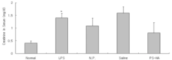
(2) Creatine
신장염을 유도하고 1시간 후에 백서의 혈 청 creatine 농도를 확인하였다. LPS군에서 는 정상군에 비하여 혈청 creatine이 유의하
게 증가하였다. NP군과 차전자약침군에서는 LPS군에 비하여 혈청 creatine 농도가 감소 하였으나 유의성은 없었다(Fig. 8).
Fig. 7. Effects of PS on serum BUN level in LPS-stimulated rats.
The male SD rats were treated as described in the materials and methods and injected intra-peritoneally with LPS (2 ㎎/
㎏). Blood samples were taken from rat hearts and serum BUN level was analysed. Data were expressed as mean ± SD (n=5).
Normal : normal SD rat LPS : LPS (2 ㎎/㎏) challenge
N.P.: LPS (2 ㎎/㎏) challenge and administration with needle prick at KI10
Saline : LPS (2 ㎎/㎏) challenge and saline(200 ㎕) injcetion at KI10
PS-HA : LPS (2 ㎎/㎏) challenge and PS-HA(10%, 200 ㎕) at KI10
* : p<0.05 compared to normal group by t-test
††† : p<0.001 compared to LPS group by ANOVA test
Fig. 8. Effects of PS on serum creatine level in LPS-stimulated rats.
The male SD rats were treated as described in the materials and methods and injected intra-peritoneally with LPS (2 ㎎/
㎏). Blood samples were taken from rat hearts and serum creatine level was analysed. Data were expressed as mean ± SD (n=5).
Normal : normal SD rat LPS : LPS (2 ㎎/㎏) challenge
N.P.: LPS (2 ㎎/㎏) challenge and administration with needle prick at KI10
Saline : LPS (2 ㎎/㎏) challenge and saline(200 ㎕) injcetion at KI10
PS-HA : LPS (2 ㎎/㎏) challenge and PS-HA(10%, 200 ㎕) at KI10
* : p<0.05 compared to normal group by t-test
(3) TNF-α
신장염을 유도하고 1시간 후에 혈청 TNF-α 농도를 확인하였다. LPS군에서는 정상군에 비하여 혈청 TNF-α가 현저하게 증가하였다. Saline군 및 차전자약침군에서 는 LPS군 및 NP군에 비하여 혈청 TNF-α 농도가 유의하게 감소하였다(Fig. 9).
Fig. 9. Effects of PS on serum TNF-alpha level in LPS-stimulated rats.
The male SD rats were treated as described in the materials and methods and injected intra-peritoneally with LPS (2 ㎎/
㎏). Blood samples were taken from rat hearts and serum TNF-alpha level was analysed. Data were expressed as mean
± SD (n=5).
Normal : normal SD rat LPS : LPS (2 ㎎/㎏) challenge
N.P.: LPS (2 ㎎/㎏) challenge and administration with needle prick at KI10
Saline : LPS (2 ㎎/㎏) challenge and saline(200 ㎕) injcetion at KI10
PS-HA : LPS (2 ㎎/㎏) challenge and PS-HA(10%, 200 ㎕) at KI10
*** : p<0.001, compared to normal group by t-test
†† : p<0.01 compared to LPS group by ANOVA test
‡‡ : p<0.01 compared to N.P. group by ANOVA test
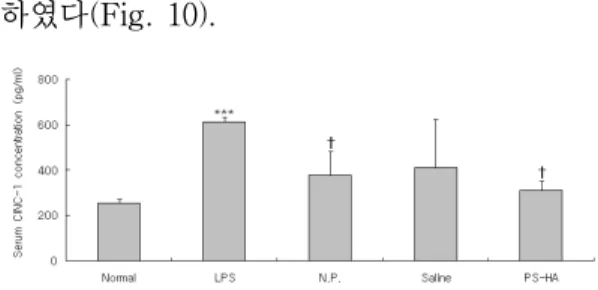
(4) CINC-1 (Cytokine-induced neutrophil chemoattractant-1)
신장염을 유도하고 1시간 후에 혈청 CINC-1 농도를 확인하였다. LPS군에서는 정상군에 비하여 혈청 CINC-1이 유의하게 증가하였다. Saline군에서는 LPS군에 비하 여 CINC-1이 감소하였으나 유의성은 없었 다. NP군 및 차전자약침군에서는 LPS군에 비하여 혈청 CINC-1 농도가 유의하게 감소
하였다(Fig. 10).
Fig. 10. Effects of PS on serum CINC-1 level in LPS-stimulated rats.
The male SD rats were treated as described in the materials and methods and injected intra-peritoneally with LPS (2 ㎎/
㎏). Blood samples were taken from rat hearts and serum CINC-1 level was analysed. Data were expressed as mean ± SD (n=5).
Normal : normal SD rat LPS : LPS (2 ㎎/㎏) challenge
N.P.: LPS (2 ㎎/㎏) challenge and administration with needle prick at KI10
Saline : LPS (2 ㎎/㎏) challenge and saline(200 ㎕) injcetion at KI10
PS-HA : LPS (2 ㎎/㎏) challenge and PS-HA(10%, 200 ㎕) at KI10
*** : p<0.001 compared to normal group by t-test
† : p<0.05 compared to LPS group by ANOVA test
4) 소변 분석
신장염을 유도하고 12시간 동안 소변을 채취하여 뇨중 creatinine과 total protein의 양을 측정하였다.
Table 3. Unine analysis in LPS-stimulated rats.
Normal LPS N.P. Saline PS-HA Creatine
(㎎/㎗) 55.51
±10.82 67.24
±5.87 44.183
±16.37 67.503
±13.43 60.01
±13.85 Total protein
(㎎/㎗) 46.7
±4.72 87.467
±20.96 32.533
±13.44 47.533
±14.74 32.867
±20.15 The male SD rats were treated as described in the materials and methods and injected intra-peritoneally with LPS (2 ㎎/
㎏). The rats were kept in the metabolic cages and the urine was collected for 12 hours. And the levels of urinary creatinine and total protein were analysed. Data were expressed as mean ± SD (n=5).
Normal : normal SD rat LPS : LPS (2 ㎎/㎏) challenge
N.P.: LPS (2 ㎎/㎏) challenge and administration with needle prick at KI10
Saline : LPS (2 ㎎/㎏) challenge and saline(200 ㎕) injcetion at KI10
PS-HA : LPS (2 ㎎/㎏) challenge and PS-HA(10%, 200 ㎕) at KI10
(1) Creatinine
신장염을 유도하고 12시간 동안 소변을 채취하여 뇨중 creatinine 양을 측정하였다.
LPS군의 creatinine 농도는 정상군에 비하 여 약간 증가하였으며, 차전자약침군과 NP 군에서는 LPS군에 비하여 약간 감소하였으 나, 유의성은 나타나지 않았다(Fig. 11).
Fig. 11. Effects of HJ on urinary creatinine level in LPS-stimulated rats.
The male SD rats were treated as described in the materials and methods and injected intra-peritoneally with LPS (2 ㎎/
㎏). The rats were kept in the metabolic cages and the urine was collected for 12 hours. And the urinary creatinine level was analysed. Data were expressed as mean ± SD (n=5).
Normal : normal SD rat LPS : LPS (2 ㎎/㎏) challenge
N.P.: LPS (2 ㎎/㎏) challenge and administration with needle prick at KI10
Saline : LPS (2 ㎎/㎏) challenge and saline(200 ㎕) injcetion at KI10
PS-HA : LPS (2 ㎎/㎏) challenge and PS-HA(10%, 200 ㎕) at KI10
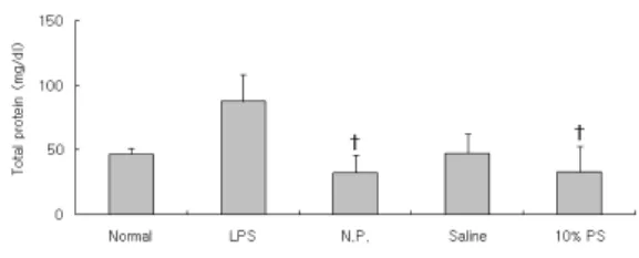
(2) Total protein
12시간 동안 소변을 채취하여 뇨중 total protein 양을 측정하였다. LPS군에서는 정상 군에 비하여 뇨중 total protein이 증가하였 으나 유의성은 없었다. NP군과 차전자약침 군의 뇨중 total protein은 LPS군에 비하여 유의하게 감소하였다(Fig. 12).
Fig. 12. Effects of HJ on total protein level in urine of LPS-stimulated rats.
The male SD rats were treated as described in the materials and methods and injected intra-peritoneally with LPS (2 ㎎/
㎏). The rats were kept in the metabolic cages and the urine was collected for 12 hours. And, total protein level in urine was analysed. Data were expressed as mean ± SD (n=5).
Normal : normal SD rat LPS : LPS (2 ㎎/㎏) challenge
N.P.: LPS (2 ㎎/㎏) challenge and administration with needle prick at KI10
Saline : LPS (2 ㎎/㎏) challenge and saline(200 ㎕) injcetion at KI10
PS-HA : LPS (2 ㎎/㎏) challenge and PS-HA(10%, 200 ㎕) at KI10
† : p<0.05 compared to LPS group by ANOVA test
5) 신장 내 염증활성
신장염을 유도하고 3시간 후에 신장을 적 출하여 신장조직을 분획한 후 TNF-alpha와 MPO의 농도를 측정하였다.
Table 4. Inflamtory activity in kidney of LPS-stimulated rats.
Normal LPS N.P. Saline PS-HA TNF-alpha
(㎎/㎗) 9775.33
±2111.531 14156.7
±1989.263 13886
±2407.278 11627.3
±1921.047 12546
±1842.308 (㎎/㎗)MPO 14.275
±6.094 96.283
±4.585 90.422
±11.783 87.717
±16.422 61.922
±4.199 The male SD rats were treated as described in the materials and methods and injected intra-peritoneally with LPS (2 ㎎/
㎏). Three hours after the LPS stimulation, rat kidney was removed and the levels of renal TNF-alpha and MPO were analysed by ELISA. Data were expressed as mean ± SD (n=5).
Normal : normal SD rat LPS : LPS (2 ㎎/㎏) challenge
N.P.: LPS (2 ㎎/㎏) challenge and administration with needle prick at KI10
Saline : LPS (2 ㎎/㎏) challenge and saline(200 ㎕) injcetion at KI10
PS-HA : LPS (2 ㎎/㎏) challenge and PS-HA(10%, 200 ㎕) at KI10
(1) TNF-α
신장염을 유도하고 3시간 후에 신장을 적 출하여 renal TNF-alpha의 농도를 측정하 였다. LPS군에서는 정상군에 비하여 renal TNF-alpha가 유의하게 증가하였다. 차전자 약침군의 renal TNF-alpha는 LPS군에 약 간 감소하였으나 유의성은 없었다(Fig. 13).
Fig. 13. Effects of HJ on renal TNF-alpha level in LPS-stimulated rats.
The male SD rats were treated as described in the materials and methods and injected intra-peritoneally with LPS (2 ㎎/
㎏). Three hours after the LPS stimulation, rat kidney was removed and renal TNF-alpha level was analysed by ELISA.
Data were expressed as mean ± SD (n=5).
Normal : normal SD rat LPS : LPS (2 ㎎/㎏) challenge
N.P.: LPS (2 ㎎/㎏) challenge and administration with needle prick at KI10
Saline : LPS (2 ㎎/㎏) challenge and saline(200 ㎕) injcetion at KI10
PS-HA : LPS (2 ㎎/㎏) challenge and PS-HA(10%, 200 ㎕) at KI10
* : p<0.05 compared to normal group by t-test
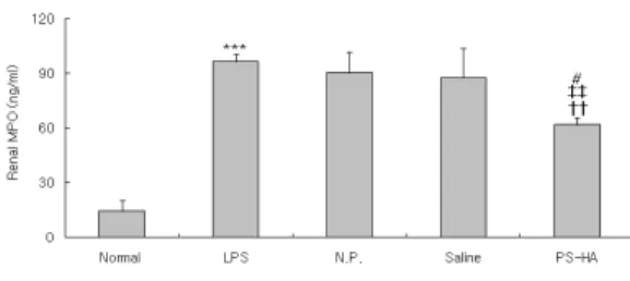
(2) MPO (myeloperoxidase)
백서에 LPS로 신장염을 유도하고 3시간 후에 신장을 적출하여 renal MPO 농도를 측정하였다. LPS군에서는 정상군에 비하여 renal MPO가 현저하게 증가하였다. N.P군 및 saline군에서는 LPS군에 비하여 renal MPO가 약간 감소하였으나 유의성은 없었 다. 차전자약침군의 renal MPO는 LPS군, NP군 및 saline군에 비하여 유의하게 감소 하였다(Fig. 14).
Fig. 14. Effects of HJ on renal MPO level in LPS-stimulated rats.
The male SD rats were treated as described in the materials and methods and injected intra-peritoneally with LPS (2 ㎎/
㎏). Three hours after the LPS stimulation, rat kidney was removed and renal MPO level was analysed by ELISA. Data were expressed as mean ± SD (n=5).
Normal : normal SD rat LPS : LPS (2 ㎎/㎏) challenge
N.P.: LPS (2 ㎎/㎏) challenge and administration with needle prick at KI10
Saline : LPS (2 ㎎/㎏) challenge and saline(200 ㎕) injcetion at KI10
PS-HA : LPS (2 ㎎/㎏) challenge and PS-HA(10%, 200 ㎕) at KI10
*** : p<0.001 compared to normal group by t-test
†† : p<0.01 compared to LPS group by ANOVA test
‡‡ : p<0.01 compared to N.P. group by ANOVA test
# : p<0.05 compared to saline group by ANOVA test
6) 조직학적 분석
신장염을 유도하고 3시간 후에 신장을 적 출하여 Hematoxilin and Eosin(H&E) 염색 법으로 조직을 염색한 후 광학현미경으로 관찰하였다. LPS군의 신장조직은 정상군에 비하여 사구체에 neutrophil의 집중과 조직 의 조밀도 저하를 나타내었다. 차전자약침군 에서는 netrophil의 집중이 대조군에 비하여 감소하였다(Fig. 15).
Fig. 15. Histological analysis of renal tissue in LPS-stimulated rats. (H&E stain)
The male SD rats were treated as described in the materials and methods and injected intra-peritoneally with LPS (2 ㎎/
㎏). Three hours after the LPS stimulation, rat kidney was removed. The renal tissue was sliced and embedded in OCT compound, and 8 ㎛ sections were stained with hematoxilin and eosin.(↑ : glomerulus, ×400)
Normal : normal SD rat LPS : LPS (2 ㎎/㎏) challenge
N.P. : LPS (2 ㎎/㎏) challenge and needle prick at KI10 Saline : LPS (2 ㎎/㎏) challenge and saline(200 ㎕/rat) injcetion at KI10
PS-HA : LPS (2 ㎎/㎏) challenge and PS(10%, 200 ㎕/rat) at KI10
Ⅳ. 고 찰
해부학적으로 신장은 한 쌍의 암적색을 띈 완두형 장기로 후복막에 위치해 있으며, 하나의 신장은 사구체와 세뇨관으로 이루어 진 신장의 기능단위인 腎元이 100만개 가량 모여 있다. 신장은 전해질과 수분을 조절하 여 순환혈액량을 유지하고 혈압을 조절하며, 암모니아 대사산물인 요소와 기타 독성물질 을 소변으로 배출함으로써 체내환경을 유지 하는 장기이므로 혈액순환이 많이 필요하여, 순환혈액의 20~25%정도가 신장에 공급된 다.
신장은 사구체에서 혈액을 여과한 후 세 뇨관에서 능동수송과 확산의 과정을 통하여 물질을 교환하기 위하여 미세한 막구조를
이루고 있으며, 능동수송에 의한 왕성한 대 사작용으로 인해 독성물질에 대한 감수성이 높다. 신장의 사구체에 발생하는 사구체신염 은 감염에 의한 면역복합체나, 항체가 사구 체기저막에 침착되어 발생한다. 전신적인 혈 관의 염증성 질환 등도 사구체신염의 원인 이 된다. 사구체기저막에 침착된 면역복합체 나 세균에 대한 항체는 자가조직을 손상시 키는 면역시스템을 활성화 시켜 사구체신염 이 촉발된다. 사구체신염은 사구체의 막구조 를 손상시켜, 혈뇨, 단백뇨 등을 유발하고, 여과기능의 손상까지 진행되면, 핍뇨와 요독 증이 발생한다.
LPS는 그람음성박테리아 세포막의 구성 성분으로 면역세포를 자극하여 cytokine을 분비시키며 증가된 혈청내 proinflammatory cytokine은 LPS에 의한 신체손상을 매개한 다. 신장의 경우 LPS는 신장의 혈행역학장 애를 유발하며 IL-1, IL-6, TNF-α 등의 cytokine을 분비시키고 IL-8, MCP-1 등의 chemokine을 분비하여 면역세포의 화학주성 을 유발한다. 또 LPS는 메산지움 세포에 많 은 양의 nitric oxide 분비를 유발시키며 메 산지움 세포와 tubular epithelial cell에 Fas 발현을 증가시킴으로서 직간접적으로 신장 손상을 초래하며16-18), 최근의 실험에서 신장 독성으로 인한 사구체신염, 급성 신부전, 간 질성신염, 신독성신염 등의 병태모델에 많이 사용되고 있다.
차전자(Plantaginis Semen)는 질경이과 (차전과:Planaraginaceae)에 속한 다년생 초 본인 질경이 또는 털질경이의 성숙한 종자 로 性은 微寒하고, 味는 甘하며, 歸經은 肝,
腎, 肺, 小腸經이며, 淸熱利尿, 滲濕通淋, 明 目, 袪痰하는 효능이 있어 水腫脹滿, 熱淋澁 痛, 暑濕泄瀉, 目赤腫痛, 痰熱咳嗽에 사용된 다6). 동의보감에 의하면 성질이 차고 맛은 달고 짜며, 독이 없으며, 기륭에 쓰고 오림 에 쓴다. 소변을 잘 나오게 하고 소변이 찔 끔찔끔 나오는 것을 통하게 한다.
陰谷(KI10)은 足少陰腎經의 合水穴, 自性 穴이며 足少陰腎, 衝脈, 陰維脈의 交會穴로, 무릎안쪽 경골뒤 큰 근육 아래 작은 근육 위에 손으로 눌러보면 맥이 뛰는 곳으로 무 릎을 꿇고 취혈한다. 祛濕通溲, 滋腎淸熱, 疎 泄厥氣, 利導下焦, 補腎培元, 調經利水의 효 능이 있어 陽萎, 月經不調, 崩漏, 小便難 등 의 治療穴이 된다5).
이에 저자는 음곡 차전자 약침이 신장염 에 미치는 영향을 알아보고자 차전자 약침 액을 시료로 정상 백서의 간세포에 대한 세 포독성을 확인하고, LPS로 유도된 신장염 병태모델 흰쥐의 음곡 상응부위에 차전자 약침을 시술한 후, 이뇨활성, 혈구 및 혈청 분석, 소변분석, 신장내 염증활성, 조직학석 분석을 시행하였다.
정상 백서의 간세포에 PS-HAS를 1%, 2.5%, 5%, 10%로 처리한 후 세포생존율을 확인한 결과 PS-HAS의 농도가 높을수록 세포의 viability가 유의하게 증가된 것을 확 인하였으므로, 10%PS-HAS를 선택하였다 (Fig 1).
LPS 투여에 따른 TNF-α의 농도 변화는, 혈청에서는 LPS 투여 1시간 후에, 신장 조 직에서는 LPS 투여 3시간 후에 정상군과
LPS투여군간에 차이가 뚜렷하게 나타났다 (Fig 2, 3). 따라서 본 실험에서 혈청 검사는 LPS 투여 1시간 후에, 신장조직내 검사는 LPS 투여 3시간 후에 시행하였다.
신장은 전해질과 수분을 조절하여 혈압을 유지하며, 암모니아 대사산물인 요소와 기타 독성물질을 소변으로 배출한다. 신장이 손상 되면, 신장의 여과 기능이 감소하여 소변량 이 감소하게 된다. 그러므로 본 실험에서는 LPS 투여후 12시간 동안 백서의 소변을 채 취하여 소변양을 측정하였다. LPS에 의해 감소된 소변량은 음곡 차전자 약침에 의해 유의하게 증가하여 정상으로 회복되었다 (Fig.4).
백혈구는 골수에서 유래하고 호중구, 호산 구, 호염구, 림프구, 단핵구 등으로 구성되며 염증이 발생하면 혈관의 내피 사이를 빠져 나가 혈관 밖으로 유주하고 화학주성에 의 해 항원과 이물에 이동하여 각각의 기능을 수행하여 염증반응을 유발한다. 본 실험에서 LPS에 의해 증가된 혈중 WBC는 음곡 차 전자약침에 의해 유의하게 감소하였다. 이는 음곡 차전자약침이 신장염 모델에서 염증반 응을 억제하였음을 의미하는 것으로 사료된 다(Fig 5~7).
BUN은 혈중에 존재하는 요소(urea)내 질 소를 나타내는 것으로, 요소는 간의 요소회 로(urea cycle)에서 단백질대사의 최종산물 로서 생성되어 혈류를 거쳐 신사구체에서 여과된 다음 세뇨관에서 일부는 재흡수 되 고 나머지는 요중으로 배설되는데, 신기능이 저하하여 사구체여과율이 감소하면 혈중 농 도가 상승하므로, 혈중 BUN은 신기능의 지
표가 된다.
혈액내의 creatinine phosphate에서 생성 된 creatine이 탈수되어 생긴 대사의 최종산 물로, creatinine량은 인체 근육량에 비례하 여 일정량이 생성되어 신장을 통하여 배설 된다. Creatinine은 오로지 신장을 통해서만 체외로 배설되고 체내에서 재순환을 하지 않아 혈중 creatinine농도는 사구체의 여과 에 따라 변하므로, creatinine 수치 역시 신 기능의 지표가 된다1).
TNF-α는 그람-음성 박테리아와 다른 감 염성 미생물에 대한 급성염증반응에 의해 활성화된 림프구에 의해서 생산되며, 내피세 포와 대식세포를 자극하여 백혈구의 주화성 과 보충을 유도하는 케모카인을 분비토록하 며, 혈액 단핵구와 T 림프구, 호중구, 혈관 내피세포, 피부섬유아세포 등의 세포에서 IL-8 유전자 발현을 유도한다. TNF-α로 유 도된 chemokine인 IL-8은 염증세포들을 활 성화하고 그들을 염증부위로 유인하는 화학 유인인자 (chemotatic factor, chemokines)의 작용을 하는데, CINC-1은 IL-8 family의 일 원이다. 최근 쥐의 염증반응 모델에서 CINC-1 수치를 측량하여 IL-8 발생을 조사 한 보고가 있다20,21). 본 실험에서는 염증반 응에서 호중구의 활성화 정도를 알아보기 위하여 CINC-1 수치를 측정하였다.
본 실험에서 혈청 BUN, creatinine, TNF-α, CINC-1의 농도를 측정한 결과, LPS에 의해 증가한 혈청 BUN, TNF-α, CINC-1이 음곡 차전자 약침에 의해 유의하 게 감소하였다(Fig. 8-11). 이는 음곡 차전 자 약침이 LPS로 유도된 신장염 병태모델
에서 염증을 억제하고 신장의 기능을 정상 화하였음을 의미하는 것으로 사료된다.
신장은 전해질과 수분을 조절하여 순환혈 액량을 유지하고 혈압을 조절하며, 암모니아 대사산물인 요소와 기타 독성물질을 소변으 로 배출하는데, 정상신장에서 적혈구와 단백 질은 신원(nephron)을 통하여 여과되지 않 는다. 그러나 사구체가 파괴되거나 투과성이 증가되면 단백질이 소변으로 나오게 된다.
본 실험에서는 백서의 소변을 채취하여 뇨중 creatinine 및 total protein을 측정하였 다. 뇨중 creatinine과 total protein 모두 LPS 투여에 의해 증가하고 음곡 차전자 약 침에 의해 감소하는 경향을 나타내었으나 유의성이 부족하여 향후 더 자세한 연구가 필요할 것으로 사려된다.(Fig 12, 13).
MPO는 호중구로부터 주로 방출되는 효 소로, MPO 분석은 염증의 지표로서 조직속 의 호중구의 수를 측량하는데 사용된다. 본 실험에서는 신장 실질내 염증활성도를 측정 하기 위해 TNF-α와 MPO의 농도를 측정한 결과 음곡차전자 약침에 의해 신장실질내 TNF-α의 농도에는 유의한 변화를 일으키 지 못하였으나, MPO 농도는 유의하게 감소 하여, 음곡 차전자 약침이 신장실질내 염증 을 억제하는데 유효한 것으로 사료되었다 (Fig 14, 15).
신장을 적출하여 신장조직을 관찰한 결과, LPS에 의한 사구체 내 neutrophil 집중과 조직 손상이 음곡 차전자 약침에 의해 억제 된 것으로 관찰되었다(Fig 16).
이상의 결과를 요약하면, 음곡 차전자 약 침은 LPS로 유도된 백서의 신장염에 대하
여 혈액내 백혈구의 증가를 억제하며, 혈청 내에서 BUN 등의 노폐물 축적을 감소시키 고, TNF-α, CINC-1 등 inflammatory cytokine을 억제하였으며, 여과율의 감소로 인한 핍뇨를 개선시켰으며, 신장의 MPO농 도를 감소시켰다. 또한, saline군에서 혈청내 WBC, BUN, TNF-α, 신장 MPO 농도가 유 의하게 감소한 것과, NP군에서 혈청내 WBC, BUN, 신장 MPO 활성과 소변 중 protein을 유의하게 감소시킨 것은, 차전자 의 약리적 작용뿐만 아니라 음곡의 경혈 특 이성도 작용한 것으로 생각된다. 따라서 음 곡에 대한 차전자약침은 신장염의 치료에 효과가 있을 것으로 생각되는 바 향후 이에 대한 지속적 연구와 임상연구가 필요할 것 으로 사려된다.
Ⅴ. 결 론
음곡(KI10)에 시술한 차전자 약침이 LPS 로 유도된 신장염 모델 rat에 미치는 영향을 알아보고자 차전자 약침액을 시료로 정상 rat의 간세포에 대한 세포독성을 확인하고 LPS 투여 후 혈청과 신장내 TNF-α의 농도 를 측정하였고, 음곡에 시술한 자전자 약침 투여 후 혈액 및 혈청 분석, 소변분석, 신장 내 염증활성, 조직학석 분석을 시행한 결과, 다음과 같은 결론을 얻었다.
혈중 WBC, 혈청 BUN, 혈청 TNF-α를 유의하게 감소시켰고, 소변량을 유의하게 증 가시켰으며, 신장실질내의 MPO를 유의하게 감소시켰다. 따라서 이상의 결과로 볼 때, 음곡 차전자 약침은 신장의 염증반응을 억
제하는 효과가 있는 것으로 사료된다.
參考文獻
1. 杜鎬京. 동의신계학. 서울 : 동양과학연구 회. 1993 : 226, 228, 247, 238-47, 326, 334-6.
2. J. CLAUDE bennett, FRED PLUM.
Cecil textbook of medicine. 20th.
philadelphia : saunders. 1996 : 552, 575-7.
3. Medzhitov R, Preston-Hurlburt P, Janeway CA Jr. A human homologue of the Drosophila Toll protein signal activation of adaptive immunity. Nature.
1997 ; 388(6640) : 394-7.
4. Aderem A, Ulevitch RJ. Toll-like receptors in the induction of the innate immune response. Nature. 2000 ; 406(6797) : 782-7.
5. Hoshino K, Takeuchi O, Kawai T, Sanjo H, Ogawa T, Takeda Y, Takeda K, Akira S. Toll-like receptor 4(TLR4)-deficient mice are hypo- responsive to lipopolysaccharide:
evidence for TLR4 as the Lps gene product. J immunol. 1999 ; 162(7) : 3749-52.
6. 임윤경, 김준표, 김태한. 대학경락경혈학 각론. 대전 : 오비기획. 2006 : 676-8.
7. 康秉秀, 高雲彩, 金圭烈, 金先熙, 盧昇鉉, 朴涌基 외. 本草學. 서울 : 永林社. 2004 : 357-8.
8. 최정배, 이경환, 김용성. 육미지황탕가황
기인삼 추출물의 Cisplatin 유도 신장독성 보호효과. 대한한방내과학회지. 2003 ; 24(3) : 579-87.
9. 황영근, 정지천. 수은으로 유발된 토끼의 신장 기능 손상에 대한 단삼(丹參)의 효 과. 대한한방내과학회지. 2000 ; 21(2) : 227-34.
10. 윤철호, 정지천, 신의섭. Bromobenzene 으로 유도된 신장기능저하에 미치는 녹 용 약침의 항산화 효과. 대한한방내과학 회지. 1999 ; 20(2) : 287-302.
11. 권선호, 송춘호. CCl4로 투여한 백서 간 손상에 미치는 차전자약침의 영향. 대한 침구학회지. 2001 ; 18(4) : 152-60.
12. 조시용, 김철홍, 윤현민, 장경전, 안창범, 송춘호. 車前子藥鍼이 急性腎不全 白鼠에 미치는 影響. 경락경혈학회지. 2005 ; 22(4) : 117-27.
12.
13. 박춘하, 채우석, 윤여충. 차전자 약침이 cisplatine에 의해 유발된 급성 신부전에 미치는 영향. 대한침구학회지. 2004 ; 21(2) : 155-66.
14. Cohen JJ, Black AJ, Wertheim SJ.
Direct effects of endotoxin on the function of the isolated perfused rat kidney. Kidney Int. 1990 ; 37(5) : 1219-26.
15. Trop M, Schiffrin EJ, Carter EA.
Effect of acute and chronic lipopolysaccharide (LPS) administration on reticuloendothelial system (RES) phagocytic activity in vivo.
Burns. 1992 ; 18(2) : 107-8.
16. Glauser MP, Zanetti G, BaumgartnerJ-D, Cohen J. Septic shock Pathogenesis. Lancet. 1991 ; 338 : 732-6.
17. Ohmori Y, Hamilton TA. A macrophage LPS-inducible early gene encodes the murine homologue of IP-10. Biochem Biophys Res Commun.
1990 ; 168 : 1261-7.
18. Tannenbaum CS, Koemer TJ, Jansen MM, Hamilton TA. Characteriation of Lipopolysaccharide-induced macrophage gene expression. J immunol. 1988 ; 140 : 3640-5.
19. 허준. 동의보감. 경남 : 동의보감출판사.
2005 : 2151.
20. Nakagawa, H, Komori, N, Shibata, F, Ikesue, A, Konishi, K, Fujioka, M, and Kato, H. Identification of cytokine- induced neutrophil chemoattractants (CINC), rat GRO/CINC-2 alpha and CINC-2 beta, produced by granulation tissue in culture: purification, complete amino acid sequences and characterization. Biochem. J. 1994 ; 301: 545–50.
21. M, Taie S, Chujo K, Asaga T, Iwanaga Y, Ono J, Maekawa N.
Urinary trypsin inhibitor reduces inflammatory response in kidney induced by lipopolysaccharide. Ueki J Biosci Bioeng. 2007 ; 104(4) : 315-20.