PGHN
Original Article
Influencing Factors to Results of the Urease Test: Age, Sampling Site, Histopathologic Findings, and Density of Helicobacter pylori
Ji-Hyun Seo, Hee-Shang Youn, Jung-Je Park*, Jung Sook Yeom, Ji Sook Park, Jin-Su Jun, Jae-Young Lim, Chan-Hoo Park, Hyang-Ok Woo, Gyung-Hyuck Ko
†, Seung-Chul Baik
‡, Woo-Kon Lee
‡, Myung-Je Cho
‡and Kwang-Ho Rhee
‡Departments of Pediatrics, *Otolaryngology, †Pathology and ‡Microbiology, Institute of Health Sciences , School of Medicine, Gyeongsang National University, Jinju, Korea
Purpose: We investigated the positivity rate and the time period to the positive color change of the urease test in children and adults and assessed the correlation of the urease test to histopathologic findings.
Methods: From 1995 to 2000, endoscopic biopsies of the antrum and body were collected from 811 children and 224 adults and subjected to urease tests and histopathology.
Results: The positivity rate of the urease test was 49.4% for 0-4 years, 48.4% for 5-9 years, 47.3% for 10-15 years, and 62.5% for 20-29 years in the antrum. The positivity rate was 85.1% in 0-4 years, 82.3% in 5-9 years, 74.7%
in 10-15 years, and 74.1% in 20-29 years for the body. In the antrum, the highest positivity rate was <1 hour for the group aged 10-29 years and 6-24 hours in the group <10 years old (p<0.0001). In the body, the highest positivity rate was <1 hour in adults and 6-24 hours in children (p<0.0001). The proportions of the positive reactions within 1 hour were similar for the antrum and the body. In the cases of more severe chronic gastritis, active gastritis, and Helicobacter pylori infiltration, a positive urease test reaction occurred more quickly (p<0.0001).
Conclusion: There were significant differences in urease tests according to age and sampling site. The discrepancy between the antrum and the body was greater in younger children. These results might be related to the low density and patchy distribution of bacteria in children and in the body. (Pediatr Gastroenterol Hepatol Nutr 2013; 16: 34∼40) Key Words: Urease test, Helicobacter pylori infection, Age, Pathology
Received:August 11, 2012, Revised:November 13, 2012, Accepted:November 30, 2012
Corresponding author: Hee-Shang Youn, Department of Pediatrics, School of Medicine, Gyeongsang National University, 15, Jinju-daero, 816beon-gil, Jinju 660-751, Korea. Tel: +82-55-750-8158, Fax: +82-55-752-9339, E-mail: [email protected]
Copyright ⓒ 2013 by The Korean Society of Pediatric Gastroenterology, Hepatology and Nutrition
This is an openaccess article distributed under the terms of the Creative Commons Attribution NonCommercial License (http://creativecommons.org/licenses/by-nc/3.0/) which permits unrestricted noncommercial use, distribution, and reproduction in any medium, provided the original work is properly cited.
INTRODUCTION
In most cases, Helicobacter pylori infection occurs pri-
marily in childhood [1]. In Korea, H. pylori infiltration, chronic gastritis of more than a moderate degree, and active gastritis have been detected even in children,
and an increase in age is gradually accompanied by a greater degree of infection [2].
The diagnostic approaches for H. pylori infection in children do not differ from those used in adults. In general, a culture of the bacteria has been considered to be the gold standard for the diagnosis of H. pylori infection, but the yield of a culture study varies be- cause H. pylori are fastidious bacteria [2]. Other wide- ly used biopsy-based tests include the histologic de- termination of H. pylori and the urease test.
Histopathologic findings for the diagnosis of H. pylori infection are assessed by a specific staining and a spe- cialized pathologist [3]. The urease test is a simple and inexpensive method that requires no special technique to perform and to read the result. Buffered urease tests require at least 1,000 organisms to gen- erate a positive reaction. Therefore, when the histol- ogy reveals only 1 or 2 organisms in the entire section, the urease test may be negative [4]. In adults, the rapid urease test is widely used, with a high sensi- tivity (70-90%) [5]. Data concerning the sensitivity of the urease test in children are controversial [6-8].
In this study, we investigated the positivity rate and the positive timing of the urease test in children and adults using three biopsy specimens from the gastric antrum and body and assessed the correla- tion of the urease test to histopathologic findings.
MATERIALS AND METHODS
Study population
After the Institutional Review Board reviewed the research protocols of the present study (GNUHIRB- 2012-07-003), 811 children and 224 young adults who underwent endoscopy of the upper digestive tract from 1995 to 2000 were enrolled. In children, the most common indication for upper gastro- intestinal endoscopy was upper abdominal pain (75.1%) followed by Henoch- chonlein purpura (H-S purpura) with abdominal pain (8.1%), frequent vomiting (5.3%), heartburn (2.6%) and suspected gastrointestinal bleeding (2.6%). Only 8 patients were diagnosed with hemorrhagic gastritis, gastric ulcers or duodenal ulcers without active bleeding (1
in 0-4 years, 4 in 5-9 years, and 3 in 10-14 years). A total of 80 patients were taking an H2 receptor antag- onist at the time of endoscopy (36 in 0-4 years, 34 in 5-9 years and 10 in 10-14 years), and 2 were on ste- roids due to nephrotic syndrome and adrenogenital syndrome. Most patients were relatively healthy without any underlying disease. A total of 85 pa- tients (10.5%) had underlying diseases such as H-S purpura (n=66), IgA nephropathy (n=10), iron-de- ficiency anemia (n=4), etc. Many young adults vol- unteered to undergo endoscopy. About a half of them (n=111, 49.6%) had no gastrointestinal symp- toms and the other half had symptoms such as epi- gastric pain (n=79, 35.3%), dyspepsia (n=26, 11.6%), vomiting (n=3, 1.4%), abdominal dis- tension (n=3, 1.4%), etc. To ensure that the present study only included healthy, young adults, subjects over the age of 30 years were excluded. In this study, the patients with active bleeding were excluded due to the fact that these patients did not undergo biopsy for urease test. All results of the urease tests and the histopathological slides were reviewed. The study populations were stratified into 4 age groups: 0-4 years (n=168), 5-9 years (n=351), 10-14 years (n=292) and 20-29 years (n=224).
Urease test
Urease tests were performed in the endoscopy room. Briefly, each of the three biopsy specimens from the antrum and the body were incubated in a 2% urea broth (urea 20 g/L, phenol red 0.04 g/L, KH2PO4 0.2 g/L, NaCl 0.5 g/L: pH 6.8), and if a change in color was noted in the following 48 hours, the biopsy was deemed to be urease test positive. The time points at which the positivity occurred were divided into 0-1 hour, 1-6 hours, 6-24 hours, and 24-48 hours. A clinical diagnosis of H. pylori infection was obtained by subjecting the gastric antral and body biopsies to urease tests and histopathology.
Histopathologic findings
Histopathology was performed after the biopsy specimens were fixed in 10% buffered formalin over- night, processed for paraffin embedding, cut into 4-5
Table 1.The Positivity Rate of the Urease Test in the Antrum and Body according to Age
Age (yr) Number Positive urease test (%)
p-value
Antrum Body
0-4 5-9 10-15 20-29 Total
168 351 292 224 1,035
83 (49.4) 170 (48.4) 138 (47.3) 140 (62.5) 531 (51.3)
143 (85.1) 289 (82.3) 218 (74.7) 166 (74.1) 816 (78.8)
<0.0001
<0.0001
<0.0001
<0.0001
<0.0001
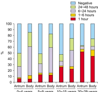
Fig. 1. The positivity rate and positive timing of the urease test both in the antrum and body according to age. The positivity rate of the urease test in the antrum was higher in 20-29 years group comparing with that in other three age groups, and the positivity rate of the urease test in the body decreased with increasing age (p<0.0001). The highest positivity timing was within 1 hour in the 20-29 years group, and within 6-24 hours in children (p<0.0001). The proportions of positive reactions within 1 hour were similar for the antrum and body in all groups.
μm thick sections, and stained with hematox- ylin-eosin (H-E). The histological results were in- terpreted using the Updated Sydney System. For this, the degrees of lymphocyte (chronicity), neu- trophil (activity), and H. pylori infiltration were clas- sified as normal, mild, moderate, or marked.
Statistical analysis
The data were analyzed using SPSS ver. 12.0 for Windows (SPSS Inc., Chicago, IL, USA). How the de- grees of the urease test and histopathologic findings in the population varied depending on age was evaluated. Statistically significant differences in the positive timing of the urease tests between age groups were determined by using the χ2 test. The re- lationship in the positive timing of the urease test be- tween the antrum and the body was determined by McNemar’s test. p-values of <0.05 were considered to be statistically significant. The inflammatory changes in the gastric antrum and body regions were correlated with the Spearman rank correlation coefficient.
RESULTS
Time to the change to positive in the urease test according to age
The cases demonstrating a positive color change within 48 hours were 49.4% for 0-4 years, 48.4% for 5-9 years, 47.3% for 10-15 years, and 62.5% for 20-29 years in the antrum. In the body, cases demonstrat- ing a positive color change within 48 hours were 85.1% in 0-4 years, 82.3% in 5-9 years, 74.7% in 10-15 years, and 74.1% in 20-29 years (Table 1). The results
of the urease tests in the antrum were significantly different compared with the results of the urease tests in the body, regardless of age (p<0.0001). The positivity rates of the urease test according to age were different for the antrum and the body (p
<0.0001).
Cases showing a positive color change within 1 hour and within 6-24 hours in the antrum were 6.0%
and 19.0% in 0-4 years, 12.5% and 16.0% in 5-9 years,
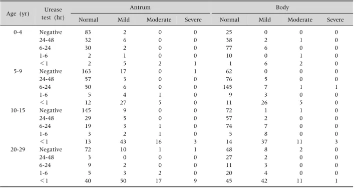
Table 2.The Distribution of Four Degrees of Infiltration of Helicobacter pylori and the Result of Urease Test according to the Degree of H. pylori Infiltration in the Gastric Antrum and Body in Four Age Groups
Age (yr) Urease test (hr)
Antrum Body
Normal Mild Moderate Severe Normal Mild Moderate Severe
0-4
5-9
10-15
20-29
Negative 24-48 6-24 1-6
<1 Negative 24-48 6-24 1-6
<1 Negative 24-48 6-24 1-6
<1 Negative 24-48 6-24 1-6
<1
83 32 30 2 2 163 57 50 5 12 145 29 19 3 13 72 3 9 5 40
2 6 2 1 5 17 3 6 4 27 9 5 3 2 43 10 0 2 3 50
0 0 0 0 2 0 0 0 1 5 0 0 1 1 16 1 0 0 2 17
0 0 0 0 1 1 0 0 0 0 0 0 0 0 3 1 0 0 0 9
25 38 77 10 1 62 76 145 9 11 72 57 74 5 14 48 27 11 20 45
0 2 6 0 6 0 5 7 3 26 1 2 7 8 37 8 2 3 4 42
0 1 0 1 2 0 0 1 0 5 1 0 0 0 11 2 0 0 0 11
0 0 0 0 0 0 0 1 0 0 0 0 0 0 3 0 0 0 0 1
25.7% and 7.9% in 10-15 years, and 51.8% and 4.9%
in 20-29 years, respectively (Fig. 1). For the body, cases showing a positive color change within 1 hour and within 6-24 hours were 5.4% and 49.4% in 0-4 years, 12.0% and 43.9% in 5-9 years, 22.3% and 27.7% in 10-15 years, and 44.2% and 6.2% in 20-29 years, respectively (Fig. 1). The speed of the positive reaction of the urease test in both the antrum and body was significantly different according to age (p
<0.0001). The positivity rate of the urease test in adults primarily occurred within 1 hour in both the antrum and in the body. The proportions of a positive reaction within 1 hour of the urease test were similar between the antrum and the body regardless of age.
The positivity rate of the urease test in children pri- marily occurred within 6-24 hours in the body. The discrepancy in the positivity rates between the an- trum and the body in children was composed from the proportion of positive reactions during 6-24 hours in the urease test.
The relation between histopathologic grades and the speed of a positive reaction to the urease test
Histopathologic evaluations were performed on 811 children and 224 adults. The proportion of mod- erate and severe degrees of chronic gastritis in the antrum increased with age, from 10.6% to 53.6%.
The proportion of moderate and severe degrees of chronic gastritis in the body also increased with age, from 11.3% to 35.2%. The proportions of active gas- tritis and H. pylori infiltration in the antrum and body increased with age (Table 2). The degrees of the his- topathologic findings in the antrum and body were positively correlated with age (Spearman’s R=0.14- 0.27, p<0.0001).
When severe chronic gastritis, active gastritis or H.
pylori infiltration were found in histopathologic eval- uations, the urease tests showed more rapid positive color changes in both the antrum and the body re- gardless of age compared with those of milder histo- pathologic grades (p<0.0001). Although bacteria
were observed in the histopathologic evaluations, a negative urease test (considered to be a false neg- ative) were observed in 2 (1.2%) in the 0-4 years group, 18 (5.1%) in the 5-9 years group, 11 (3.8%) in the 10-15 years group, and 22 (9.8%) in the 20-29 years group.
DISCUSSION
In the present study, the positivity rate and the positive timing of the urease test differed according to age. The positivity rate of the urease test using an- tral biopsy specimens increased with increasing age, and the positivity rate of the urease test using body biopsy specimens decreased with increasing age. The positive urease test had a high concordance with both the density of bacteria and the severity of gas- tritis [8]. The accuracy of the urease test depends pri- marily on the H. pylori density in the gastric sample, which is generally lower in children than in adoles- cents and adults [9]. In this study, the severity of ac- tive gastritis and the density of H. pylori in the an- trum and body increased with age, and this outcome suggested that the histopathologic findings influ- ence the positivity rate and the positive time in the gastric antrum but not in the gastric body.
In children, a low degree of H. pylori colonization has been noted [9], and the degree of colonization was significantly lower in the body and cardia than in the antrum [10]. These discrepancies could be at- tributed to sampling errors because of the presence of very low numbers of H. pylori in the tissue samples or because of a patchy distribution of the organism in the stomach’s mucosa [11]. In this study, the pro- portions of the positive color change within 1 hour in the antrum and body were similar in all age groups, and no differences in the speed of the positive re- action in the antrum and the body were observed in the adults. The discrepancy of the urease test be- tween the antrum and the body in children was greater than that in adults, and this outcome re- sulted from the differences in the proportions of the positive color changes during 6-24 hours. These re- sults suggested that the H. pylori infection persisted
from early childhood and that the density of the bac- teria increased with age.
In most studies, biopsy specimens were taken from the antrum because this area of the heaviest colonization for H. pylori may be at the lesser curve at the angulus in the prepyloric region [12]. The pooled sensitivity of the urease test increased when samples were obtained from both the antrum and the body [13,14]. In this study, the positivity rate of the urease test in both the antrum and the body was higher comparing with only the antrum. Increasing the number of gastric antral biopsies significantly im- proves the sensitivity of the rapid urease test and hastens the time required for the test to become pos- itive for the diagnosis [11]. In children aged 1-5 years, the positivity rate of urease test using three bi- opsy specimens from antrum and body with 48 hour-observation time (86.3%) was higher than us- ing one biopsy specimen from antrum and body with 24 hour-observation period (9.8%) [15]. Comparing the inflammatory changes of the antrum and the body in H. pylori infected children showed a poor cor- relation with the antrum and the body’s density [10]. The degree of the colonization of H. pylori ac- cording to the anatomy of stomach (antrum, body, and cardia) differed in several reports [12-14], and the degree of colonization may be related to the dif- ferences in either the migration pattern from the an- trum to the cardia or to the focal bacteria distribution [10]. In the present study, the histology of the an- trum and the body showed a similar severity of gas- tritis and bacterial density. However, the result of uresase test in the antrum was different in the body.
According to the biopsy site in children, the different positive timings of the urease test might be ex- plained by the differences in the density and in the degree of the patchy distribution of bacteria. Debate continues regarding the optimum site and the num- ber of gastric biopsies for the diagnosis of H. pylori.
A false-negative urease test was observed in 3.0%
(31/1,037) for the antrum and 1.9% (20/1,037) for the body, and the false-negative urease test results in- creased with age. These findings are different from other reports that false negatives were higher below
the age of 5 years compared with 6 years old and older [16]. False-negative urease tests were associated with the use of acid suppression medication such as proton pump inhibitor (PPI), bleeding, and a very low density of H. pylori [14,15]. The hypochlorhydric subjects had less dense H. pylori colonization, body- predominant colonization and gastritis [17]. The use of acid suppression medication, in particular PPIs, has been shown to reduce H. pylori density and colo- nization and to suppress the urease activity of the bacterium, thus reducing the accuracy of the test [18]. Another factor related to false-negative urease test was to sampling errors because of a patchy dis- tribution of the organism in the stomach’s mucosa [19]. However, the exact reason of false-negative ure- ase tests in this study could not be proved because this study retrospectively reviewed the results of the urease tests and histology only, and no clinical his- tory of volunteers was evaluated.
There are some limitations to the present study.
First, we observed the timing of the positive urease test for 48 hours. The buffered urease test received regulatory approval to be read at 24 hours. Although the approved time for reading is different according to the type of urease test, the sensitivity increased over time [20]. A 90% positivity of the CLO-test after 24 hours was reported in 42 infected children, as demonstrated by the Giemsa staining of antral speci- mens [18]. Second, another limitation of this study was that the histologic diagnosis of H. pylori was based on the H-E stain alone. However, the sensi- tivity of H-E stains for detecting H. pylori were similar to that of the Giemsa stains [21]. Another limitation is that we were unaware of the volunteers’ clinical information, such as medications and underlying diseases. Although they were volunteers, the pro- portion of moderate and severe degrees of chronic gastritis in the antrum was highest in young adults.
In conclusion, there were significant differences in the positivity rate and the positive timing of urease tests according to age. The discrepancy between the antrum and the body was greater in younger chil- dren despite the similarity in all age groups of the proportion of the positive color change within 1 hour
in the antrum and body. These results might be re- lated to the low density and patchy distribution of bacteria in children and in the body. In children, the urease test may be more accurate when three or more specimens of both the antrum and body biopsies are used. Further studies concerning the precise time to observe the color change and the exact number of gastric biopsy specimens are needed for urease tests to accurately diagnose H. pylori infection in children.
ACKNOWLEDGEMENTS
This study was supported by a grant from the National R and D Program for Cancer Control of the Ministry of Health & Welfare of the Republic of Korea (0820050). The biospecimens used in this study were provided by Gyeongsang National University Hospital, which is a member of the National Biobank of Korea that is funded by the Ministry of Health and Welfare.
REFERENCES
1. Malaty HM, El-Kasabany A, Graham DY, Miller CC, Reddy SG, Srinivasan SR, et al. Age at acquisition of Helicobacter pylori infection: a follow-up study from in- fancy to adulthood. Lancet 2002;359:931-5.
2. Ko GH, Park CK, Choi CS, Park HB, Lee JH, Lee HJ, et al. Helicobacter pylori infection and histopatho- logical features of gastric mucosa. Korean J Pathol 1996;30:199-209.
3. Ohkusa T, Miwa H, Endo S, Okayasu I, Sato N.
Helicobacter pylori is a fragile bacteria when stored at low and ultra-low temperatures. J Gastroenterol Hepatol 2004;19:200-4.
4. Dixon MF, Genta RM, Yardley JH, Correa P.
Classification and grading of gastritis. The updated Sydney System. International Workshop on the Histopathology of Gastritis, Houston 1994. Am J Surg Pathol 1996;20:1161-81.
5. Midolo P, Marshall BJ. Accurate diagnosis of Helico- bacter pylori. Urease tests. Gastroenterol Clin North Am 2000;29:871-8.
6. Graham DY. Helicobacter pylori and the endoscopist:
whether to diagnose. Gastrointest Endosc 1991;37:577-9.
7. Madani S, Rabah R, Tolia V. Diagnosis of Helicobacter pylori infection from antral biopsies in pediatric pa-
tients is urease test that reliable? Dig Dis Sci 2000;45:1233-7.
8. Dondi E, Rapa A, Boldorini R, Fonio P, Zanetta S, Oderda G. High accuracy of noninvasive tests to diag- nose Helicobacter pylori infection in very young children. J Pediatr 2006;149:817-21.
9. Elitsur Y, Hill I, Lichtman SN, Rosenberg AJ. Prospec- tive comparison of rapid urease tests (PyloriTek, CLO test) for the diagnosis of Helicobacter pylori infection in symptomatic children: a pediatric multicenter study.
Am J Gastroenterol 1998;93:217-9.
10. Drumm B. Helicobacter pylori in the pediatric patient.
Gastroenterol Clin North Am 1993;22:169-82.
11. Carelli AP, Patrício FR, Kawakami E. Carditis is re- lated to Helicobacter pylori infection in dyspeptic chil- dren and adolescents. Dig Liver Dis 2007;39:117-21.
12. Siddique I, Al-Mekhaizeem K, Alateeqi N, Memon A, Hasan F. Diagnosis of Helicobacter pylori: improving the sensitivity of CLOtest by increasing the number of gastric antral biopsies. J Clin Gastroenterol 2008;42:
56-60.
13. Woo JS, el-Zimaity HM, Genta RM, Yousfi MM, Graham DY. The best gastric site for obtaining a pos- itive rapid ureas test. Helicobacter 1996;1:256-9.
14. Tang JH, Liu NJ, Cheng HT, Lee CS, Chu YY, Sung KF, et al. Endoscopic diagnosis of Helicobacter pylori in- fection by rapid urease test in bleeding peptic ulcers: a prospective case-control study. J Clin Gastroenterol 2009;43:133-9.
15. Park JS. Comparison of positive rate of urease tests
with different number of biopsy specimens and differ- ent biopsy sites according to the age [Unpublished doc- toral dissertation]. Jinju: Gyeongsang National Uni- versity; 2005.
16. Gisbert JP, Abraira V. Accuracy of Helicobacter pylori diagnostic tests in patients with bleeding peptic ulcer:
a systematic review and meta-analysis. Am J Gastroenterol 2006;101:848-63.
17. Roma-Giannikou E, Roubani A, Sgouras DN, Panayio- tou J, van-Vliet C, Polyzos A, et al. Endoscopic tests for the diagnosis of Helicobacter pylori infection in chil- dren: Validation of rapid urease test. Helicobacter 2010;15:227-32.
18. Bravo LE, Realpe JL, Campo C, Mera R, Correa P.
Effects of acid suppression and bismuth medications on the performance of diagnostic tests for Helicobacter py- lori infection. Am J Gastroenterol 1999;94:2380-3.
19. El-Omar EM, Oien K, El-Nujumi A, Gillen D, Wirz A, Dahill S, et al. Helicobacter pylori infection and chronic gastric acid hyposecretion. Gastroenterology 1997;113:
15-24.
20. Oderda G, Dell'Olio D, Morra I, Ansaldi N. Rapid urease test (CLO-Test) for early detection of Campylobacter pylori infection in children. Am J Gastroenterol 1988;83:792.
21. Engstrand L, Rosberg K, Hübinette R, Berglindh T, Rolfsen W, Gustavsson S. Topographic mapping of Helicobacter pylori colonization in long-term-infected pigs. Infect Immun 1992;60:653-6.