EVALUATION OF BONE RESPONSE BY RESONANCE FREQUENCY ANALYSIS OF ANODIZED IMPLANTS
Hyun-Ki Roh, D.D.S., M.S.D., Seong-Joo Heo, D.D.S., Ph.D., In-Chul Rhyu, D.D.S., Ph.D.*
Department of Prosthodontics, Graduate School, Seoul National University
* Department of Periodontics, Graduate School, Seoul National University
Statement of problem.Resonance frequency analysis has been increasingly served as a non- invasive and objective method for clinical monitoring of implant stability. Many clinical studies must be required for standardized data using RFA.
Purpose. This study was performed to evaluate RFA value changes in two anodized implant groups.
Material and method. Among a total of 24 implants, twelve screw shaped implants as a test group (H2-R8.5) were manufactured, which had a pitch-height of 0.4 mm, an outer diameter of 4.3 mm, a length of 8.5 mm, and external hexa-headed, were turned from 5 mm rods of com- mercially pure titanium (ASTM Grade IV, Warantec Co., Seoul, Korea), and another twelve implants as a control group were Bra。nemark Ti-Unite MK4 (diameter 4.0 mm, length 8.5 mm). Each group was installed in tibia of rabbit. Two implants were placed in each tibia (four implants per rab- bit). Test two implants were inserted in right side and control two in left side. ISQ values were measured using OsstellTM(Integration Diagnostics Ltd. Sweden) during fixture installation, and 12 weeks later and evaluated the RFA changes.
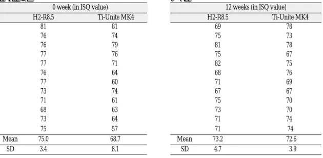
Results. Mean and SD of baseline ISQ values of test group were 75.0 ± 3.4 and 68.7 ± 8.1 for control group. Mean and SD of ISQ values 12 weeks after implant insertion were 73.2 ± 4.7 for test group and 72.6 ± 3.9 for control group.
There were no statistically significant differences between groups in ISQ values after 3months (P>0.05). From the data, RFA gains after 3 months were calculated, and there was sta- tistically significant difference between groups (P<0.05).
Conclusion. Although there were RFA changes between groups, implant stability after exper- imental period shows alike tendency and good bone responses.
Key Words
Resonance frequency analysis (RFA), Implant stability, Anodized implant surface
J Korean Acad Prosthodont : Volume 43, Number 6, 2005
F
or successful implant therapy, it is essential to achieve well-maintaining osseointegration.Osseointegration is a continuing structural and functional coexistence, possibly in a symbiotic man- ner, between differenciated, adequately remod- eling, biologic tissues and strictly defined and con- trolled synthetic components, providing long lasting, specific clinical functions without rejec- tion mechanics. The importance of implant sur- face properties for successful osseointegration was first pointed out by Albrektsson et al.1Surface properties are important for and may be used to facilitate tissue integration. However, a number of questions have been posed regarding the important role of the titanium implant surface properties during ‘dynamic build-up’of the osseointegration process. Interest in titanium implant surface oxide properties has increased with the development of methods to characterize such surfaces. Moreover, the possibility of surface modification of titanium implants to improve tissue responses is an outstanding feature in metal implantology research. In the air at room temperature, the surface of titanium is covered spontaneously by an oxide layer, which is 1.5-10 nm in thickness.2It was defined that the oxide lay- er has low level of electronic conductivity,3great thermodynamic stability,4and low ion-forma- tion tendency in aqueous environments.5These properties may be the reasons for the excellent bio- compatibility of titanium implants.
Recently, an electrochemical procedure for modifying the Ti surface was proposed, which has since attracted much attention. By applying a positive voltage to a Ti specimen immersed in an electrolyte, anodic oxidation (or anodization) of Ti occurs forming a TiO2surface layer. When the applied voltage is increased to a certain point, a micro-arc occurs resulting from dielectric break- down of the TiO2layer. At the moment the dielec-
tric breakdown occurs, Ti ions in the implant and OH ions in the electrolyte move in opposite directions very quickly to form TiO2again. This process is generally referred to as micro-arc oxi- dation (MAO) or plasma electrolysis.6The new- ly formed TiO2layer is both porous and firmly adhered to the substrate, which is beneficial for the biological performance of the implants.
Recent studies on the biological response of Ti implants demonstrated the MAO process con- stitutes one of the best methods of modifying the implant surface.7However, further research is necessary for the complete characterization of the oxide layer and identification of the optimum conditions for the MAO process.
Resonance frequency measurements can be related to the stiffness of an implant in the sur- rounding tissues and also to the level of the sur- rounding bone. Primary implant stability is relat- ed to bone quality, that is, the density of the surrounding bone and the height of the bone level and surgical technique. Secondary implant stability can be increased by bone formation and remodeling at the implant/bone interface. Further investigations using resonance frequency analy- sis are required to study the structural changed at the implant-tissue interface during healing and the influence of variables including boned quality and quantity. This may enable desirable levels of stability at fixture placement and the degree of healing prior to loading to be established to ensure a successful long-term outcome.
Nowadays, resonance frequency analysis (RFA) technique has been increasingly served as a sen- sitive and objective tool for clinical monitoring of implant stability.8,9However, for the precise eval- uation of individual implant stability or com- parison with other implants in various clinical con- ditions, standardized baseline data using RFA are urgently required.10Therefore, more clinical stud- ies are needed to elucidate resultant implant sta-
bilities in such specific conditions as identification of implant status at risk for implant failure, indi- vidualization of healing periods after implant placement and so on.
In present study, it was planned and performed that RFA value changes with time would be observed after implants placement in rabbits.
The aims of this study were to evaluate the RFA values of two anodized implant groups with different primary stability immediately after implant placement and 12 weeks later.
MATERIALS AND METHODS
2.1. Implant preparation : design and surface oxide
Among a total of 24 implants, twelve screw shaped implants as a test group (H2-R8.5) were manufactured, which had a pitch-height of 0.4 mm, an outer diameter of 4.3 mm, a length of 8.5 mm, and external hexa-headed, were turned from 5 mm rods of commercially pure titanium (ASTM Grade IV, Warantec Co., Seoul, Korea), and another twelve implants as a control group were Bra。nemark Ti-Unite MK4 (diameter 4.0 mm, length 8.5 mm). Surface oxides used in the test group were prepared with the use of a DC pow- er supply in electrolyte solution.
2.2. Micro-arc Oxidation (MAO)
Test implants (H2-R8.5) were prepared using MAO methods at the galvanostatic mode.
MAO of the test group was carried out in an aqueous electrolyte by applying a pulsed DC field to the specimen with a frequency of 660 Hz. The electrolyte was prepared by dissolving 0.15 mol calcium acetate monohydrate {Ca(CH3COO)2∙
H2O} and 0.02 mol calcium glycerophosphate (CaC3H7O6P) in de-ionized water. High range
of DC 300 Voltage were applied to the speci- mens, with each treatment lasting 3 minutes.
All MAO processing was carried out in a water- cooled bath made of stainless steel, using a stain- less steel plate (100×60×1 mm) as the counter electrode.
2.3 Animals and surgical technique
6 mature (average age 10 months old weighing 3-3.5kg) New Zealand white rabbits of both sex- es were included in this study. During surgery, ani- mals were anaesthetized with intramuscular injections of Ketamine 10mg/kg (Yu-han Co., Seoul, Korea) and Rompune 0.15mg/kg (Bayer Korea Co., Seoul, Korea). Prior to surgery, the shaved skin was carefully washed with a mixture of iodine and 70% ethanol. Local anesthesia with 1.0ml of 5% lidocaine including 1:100,000 epi- nephrine (Yu-han Co., Seoul, Korea) was admin- istered, at the tuberosity tibiae part of the bone where the incision was planned, under aseptic con- ditions. The skin and fascial layers were opened and closed separately. The periosteal layer was gen- tly pulled away from the surgical area and was not re-sutured. During all surgical drilling sequences, low rotary drill speeds not exceeding 1000rpm and profuse saline cooling were used. Drilling and fix- ture insertion was performed following surgical protocol of the Oneplant system�(Warantec Co., Seoul, Korea).
Two implants were placed in each tibia (four implants per rabbit), which penetrated one cor- tical layer only. Test two implants were inserted in right side and control two in left side, separated in 10 mm each other. The animals were kept in sep- arate cages and immediately after surgery, were allowed to be fully weight-bearing. After a follow- up period of 12 weeks, the animals were sacrificed in CO2chamber.
2. 4. Resonance frequency measurements
Directly after implant insertion, the base line RFA was monitored on the implants.
At the day of sacrifice, i.e. 3 month after implant insertion, the RFA was again tested. This method is a non-destructive technique that demonstrates the implant stability in terms of interfacial stiff- ness (Hz). The frequency response of the sys- tem was measured by attaching the transducer to a screw implant.
OsstellTM(Integration Diagnostics Ltd. Sweden) was used for implant stability measurements in this study. The transducers were orientated per- pendicular to the long axis of tibia and were tightened by hand. Results were represented as an ISQ (Implant Stability Quotient, 1-100).
2.5. Statistics
Data are presented as means ± SD. One-way analysis of variance (ANOVA) was used for sta- tistical analysis of the data. Differences were considered statistically significant at P<0.05.
RESULTS
Mean and SD of baseline ISQ values of test group were 75.0 ± 3.4 and 68.7 ± 8.1 for control group. Mean and SD of ISQ values 12 weeks after implant insertion were 73.2 ± 4.7 for test group and 72.6 ± 3.9 for control group.(Table I, II and III)
There were no statistically significant differ- ences between groups in ISQ values after 3 months.
Table II. RFA for H2-R8.5 and Ti-Unite after 12 weeks
12 weeks (in ISQ value)
H2-R8.5 Ti-Unite MK4
69 78
75 73
81 78
75 67
82 75
68 76
71 69
67 67
75 70
73 70
71 74
71 74
Mean 73.2 72.6
SD 4.7 3.9
Table I. RFA for H2-R8.5 and Ti-Unite at fixture installation
0 week (in ISQ value)
H2-R8.5 Ti-Unite MK4
81 81
76 74
76 79
77 76
77 71
76 64
77 60
73 74
71 61
68 63
73 64
75 57
Mean 75.0 68.7
SD 3.4 8.1
Table III. RFA change for H2-R8.5 and Ti-Unite at each period (Mean and SD in ISQ value)
Implant Fixture installation 12weeks RFA gain
H2-R8.5 75.0 ± 3.4 73.2 ± 4.7 -1.8 ± 5.7
Ti-Unite MK4 68.7 ± 8.1 72.6 ± 3.9 3.9 ± 8.1
(P>0.05) (Fig. 1) From the data, RFA gains after 3 months were calculated, and there was statistically significant difference between groups. (P<0.05) (Fig. 2)
DISCUSSION
To determine the ability of the surface proper- ties of oxidized implants to influence bone response, the bone response was measured with the RFA value.
Rough surface implant showed great success rate clinically, and took the place of prototype implant - machined implant. Many surface treating tech- niques were developed, and being developed now - sand blasting, acid etching, ion deposi- tion, anodic oxidation etc.
In this study, among the above surface modi- fications, we focused on anodic oxidation tech- nique. Ti-Unite (Nobel biocare) implant is produced under static current method. In this case, voltage is changing continuously, generally growing up as surface oxide getting thicker. We used static volt- age technique at 300 voltage, and some fluctua- tion was used for thicker oxide layer.
Implant primary stability is not only depends on bony quality and quantity but also depend on sur-
gical technique used.11 Bony housing used in this experiment was tibia of rabbit, therefore the physical properties of implant bed were simi- lar condition. Surgical protocols of implant inser- tion and implant macrostructure provide a mechanical engagement and hoop stress on bone- implant interface and these make primary stability of implant. We set up different stability condition from the start according to manufacture’s guide.
After 12 weeks, there were no significant differ- ences in RFA values between groups, but slight tendency of high level ISQ of H2-R8.5 test groups were expressed.
Test groups showed drop of ISQ after 12 weeks.
After 3 months, all implants had enough stabil- ity in terms of secondary stability. During 12 weeks, there was remodeling of bone. According to the difference of magnitude of hoop stress, the phase of bone response might be different. It was expressed by ISQ change between groups. At clinical immediate loading condition, which type of primary stability will benefit and guarantee bony remodeling and conversion to secondary bio- logic stability is in question. May be extra ISQ val- ues guide and protect from environmental force.
In summary, the results of the present study indi- cated that all anodized implant surfaces are com- Fig. 1. RFA at implant placement and after 12 weeks. Fig. 2. RFA change after 12 weeks.
patible to the biologic action of bone and above crit- ical level of implant stability, increase or decrease of ISQ value according to conversion from primary stability to secondary stability might be have little significance in implant survival.
CONCLUSIONS
Within the limitations of this study, the fol- lowing conclusions were drawn:
1. There were no statistically significant differences between groups in RFA values after 12 weeks (P>0.05).
2. The RFA gains after 12 weeks were statistically significant difference between groups (P<
0.05). In H2-R8.5 group, there was drop of RFA value and in Ti-Unite MK4 group, there was increase of RFA value.
REFERENCES
1. Albrektsson T, Branemark PI, Hansson HA, Kasemo B, Larsson K. The interface zone of inor- ganic implant in vivo ; Titanium implants in bone.
Ann Biomed Eng 1983;11:1-27.
2. Kasemo B, Lausmaa J. Aspect of surface physics on titanium implants. Swed Dent J 1983;28(Suppl.):19- 36.
3. Zitter H, Plenk HJ. The electrochemical behav- ior of metallic implant materials as indicator of their
biocompatibility. J Biomed Mater Res 1987;21:881- 896.
4. Solar RJ, Pollack SR, Korostoff E. In vitro corrosion testing of titanium surgical implant alloys: an ap- proach to understanding titanium release from im- plants. J Biomed Mater Res 1979;13:217-250.
5. Tengvall P, Lindstrom I. Physico-chemical consid- erations of titanium as a biomaterial. Clin Mater 1992;9:115-134.
6. Ishizawa H, Ogino M. Formation and characteri- zation of anodic titanium oxide films containing Ca and P. J Biomed Mater Res 1995;29:65-72.
7. Sul YT, Johansson CB, Jeong YS, Albrektsson T. The electrochemical oxide growth behavior on tita- nium in acid and alkaline electrolytes. Med Eng and Phys 2001;23:329-46.
8. Meredith N. Assessment of implant stability as a pognotic determinant. Int J Prosthodont 1998;11:491- 501.
9. Sennerby L, Meredith N. Resonance Frequency Analysis : Measuring implant stability and os- seointegration. Compendium 1998;19:493-502.
10. Park C. A study of the measurement of the implant stability using resonance frequency analysis. PhD thesis, 2000, Dankook University, Korea.
11. Albrektsson, T., Branemark, P.I., Hansson, H., &
Lindstrom, J. (1981) Osseointegratd titanium im- plants. Requirements for ensuring a long-lasting direct bone-to-implant anchorage in man. Acta Orthopaedica Scandinavica, 52, 155.
Reprint request to:
SEONG-JOOHEO, D.D.S., Ph.D.
DEPT. OFPROSTHODONTICS.COLLEGE OFDENTISTRY SEOULNATIONALUNIVERSITY
28-1 YEONGUN-DONG,CHONGNO-GU. 110-749, SEOULKOREA [email protected]