INTRODUCTION
Isocyanate chemicals are currently the most common causes of occupational asthma worldwide and in Korea as well with a prevalence of 2.9% to 13% in exposed workers (1). Long- term follow-up studies of subjects with isocyanate asthma have suggested that asthmatic symptoms may persist in some sub- jects for months or years after work withdrawal (2-6). Our previous study also demonstrated that 25 (61%) among 41 patients had persistent asthma symptoms after 5 yr’ follow- up period, despite anti-asthmatic medications and complete avoidance from exposure (7).
Vascular endothelial growth factor (VEGF) is a multi-func- tional cytokine, which plays a role in chronic airway inflam- mation, angiogenesis and airway remodeling in bronchial asthma (8-11). VEGF could increase vascular permeability, which results in leak of plasma protein into the extravascular space (12). However, the mechanisms of VEGF in toluene diisocyanate (TDI)-induced asthma are unknown. In a murine model of TDI-induced asthma (13), VEGF level was increased in bronchial alveolar lavage (BAL) fluid after TDI challenges.
Pre-treatment of VEGF inhibitors could reduce influx of
inflammatory cells, airway hyperresponsiveness (AHR) and VEGF secretion in BAL fluid, indicating a contribution of VEGF in TDI-induced asthma. Although there have been a few studies suggesting a pathogenic contribution of VEGF in asthma pathology (12, 14, 15), to the best of our knowledge, this is the first study to evaluate a possible involvement of VEGF in both airway secretion and serum in acute and chron- ic airway inflammation of TDI induced asthma patients.
MATERIALS AND METHODS Subjects
Twenty-four subjects with TDI-induced asthma were en- rolled. All subjects were withdrawn from the workplace to avoid further exposure to TDI and received anti-asthmatic medications according to symptom severity. The subjects were classified into two groups : group I included 11 newly diag- nosed asthma patients, and group II included 12 chronic TDI- asthma patients who had been followed for ≥4 yr with anti- asthmatic medications. They also had symptoms of asthma
Jeong-Hee Choi, Yu-Jin Suh, Soo-Keol Lee, Chang-Hee Suh, Dong-Ho Nahm, Hae-Sim Park
Department of Allergy and Rheumatology, Ajou University School of Medicine, Suwon, Korea
Address for correspondence Hae-Sim Park, M.D.
Department of Allergy and Rheumatology, Ajou University School of Medicine, San-5, Wonchon-dong, Yeongtong-gu, Suwon 442-749, Korea
Tel : +82.31-219-5196, Fax : +82.31-219-5154 E-mail: [email protected]
*This study was funded by a grant of the Korean Health 21 R&D project, Ministry of Health & Welfare, Republic of Korea (01-PJ10-PG6-01GN14-0007).
359
Acute and Chronic Changes of Vascular Endothelial Growth Factor (VEGF) in Induced Sputum of Toluene Diisocyanate (TDI)-induced Asthma Patients
Vascular endothelial growth factor (VEGF) is a multi-functional cytokine involved in inflammation, repair and angiogenesis in asthmatic airway. This study aimed to eval- uate the role of VEGF in immediate bronchoconstriction induced by TDI inhalation, and in chronic TDI-asthma patients. 11 newly diagnosed TDI-asthma patients (group I), 12 chronic TDI-asthma patients with persistent asthma symptoms followed for >4 yr and 15 unexposed healthy controls were enrolled. In group I, induced sputum and serum were collected before and 7 hr after placebo- and TDI-bronchoprovocation test (BPT). In group II, induced sputum and serum were collected every 2 yr. VEGF levels were measured by ELISA. There were no significant differences in sputum and serum VEGF levels between patients and controls. Before and after placebo- and TDI-BPT, no significant changes were noted in sputum and serum VEGF levels of group I. In group II patients, sputum VEGF showed variable changes at 1-yr, then decreased significantly at 2-yr (p<0.05), while serum VEGF showed variable changes at 2-yr, which decreased significantly at 4-yr (p<0.05). These results suggest that VEGF may play a minor role in immediate bronchoconstriction after TDI-BPT. In chronic TDI-asthma, VEGF may be involved to 2 yr after the diagnosis and the con- tribution may decrease after then.
Key Words : Vascular Endothelial Growth Factor; Toluene 2,4-Diisocyanate; Asthma, Exercise-Induced;
Bronchoprovocation Test; Sputum
Received : 24 February 2004 Accepted : 12 March 2004
requiring anti-asthma medications with complete avoidance.
The diagnosis of occupational asthma had been confirmed by positive responses to bronchoprovocation tests to TDI (TDI- BPT). Age matched unexposed healthy subjects were enrolled as a control group. Atopy was determined by a positive skin prick test response to at least one common inhaled allergen.
Sera from all subjects were collected at diagnosis and every 2 yr; subjects stopped using inhaled or oral steroids at least 4 weeks before the study. They underwent an interview, chest radiography, skin prick tests with common inhaled allergens, lung function measurement and inhalation challenge with both methacholine, and TDI (80:20=2.4 form: 2.6 form, Aldrich, Milwaukee, WI, U.S.A.). All subjects gave their informed consent and the protocol was accepted by the Institu- tional Review Board of Ajou Medical Center, Suwon, Korea.
Bronchial challenge test with methacholine and TDI
The methacholine bronchial challenge test was done accord- ing to the method previously described (7). Briefly, aerosols were generated by a DeVilbiss 646 nebulizer connected to a DeVilbiss dosimeter driven by compressed air (DeVilbiss Co., Doylesttown, PA, U.S.A.). Five inhalations of normal saline at 5-min intervals were taken followed by a series of succes- sively doubled doses of methacholine (0.075-25 mg/mL) until a 20% fall in FEV1was observed or the maximum dose given.
FEV1 was measured 5-min after the beginning of each set of inhalations of aerosolized methacholine. The methacholine PC20level was determined by interpolation from the dose- response curve. The TDI-BPT was performed according to a standardized protocol (7). On the first day, placebo challenge was performed for one day before the TDI challenge. On the next day, the subjects were exposed to pure TDI monomers (Aldrich, U.S.A.) in a small closed room for 5-15 min until asthmatic symptoms were induced and FEV1decreased more than 20% from the baseline value. The concentration of TDI measured by TLD-1, a toxic gas detector with Cheakey (HAD Scientific, Lincolnshire, IL, U.S.A.) was less than 20 ppb. FEV1 and FEF25-75%were measured with a spirometer (MultiSPIRO SX/PC, Tempe, AZ, U.S.A.) immediately before exposure and every hours for 7 hr after the exposure.
Sputum was induced using a previously described method (14) and collected just before (0 min) and after completing the placebo- and TDI-BPT (420 min after the TDI exposure).
Collected sputum samples were immediately processed as reported previously (16). The volumes of induced sputum were determined, and equal volumes of phosphate-buffered saline-0.5% Tween (PBST) were added. The samples were then mixed by vortex mixer for 1 min, centrifuged for 20 min.
at 3,000 rpm, and kept frozen at -20℃.
VEGF levels were measured in induced sputum from group I and II subjects. Sputum was collected twice before the chal- lenge as baseline value, and at 7 hr after the TDI- and placebo- challenge tests.
Sera collection before and after TDI-BPT were completed in 9 subjects of group I.
VEGF level in both sputa and sera were measured by ELISA kit (R&D system, Minneapolis, MN, U.S.A.). Both assays were performed according to the manufacturer’s guidelines.
The protein content within the sputum was measured by the Bradford method, and the VEGF levels in the sputum were presented as ratios to the protein content.
Statistical analysis
ANOVA were done to compare clinical data of two groups of the study subjects and control. Mann Whitney U test was applied to compare clinical parameters between group I and II. The Wilcoxon-Signed rank tests were applied to compare the changes of VEGF in serum and induced sputum in both groups. p-value of 0.05 or less was regarded as significant.
RESULTS
Clinical characteristics of the study subjects
The study subjects and controls were well matched for age (Table 1). Both group I and II subjects had similar lung func- tion measurements at baseline. No significant differences were noted in atopy prevalence, duration of TDI exposure in work-
Group I (n=11) Group II (n=12) Controls (n=15) p value
Age (yr) 42.0±10.8 44.8±6.87 40.6±4.23 NS
FEV1 82.8±25.3 79.1±23.7 NA NS*
Atopy* (Presence/Absence) 4/8 4/8 NA NS*
Duration of exposure (months) 92.5±25.5 84.0±8.7 NA NS*
Asthma symptoms before 20.3±15.2 33.0±38.2 NA NS*
diagnosis (months)
VEGF/protein in sputum (baseline, pg/mg) 823.76±676.6 978.66±247.52 1019.3±432.5 NS VEGF in serum (baseline, pg/mL) 122.76±101.75 307.08±241.80 186.91±132.13 NS*
Table 1.Comparison of VEGF levels in induced sputum and sera between patients and controls
NA, Not available; NS, No statistical significance by ANOVA. NS*: No statistical significance between group I and II.
place, and asthma symptom duration before the diagnosis (p>0.05). Eight of group I had immediate bronchoconstric- tions after TDI-BPT and three had dual asthmatic responses.
Changes of VEGF in serum and induced sputum of TDI and placebo challenges
There was a wide variation in the level of VEGF in serum and sputum in group I subjects. VEGF level of group I tended to be lower than group II, but no significant differences were noted among the study subjects as shown in Table 1 (p>0.05).
Fig. 1A demonstrated the changes of VEGF level in induced sputum of group I patients before and after TDI-BPT. There were no significant differences in VEGF levels in induced sputum after TDI-BPT as well as in placebo challenges (place- bo challenge data was not shown, p>0.05, respectively). Fig.
2B showed changes of VEGF level in serum before and after
TDI-BPT. No significant differences were also noted after TDI-BPT and placebo challenges (placebo challenge data was not shown, p>0.05, respectively).
Changes of VEGF in sera and induced sputa during long term follow-up period
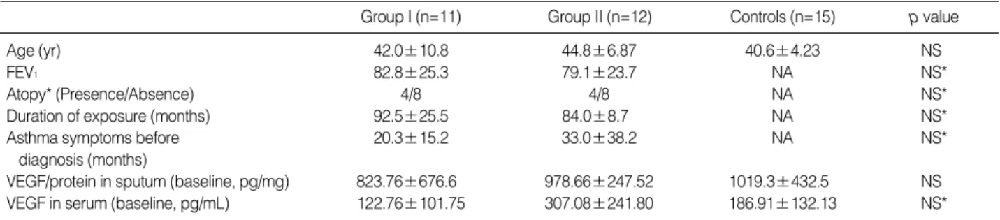
Fig. 2A shows the changes of VEGF level of group I sub- jects in induced sputum during 2 yr follow-up period. Spu- tum VEGF level showed variable changes at 1-yr, which de- creased significantly at 2-yr after the diagnosis (p=0.03). Fig.
2B shows changes of serum VEGF levels during 4 yr follow- up period. Serum VEGF level showed variable changes at 2-yr after the diagnosis (p=0.27), which decreased significantly at 4-yr (p=0.004). There were no significant correlations between VEGF levels in both sputum and sera with clinical parame- ters including FEV1values and airway hyperresponsiveness to methacholine (data not shown). Also no significant corre- lation was found between VEGF level and neutrophil and eosinophil count in the sputum (data not shown).
DISCUSSION
The pathogenic mechanism of isocyanate-induced occupa- tional asthma is complicated because several immunological and non-immunological mechanisms are involved (1). The previous study (2-6) for analyzing prognostic factors in iso- cyanate induced asthma patients demonstrated that more than 50% of the study subjects had persistent asthma symp- toms, indicating that isocyanate, especially TDI-induced asth- ma had a poor outcome with abnormal airway function. The underlying mechanism which explains the poor prognosis is still unknown. The pathologic findings to suggest airway
VEGF/protein, pg/mg
1,600
1,400
1,200
1,000
800
600
400
200 Initial diagnosis 1 yr 2 yr
p=0.27 p=0.05
p=0.03
A
Fig. 2.Changes of sputum (A) and sera (B) VEGF levels in 12 TDI-induced asthma patients (group II). VEGF levels in induced sputum are presented as the ratio to protein concentation.
VEGF, pg/mL
1,000
800
600
400
200
0 Initial diagnosis 2 yr 4 yr
p=0.27 p=0.01
p=0.004
B
VEGF/protein, pg/mg
2,500 2,000
1,500
1,000 500
0 0 420 min
Time after TDI challenges
Time after TDI challenges
VEGF, pg/mL
250 200 150
100 50
0 0 420 min
p=0.70 p=1.0
A B
Fig. 1.Changes of VEGF after TDI bronchoprovocation test in in- duced sputum (A) and serum (B) of TDI asthma patients. No signif- icant changes are noted in both samples (p>0.05, respectively).
remodeling with increased expression of TGF- was noted in the airway mucosa of TDI-asthma patients, especially those who had been complaining of persistent asthma symptoms (1). These findings may suggest that airway remodeling may be progressed from an early stage in TDI-asthma patients, which result in persistent asthmatic symptoms despite anti- asthmatic medications and complete avoidance.
VEGF is an endothelial cell-specific mitogen that has been shown to play a key role in angiogenesis. Furthermore, VEGF is a potent enhancer of microvascular permeability (17). VEGF can be produced by a wide variety of inflammatory cells includ- ing macrophages, neutrophils, and eosinophils and involved in their activation (12, 13, 16). A mouse model of TDI-asth- ma study (13) suggested a possible contribution of VEGF in airway obstruction, migration of inflammatory cells and air- way hyperresponsiveness development. However, in the pre- sent study using human samples of TDI-induced asthma patients, VEGF levels showed no significant changes after TDI-BPT in both sputum and serum. These findings sug- gested that VEGF might have a minor role in immediate bronchoconstriction after TDI exposure in human asthma.
Several investigators suggested contribution of VEGF in chron- ic asthma and airway remodeling in chronic non-occupation- al asthma patients. Hoshino et al. (14) demonstrated that VEGF expression was increased in the asthmatic airway of chronic asthma patients, which was correlated with airway caliber and airway hyperresponsiveness. The other study pre- sented increased VEGF level in induced sputum in compar- ison to normal controls with imbalance to endothelin (16).
In this study, we found no differences in VEGF level in in- duced sputum of TDI asthma patients and normal controls, which was presented in two ways: VEGF level itself and the ratio of VEGF level to protein concentration. Normal controls enrolled in this study were defined as non-smoker, non-atopic healthy controls. And no difference was found between two groups of asthmatic patients. Correlation coefficient was poor between VEGF an eosinophil cationic protein level in induced sputum (data was not shown). To clarify these differences, further studies will be needed in a larger number of asthmat- ic patients and controls. However, new findings in this study was serial observation of VEGF level in the induced sputum and sera, which demonstrated significant decreases of VEGF level in airway secretion 2 yr earlier than those in serum. Re- modeling as a cogent mechanism in chronic asthma must also take into account matrix proteins, growth factor, insulin growth factor and possibly anti-apoptotic factors. Thus stud- ies dealing with single factor such as VEGF are not likely to explain in the complexity of remodeling. As VEGF was detec- table in the airway secretion, we can speculate that VEGF may be involved in airway inflammation/angiogenesis up to 2 yr after the diagnosis in chronic TDI-asthma patients, and the contribution of VEGF may decrease after then.
In conclusion, the importance of VEGF contribution in acute bronchoconstriction caused by TDI seems to be low in
TDI induced asthma patients. However, VEGF involvement in chronic TDI asthma was suggested up to 2 yr after the diagnosis.
REFERENCES
1. Park HS, Cho SH, Hong CS, Kim YY. Isocyanate-induced occupa- tional asthma in far-east Asia: pathogenesis to prognosis. Clin Exp Allergy 2002; 32: 198-204.
2. Tarlo SM, Banks D, Liss G, Broder I. Outcome determinants for iso- cyanate induced occupational asthma among compensation claimants.
Occup Environ Med 1997; 54: 756-61.
3. Lemiere C, Cartier A, Dolovich J, Chan-Yeung M, Grammer L, Ghez- zo H, L’Archeveque J, Malo JL. Outcome of specific bronchial respon- siveness to occupational agents after removal from exposure. Am J Respir Crit Care Med 1996; 154: 329-33.
4. Paggiaro PL, Vagaggini B, Dente FL, Bacci E, Bancalari L, Carrara M, Di Franco A, Giannini D, Giuntini C. Bronchial hyperrespon- siveness and toluene diisocyanate. Long-term change in sensitized asthmatic subjects. Chest 1993; 103: 1123-8.
5. Park HS, Nahm DH. Prognostic factors for toluene diisocyanate-in- duced occupational asthma after removal from exposure. Clin Exp Allergy 1997; 27: 1145-50.
6. Piirila PL, Nordman H, Keskinen HM, Luukkonen R, Salo SP, Tuomi TO, Tuppurainen M. Long-term follow-up of hexamethylene diiso- cyanate- diphenylmethane diisocyanate-, and toluene diisocyanate- induced asthma. Am J Respir Crit Care Med 2000; 162: 516-22.
7. Park HS, Lee SK, Kim HY, Nahm DH, Kim SS. Specific immunoglob- ulin E and immunoglobulin G antibodies to toluene diisocyanate-hu- man serum albumin conjugate: useful markers for predicting long-term prognosis in toluene diisocyanate-induced asthma. Clin Exp Allergy 2002; 32: 551-5.
8. Hoshino M, Takahashi M, Aoike N. Expression of vascular endothe- lial growth factor, basic fibroblast growth factor, and angiogenin immunoreactivity in asthmatic airways and its relationship to angio- genesis. J Allergy Clin Immunol 2001; 107: 295-301.
9. Demoly P, Maly FE, Mautino G, Grads S, Gougat C, Sahla H, Godard P, Bousquet J. VEGF levels in asthmatic airways do not correlate with plasma extravasation. Clin Exp Allergy 1999; 29: 1390-4.
10. Dvorak, HF, Brown LF, Detmar M, Dvorak AM. Vascular perme- ability factor/vascular endothelial growth factor, microvascular hyper- permeability, and angiogenesis. Am J Pathol 1995; 146: 1029-39.
11. Folkman J, Shing Y. Angiogenesis. J Biol Chem 1992; 267: 10931-4.
12. Mc Donald DM. Angiogenesis and remodeling of airways vascula- ture in chronic inflammation. Am J Respir Crit Care Med 2001; 164:
S39-45.
13. Lee YC, Kwak YG, Song CH. Contribution of vascular endothelial growth factor to airway hyperresponsiveness and inflammation in a murine model of toluene diisocyanate-induced asthma. J Immunol 2002; 168: 3595-600.
14. Hoshino M, Nakamura Y, Hamid QA. Gene expression of vascular endothelial growth factor and its receptors and angiogenesis in bro- nchial asthma. J Allergy Clin Immunol 2001; 107: 1034-8.
. .
15. Asai K, Kanazawa H, Otani K, Shiraishi S, Hirata K, Yoshikawa J.
Imbalance between vascular endothelial growth factor and endostatin levels in induced sputum from asthmatic subjects. J Allergy Clin Im- munol 2002; 110: 571-5
16. Park HS, Jung KS, Kim HY, Nahm DH, Kang K. Neutrophil acti- vation following TDI bronchial challenges to the airway secretion
from subjects with TDI-induced asthma. Clin Exp Allergy 1999; 29:
1395-401.
17. Senger DR, Galli SJ, Dvorak AM, Perruzzi CA, Harvey VS, Dvorak HF. Tumor cells secrete a vascular permeability factor that promotes accumulation of ascites fluid. Science 1983; 219: 983-5.