© 2011 The Korean Academy of Medical Sciences.
This is an Open Access article distributed under the terms of the Creative Commons Attribution Non-Commercial License (http://creativecommons.org/licenses/by-nc/3.0) which permits unrestricted non-commercial use, distribution, and reproduction in any medium, provided the original work is properly cited.
pISSN 1011-8934 eISSN 1598-6357
Development of Acute Megakaryoblastic Leukemia with
Isochromosome (12p) after a Primary Mediastinal Germ Cell Tumor in Korea
The association of hematological malignancies with a mediastinal germ cell tumor (GCT) is very rare. We report one case of a young adult male with primary mediastinal GCT who subsequently developed acute megakaryoblastic leukemia involving isochromosome (12p).
A 25-yr-old man had been diagnosed with a mediastinal GCT and underwent surgical resection and adjuvant chemotherapy. At 1 week after the last cycle of chemotherapy, his peripheral blood showed leukocytosis with blasts. A bone marrow study confirmed the acute megakaryoblastic leukemia. A cytogenetic study revealed a complex karyotype with i(12p). Although additional chemotherapy was administered, the patient could not attain remission and died of septic shock. This case was definitely distinct from therapy-related secondary leukemia in terms of clinical, morphologic, and cytogenetic features. To our knowledge, this is the first case report of a patient with mediastinal GCT subsequently developing acute megakaryoblastic leukemia involving i(12p) in Korea.
Key Words: Mediastinal Germ Cell Tumor; Acute Megakaryoblastic Leukemia;
Isochromosome (12p) Nae Yu1, Hye Ryoun Kim1,
Young-Joo Cha1, Eun Kyung Park2 and Jeong Wook Kim2
Departments of 1Laboratory Medicine and 2Internal Medicine, Chung-Ang University College of Medicine, Seoul, Korea
Received: 14 March 2011 Accepted: 8 June 2011 Address for Correspondence:
Hye Ryoun Kim, MD
Department of Laboratory Medicine, Chung-Ang University College of Medicine, 29 Heukseok-ro, Dongjak-gu, Seoul 156-755, Korea
Tel: +82.2-6299-2718, Fax: +82.2-6298-8630 E-mail: [email protected]
DOI: 10.3346/jkms.2011.26.8.1099 • J Korean Med Sci 2011; 26: 1099-1102
CASE REPORT
Oncology & Hematology
INTRODUCTION
Germ cell tumors (GCTs) account for 2% of human malignan- cies, but are the most common tumors in males 15-35 yr old (1).
Mediastinal GCTs occur predominantly within the anterior me- diastinum, which account for 1%-4% of mediastinal tumors, and that have different clinical characteristics from testicular GCTs (2, 3). The association between hematological malignancies and mediastinal GCT was first reported in 1983 (4) and more than 50 cases have been published since (5-9). Most often, the mega- karyocytic lineage of hematopoiesis is involved in hematologic malignancy, resulting in acute megakaryoblastic leukemia, my- elodysplsia with abnormal megakaryocytes, or idiopathic throm- bocytopenia, essential thrombocythemia. Other hematologic diagnoses included acute lymphocytic or acute myeloid leuke- mia (AML) and, in rare cases, malignant histiocytosis or system- ic mastocytosis (4-7, 9). A total of 64 cases of hematologic malig- nancies with mediastinal GCT cases have been published (1, 4-9) and only one case has reported bone marrow involvement in a mediastinal GCT case in Korea (10). Here we present a case in which a patient developed acute megakaryoblastic leukemia involving i(12p) after a primary mediastinal GCT.
CASE DESCRIPTION
A 25-yr-old man presenting with chest pain was admitted to
Chung-Ang University Hospital (Seoul, Korea) because of an ab- normal mass in the anterior mediastinal area. He had no remark- able past medical and familial history. He had been diagnosed with a malignant mediastinal GCT (immature teratoma 80%, embryonal carcinoma 10%, seminoma 5%, yolk sac tumor 5%) in January 2010. Peripheral blood examination showed the fol- lowing: Hb level, 14.5 g/dL; leukocyte count, 10,770/μL (neutro- phil 70%, lymphocyte 21%, no blasts); and platelet count, 257,000/
μL. Serum alpha-fetoprotein (AFP), beta-human chorionic go- nadotropin (hCG), and lactate dehydrogenase (LDH) were 11,680 ng/mL, 0.847 mIU/mL, and 187 IU/L, respectively. After surgi- cal resection of the tumor, the patient received four cycles of ad- juvant chemotherapy with bleomycin, etoposide, and cisplatin between February and April 2010. During routine follow-up in August 2010, a persistent tumor lesion was found and the patient received three cycles of adjuvant chemotherapy with pacilitaxel, ifosfamide, and cisplatin between August and October 2010. A peripheral blood examination performed in October 2010 showed pancytopenia without blasts.
After 1 week of the last chemotherapy cycle, the patient visit- ed the emergency room with a fever. Peripheral blood examina- tion showed the following: Hb level, 5.9 g/dL; leukocyte count, 12,920/μL with 16% neutrophils, 22% lymphocytes, 57% blasts;
5 nucleated red cells per hundred white blood cells; and platelet count, 6,000/μL (Fig. 1A). Serum AFP and LDH levels were 234 ng/mL and 2,964 IU/L, respectively. Based on these findings,
Yu N, et al. • Development of AML with i(12p) after a GCT in Korea
1100 http://jkms.org DOI: 10.3346/jkms.2011.26.8.1099
acute leukemia was suspected and a bone marrow examination was performed in November 2010. In the bone marrow aspira- tion, about 40.4% of all nucleated cells were blasts. The blasts were medium to large-size with round, slightly irregular nuclei and one to three nucleoli. The cytoplasm of blasts was basophil- ic and might show distinct bleb or pseudopod formation (Fig.
1B). The dysplastic features of a megakaryocytic lineage (e.g. mi- cromegakaryocytes and hypolobulation of the nucleous) were found in more than 30% of 30 megarkaryocytes (Fig. 1C). Cyto- chemical staining showed that the cells had dot-like positivity for periodic acid-Schiff (Fig. 1D) and acid phosphatase, in con- trast, negativity for myeloperoxidase, α-naphthyl acetate ester- ase and α-naphthyl butyrate esterase. The bone marrow biopsy showed about 90%-100% cellularity (Fig. 1E). Immunopheno- typing showed positivity for CD13, CD33, CD117, MPO, CD7, CD61, and HLA-DR, and was negative for CD2, CD3, CD5, CD19, CD20, cCD22, cCD79a, CD10, CD14, and TdT. Cytogenetic ex- amination revealed a 46,XY,add(5)(q11.2),del(10)(p11.2), -17, der(18)t(17;18) (q11.2;q21), +i(12p)[30] karyotype (Fig. 2). FISH analysis using AML1/ETO, BCR/ABL, MLL, PML/RARA, and CBFB gene probes (Vysis, Downers Grove, IL, USA) demonstrat- ed no abnormalities. After conferring a diagnosis of acute mega- karyoblastic leukemia, chemotherapy with idarubicin and ara- C was administered and the patient showed pancytopenia. The patient was then treated with mitoxantrone and ara-C, and bone marrow examination was performed that showed severe hypo- cellular marrow and reactive increase of histiocytes and plasma cells. Despite the additional chemotherapy, the disease not go
into remission and the patient died of septic shock 1 month af- ter the diagnosis of leukemia had been made. A summary of the clinical course is shown in Table 1.
DISCUSSION
This case concurs with previous evidence of an association be- tween hematological malignancies and mediastinal GCTs in young males (5-9). Hematological malignancies associated with mediastinal GCTs must be distinguished from therapy-related secondary leukemia. Leukemia associated with the use of alkylat- ing agents usually occurs after an average interval of 5-7 yr, often
A B C
D E
Fig. 1. (A) Peripheral blood smear showing blasts (57% of nucleated cells). (B) Bone marrow smear revealed many medium-sized myeloblasts (about 40.4% of all nucleated cells). (C) Dysplasic megakaryocyte. (D) PAS positivity (granular pattern). Wright-Giemsa stain, × 1,000 magnification. (E) Bone marrow biopsy showing compact marrow with increased dysplastic megakaryocytes (H&E, × 400 magnification).
1
6
13
19
2
7
14
20
4
9
15
21
10
16
22
11
17
X 3
8
5
12
18
Y Fig. 2. Giemsa-banded karyotype showing 46,XY,add(5)(q11.2),del(10)(p11.2), -17,der(18) t(17;18)(q11.2;q21),+i(12p)[30].
Yu N, et al. • Development of AML with i(12p) after a GCT in Korea
http://jkms.org 1101
DOI: 10.3346/jkms.2011.26.8.1099
preceded by a preleukemic period with myelodysplasia and fre- quently progressing to AML with French-American-British clas- sification (FAB) subtypes M1 or M2 (11). Topoisomerase II in- hibitor-related secondary leukemia is generally diagnosed 2-3 yr after chemotherapy and most commonly exhibit FAB M4 or M5 phenotypes. While the latter type of leukemia is frequently associated with translocations of the long arm of chromosome 11 (11q23) (12-15), leukemia that appears after treatment with alkylating agents displays alterations in chromosome 5 or 7 in a high proportion of patients (60%-90%) (5).
In contrast, leukemia associated with mediastinal germ cell tumors appears to have no consistent cytogenetic abnormali- ties (9). However, in some cases, immunohistochemical and cy- togenetic evidence, especially presence of i(12p) chromosome, had previously suggested the clonal relationship between the mediastinal GCTs and the hematologic malignancy (16). Also, two cases of leukemia have been reported in which i(12p) was found and both of these leukemia patients had mediastinal GCTs (17). The most common karyotypic abnormality in a mediasti- nal GCT and in the associated leukemic blasts was i(12p), sug- gested that i(12p) was might be a potential predictive marker for development of acute megakaryoblastic leukemia. Although our patient was treated with an alkylating agent (ifosfamide), the interval period between mediastinal GCT and the development of acute leukemia was only 10 months, the leukemia phenotype expressed in bone marrow and peripheral blood was that of acute megakaryoblastic leukemia, and a characteristic chromosomal abnormality i(12p) was also found in this case. Therefore, the clinical, morphologic, and cytogenetic features of the leukemia in our case are definitely different from therapy-related acute leukemia although the patient received seven cycles of chemo- therapy.
The most possible explanation for the association between mediastinal GCTs and acute megakaryoblastic leukemia was suggested by Nichols et al. (9) who found that hematologic neo- plasm is a consequence of multipotential differentiation of ma- lignant germ cells. Presumptive corroboration of this theory is
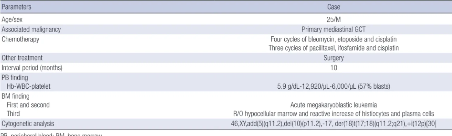
Table 1. Summary of the clinical data for the patient with acute megakaryoblastic leukemia and a primary mediastinal GCT
Parameters Case
Age/sex 25/M
Associated malignancy Primary mediastinal GCT
Chemotherapy Four cycles of bleomycin, etoposide and cisplatin
Three cycles of pacilitaxel, ifosfamide and cisplatin
Other treatment Surgery
Interval period (months) 10
PB finding
Hb-WBC-platelet 5.9 g/dL-12,920/µL-6,000/µL (57% blasts)
BM finding First and second
Third Acute megakaryoblastic leukemia
R/O hypocellular marrow and reactive increase of histiocytes and plasma cells Cytogenetic analysis 46,XY,add(5)(q11.2),del(10)(p11.2),-17, der(18)t(17;18)(q11.2;q21),+i(12p)[30]
PB, peripheral blood; BM, bone marrow.
provided by the finding of i(12p) as a marker chromosome in the cytogenetic analysis of bone marrow in a patient with a medias- tinal GCT and leukemia.
Hematologic neoplasms with primary mediastinal GCT pos- sess a very aggressive clinical course. Patients tend to die before treatment, do not respond to anti-leukemic therapy, or achieve only short remissions. Allogenic bone marrow transplantation may be the only curative strategy despite one report with two pa- tients who failed to respond to this treatment (5). However, a sub- group of patients with platelet disorders seemed to have a slight- ly better prognosis (4, 6). The patient in our case also showed very aggressive clinical course and little response to chemother- apy. His survival period after AML diagnosis was only 2 months.
There has been one report showing and association between mediastium and hematological diseases in Korea which present- ed a case of a primary mediastinal GCT with bone marrow in- volvement (10). However, this case did not present either acute megakaryoblastic leukemia or leukemia involving i(12p); rather, this group reported acute undifferentiated leukemia with triso- my 8 and concurrent mediastinal GCT. It remains unclear why some patients with non-seminomatous mediastinal GCTs de- velop a hematologic disorder whereas the majority of patients are unaffected. Based on cytogenetic findings (high incidence of i[12p]) and there is a short interval between the treatment of GCT and being diagnosed with leukemia, no relationship appar- ently exists between the treatment of GCTs and the development of leukemia (5).
This is the first report of a patient with a mediastinal GCT who subsequently developed acute megakaryoblastic leukemia with i(12p) in Korea. Future studies on pathogenesis of the association between these diseases are needed. Furthermore, improved strat- egies for treating hematological disorders in patients with medi- astinal GCTs should be another area of further investigation.
REFERENCES
1. Ikdahl T, Josefsen D, Jakobsen E, Delabie J, Fosså SD. Concurrent medi-
Yu N, et al. • Development of AML with i(12p) after a GCT in Korea
1102 http://jkms.org DOI: 10.3346/jkms.2011.26.8.1099
astinal germ-cell tumour and haematological malignancy: case report and short review of literature. Acta Oncol 2008; 47: 466-9.
2. Takeda S, Miyoshi S, Ohta M, Minami M, Masaoka A, Matsuda H. Pri- mary germ cell tumors in the mediastinum: a 50-year experience at a sin- gle Japanese institution. Cancer 2003; 97: 367-76.
3. Weidner N. Germ-cell tumors of the mediastinum. Semin Diagn Pathol 1999; 16: 42-50.
4. Garnick MB, Griffin JD. Idiopathic thrombocytopenia in association with extragonadal germ cell cancer. Ann Intern Med 1983; 98: 926-7.
5. Hartmann JT, Nichols CR, Droz JP, Horwich A, Gerl A, Fossa SD, Beyer J, Pont J, Fizazi K, Einhorn L, Kanz L, Bokemeyer C. Hematologic disorders associated with primary mediastinal nonseminomatous germ cell tumors.
J Natl Cancer Inst 2000; 92: 54-61.
6. Helman LJ, Ozols RF, Longo DL. Thrombocytopenia and extragonadal germ-cell neoplasm. Ann Intern Med 1984; 101: 280.
7. Ladanyi M, Roy I. Mediastinal germ cell tumors and histiocytosis. Hum Pathol 1988; 19: 586-90.
8. Nichols CR, Hoffman R, Einhorn LH, Williams SD, Wheeler LA, Garnick MB. Hematologic malignancies associated with primary mediastinal germ-cell tumors. Ann Intern Med 1985; 102: 603-9.
9. Nichols CR, Roth BJ, Heerema N, Griep J, Tricot G. Hematologic neopla- sia associated with primary mediastinal germ-cell tumors. N Engl J Med 1990; 322: 1425-9.
10. Lim JG, Lee MK, Choi JR, Park Q, Song KS, Yang CH. Bone marrow in- volvement of primary mediastinal germ cell tumor: a case report. Korean J Clin Pathol 1999; 19: 496-9.
11. Bennett JM, Catovsky D, Daniel MT, Flandrin G, Galton DA, Gralnick HR, Sultan C. Proposals for the classification of the acute leukaemias. French- American-British (FAB) co-operative group. Br J Haematol 1976; 33: 451-8.
12. Bokemeyer C, Schmoll HJ. Treatment of testicular cancer and the devel- opment of secondary malignancies. J Clin Oncol 1995; 13: 283-92.
13. Felix CA. Secondary leukemias induced by topoisomerase-targeted drugs.
Biochim Biophys Acta 1998; 1400: 233-55.
14. Ratain MJ, Rowley JD. Therapy-related acute myeloid leukemia second- ary to inhibitors of topoisomerase II: from the bedside to the target genes.
Ann Oncol 1992; 3: 107-11.
15. Whitlock JA, Greer JP, Lukens JN. Epipodophyllotoxin-related leukemia.
Identification of a new subset of secondary leukemia. Cancer 1991; 68:
600-4.
16. Brahmanday GR, Gheorghe G, Jaiyesimi IA, Orazi A, Zekman R, Parikh R, Wills SM, Einhorn LH. Primary mediastinal germ cell tumor evolving into an extramedullary acute megakaryoblastic leukemia causing cord compression. J Clin Oncol 2008; 26: 4686-8.
17. Duncan AM. Isochromosome of chromosome 12: clinically useful marker for male germ cell tumors. J Natl Cancer Inst 1990; 82: 1433.