책임저자:김세준, 대전시 중구 대흥로 64
가톨릭대학교 의과대학 대전성모병원 외과학교실, 301-723 Tel: 042-220-9520, Fax: 042-220-9565
E-mail: [email protected]
접수일 : 2013년 7월 9일, 심사일 : 2013년 10월 2일 게재승인일 : 2013년 10월 2일
본 연구는 2012년도 대한이식학회 젊은 연구자 연구비 지원으로 수 행되었음(수혜자: 김세준).
인간 지방유래 줄기세포 이식이 마우스 동종이형 피부 이식편의 생존 및 면역계에 미치는 영향 분석
가톨릭대학교 의과대학 대전성모병원 외과학교실
1, 병리학교실
2, 마취통증의학교실
3이상철1ㆍ설혜정2ㆍ이상묵3ㆍ김세준1
Effects of Human Adipose-Tissue Derived Stem Cell Infusion on the Immunological Consequences in Skin Allograft Mice
Sang Chul Lee, M.D.
1, Haejoung Sul, M.D.
2, Sang-Mook Lee, M.D.
3and Say-June Kim, M.D.
1Departments of Surgery
1, Pathology
2, and Anesthesiology
3, Daejeon St. Mary's Hospital,
The Catholic University of Korea College of Medicine, Daejeon, Korea
Background: Many in vitro experiments have demonstrated the immunosuppressive properties of mesenchymal stem cells (MSCs). However, such properties have not yet been fully established in an in vivo setting. The purpose of this study was to determine immunosuppressive and anti-inflammatory properties of MSCs in a preclinical animal model in order to pave the way for replacement of conventional immunosuppressive therapy.
Methods: Male C57BL/6 mice and male BALB/c mice were chosen as skin graft donors and recipients, respectively. After performance of full-thickness skin transplantation on the back of mice, adipose tissue derived stem cells (1.0×10
6/0.1 mL) stained with 4, 6-diamidino-2-phenylindole were transplanted into adipose tissue derived stem cell (ASC)-infused mice and phosphate buffered saline (PBS; 0.1 mL) was infused into PBS-infused mice. Immunological properties and graft survival were accessed and compared.
Results: The serum levels of proinflammatory interleukin (IL)-6 showed a decrease in ASC-infused mice compared to PBS-infused mice ( P <0.005). In addition, interferon-λ, IL-10, and tumor necrosis factor-α mRNA levels in the skin graft showed a decrease in ASC-infused mice, although without statistical significance. In ASC-infused mice, donor specific hyporesponsiveness was identified in a mixed lymphocyte reaction assay at 30 days after transplantation. In addition, ASC-infusion resulted in markedly prolonged skin allograft survival compared with PBS-infusion ( P <0.001).
Conclusions: Administration of ASC not only induced anti-inflammation and immunosuppression, but also resulted in prolonged graft survival, suggestive of their potent immunosuppressive properties. Therefore, conduct of further and more exquisite studies will be required in order to determine the role of MSC in the solid organ transplantation field in order to avoid adverse effects and toxicities caused by chemical immunosuppressive regimens.
Key Words: Adipose tissue-derived stem cells, Immunological tolerance, Mesenchymal stromal cells, Skin transplantation, Immunosuppression
중심 단어: 지방유래 줄기세포, 면역학적 관용, 중간엽 줄기세포, 피부이식, 면역억제
서 론
면역학적 관용은 면역억제제 복약 없이도 장기간 이식 편의 생존이 가능한 상태로 정의할 수 있다(1). 이러한 면역학적 관용의 획득은 인간 고형장기 이식영역에 있어 서 궁극적인 목표이다. 공여자와 수혜자간의 면역학적 장벽을 극복하고자 이용되는 다양한 화학적 면역억제제 는 불가피하게 많은 독성 및 부작용을 유발할 뿐만 아니 라 만성 거부반응을 예방하지 못해 이식편의 소실을 유 발할 수 있다. 따라서 이와 같은 화학적 면역억제제의 단
점을 극복하고자 하는 많은 시도가 있었으며, 특히 최근 에는 활발하게 연구가 진행 중인 줄기세포 연구를 기반 으로 한 생물학적 면역억제요법으로 면역학적 장벽을 극 복하고자 하는 시도가 실험실 및 동물실험 수준에서 이 루어지고 있다(2-13).
여러 줄기세포 중 중간엽 줄기세포(mesenchymal stem cells, MSCs)는 태아 및 성인의 결합조직으로부터 얻어지 는 줄기세포로 자기 재생 능력(self-renewal capacity) 뿐 만 아니라 근육, 지방, 간질, 인대, 연골 및 뼈 등 다양한 조직으로의 분화 능력을 지닌다(14,15). 다양한 in vitro 실험실 연구에서 이러한 MSC가 T세포, B세포 뿐만 아니 라 항원제시 세포 등 모든 면역 담당세포에 영향을 미치 는 것이 밝혀졌다(14-24). 간단히 언급하면, MSC는 분열 촉진인자(mitogenic stimuli)에 대한 T세포 반응을 억제 하며 직접적으로 T세포의 증식을 억제한다(5,11). 또한 MSC는 B세포의 증식 및 B세포가 항원제시세포로 분화 되는 것을 억제한다(25). 더 나아가 MSC는 단핵구가 성 숙한 가지세포로 분화되는 것을 억제하여 항원제시세포 의 면역기능 또한 약화시킨다(26).
면역학적 관용 유도에서 MSC를 주목하는 또 다른 이 유는 MSC를 동종이형간 혹은 이종 간에 배양 및 이식을 시행한 후에도 동종이형반응(allogenic response)과 같은 면역반응을 유발하지 않기 때문이다(17). 이와 같은 MSC 의 저면역성은 MSC가 1형 조직적합성 항원은 발현하지만 2형 조직적합성 항원은 발현하지 않기 때문으로 생각된 다. 즉, MSC를 동종이형 및 이종 간에 이식한 경우 MSC 가 1형 조직적합성 항원을 발현하여 숙주의 자연살상세포 매개 세포파괴로부터 MSC를 보호하게 되며, 또한 MSC가 2형 조직적합성 항원을 발현하지 않음으로써 MSC를 ef- fector CD4 T세포에 의한 이식으로 피할 수 있게 된다.
이와 함께 Fas리간드 및 effector T세포의 유도보조인자 (costimulatory factors; B7-1, B7-2, 혹은 CD40)가 MSC에 서 발현되지 않는 것도 MSC의 저면역성을 유발하리라고 생각된다(17).
이와 같은 MSC의 면역조절기능을 지지하는 많은 in vitro 연구들에 비해, MSC의 면역성을 입증하는 in vivo 논문은 많지 않다(4,7,13,27). Bartholomew 등(4)은 처음 으로 동종이형 MSC의 주입이 면역학적으로 적격한(im- munocompetent) 개코원숭이에서 제3자 피부 이식편 생 존을 증가시켰다고 보고하였다. 이 연구 이후 MSC의 면 역조절기능을 면역관련질환의 치료에 이용하고자 하는 노력이 있어왔다. 대체적으로 MSC의 in vitro에서의 면역 억제기능을 지지하는 연구가 많지만, in vivo 실험의 경 우 MSC의 주입이 이식편의 생존율 증가와 직접적인 관
련이 없다는 보고도 있는 등(18) 실험결과가 전반적으로 일치되지는 않는다. 이에 저자들은 지금까지의 연구 결 과를 바탕으로 본 연구를 통해 MSC의 면역학적 관용 유 도에 있어서의 역할을 전임상 마우스의 피부 전층 이식 모델을 통해 밝히고자 하였다.
대상 및 방법 1) 실험 동물
8주령 수컷 C57BL/c 마우스(Damool Science, Daejeon, Korea)를 피부 이식편의 공여쥐로 이용하였고, 8주령 수 컷 BALB/c 마우스(Damool Science)를 피부 이식편의 수 혜쥐로 이용하였다. 실험 동물군을 phosphate buffered saline (PBS; pH 7.4) 주입군과 adipose tissue derived stem cell (ASC) 주입군으로 나누어 피부 이식 1시간 내에 PBS와 ASC를 각각 꼬리정맥을 통해 경정맥 주입하였다.
실험 동물군에게 모두 피부 이식을 시행했으며 피부 이식 1시간 내에 꼬리정맥을 통해 주입하는 시료의 차이 에 따라 PBS (pH 7.4) 주입군과 ASC 주입군으로 나누었다.
각 실험군은 26마리씩으로 구성된다. 각 실험군에서 10 마리씩은 이식편 생존을 확인하기 위한 아군(subgroup) 으로 정했으며, 또한 각 실험군에서 16마리씩은 정해진 시기에 희생시켜 조직 및 혈액을 얻기 위한 아군으로 정 하였다. 이식 후 각각의 마우스의 이식편 손상을 막기 위 해 각각 1마리씩 쥐장에 분리 배치되었으며 음식과 물에 자유롭게 접근할 수 있게 했다.
2) 지방유래 줄기세포의 추출 및 배양
인간 ASC는 Hurim Biocell Co. (Seoul, Korea)로부터 기증받았다. 기증받은 ASC는 지방흡입술 과정 중 획득된 피하지방으로부터 분리하여 3번의 계대 배양 후의 상태 였다. ASC를 해동한 뒤 37oC/5% CO2 조건하에서 Mesen Pro RS basal medium (Gibco, Saint Aubin, France)에 성장보조인자 및 항생제(amphotericin B, streptomycin 및 penicillin)를 첨가하여 배양하였다. 세포가 80% con- fluence에 도달하면, 37oC에서 0.25% trypsin/ethylene- diaminetetraacetic acid (EDTA) 처리하고 원심분리하였 다. 새로운 배양액에 재부유(resuspension)한 세포를 새 로운 플라스크에 옮긴 뒤 다시 배양하였다. 이러한 방식 으로 3회 계대 배양된 ASC (총 6계대 세포)를 이용하여 실험을 진행하였다.
3) 흐름세포 측정법(flow cytometry analysis)
지방으로부터 분리된 세포가 MSC계열에 속하는지 알
아보고자 세포표면 항원의 발현을 흐름세포 측정법으로 측정하였다. 3회 계대 배양된 세포를 형광물질이 접합된 (fluorescein isothiocyanate-conjugated) anti-CD44, an- ti-CD45, anti-CD29 항체와 그에 따른 control isotype (mouse immunoglobulin G isotype control antibodies, BD Biosciences, Heidelberg, Germany)을 4oC에서 반응 시켰다. 세포는 세척 후 ACSCalibur (FACSCalibur) kit (BD Biosciences)를 이용하여 측정하였다.
4) 지방유래 줄기세포의 표식(labeling)
조직 내로 이식한 ASC를 식별하기 위해 마우스로 주입 하기 전 ASC의 핵을 4’,6-diamidino-2-phenylindole (DAPI, Sigma-Aldrich, Yongin, Korea)을 이용하여 염색하였다.
배양액에 DAPI를 첨가하여 최종 농도가 50 mg/mL가 되 게 하였다. DAPI 염색은 37oC에서 5% CO2 하에서 30분 동안 시행하였으며 PBS로 6회 세척하였다. ASC은 37oC 에서 0.25% trypsin/EDTA 처리(digestion)한 뒤, 원심분 리하여 PBS에 2×10⁸ cells/mL의 농도로 재부유하였다.
5) 피부 및 지방유래 줄기세포 이식
Tiletamine-zolazepam (Zoletil) 30 mg/kg을 복강 내로 주입하여 마우스를 마취시킨 후 피부 이식을 시행하였다.
3.0×3.0 cm 전 층(표피층과 진피층) 피부 이식편을 공여 쥐(C57BL/c mice)의 등(back) 정중선에서 panniculus car- nosus를 포함하도록 획득한 뒤 완전히 펴서 이식 전까지 4oC의 PBS가 담겨있는 petri dish에 놓았다. 수혜쥐에서도 등에서 같은 크기의 전층 피부층을 제거하였다. 공여쥐의 피부 이식편을 방향에 맞게 옮긴 뒤 silk 4-0 (Echicon, Cincinnati, OH, USA)를 이용한 단순 단속봉합으로 수혜 쥐의 등에 고정시켰다. 피부 이식을 시행한 뒤 1시간 내에 꼬리정맥을 이용하여 각각 PBS 0.1 mL 혹은 ASC 1.0×
106개 ASC/0.1 mL의 세포농도로 이식을 시행하였다.
1.0×10⁶의 농도는 세포 치료제로서의 사용 시 최적의 효과를 보일 것으로 예상되는 농도로, 피부 이식편을 이 용한 다른 연구 및 본 연구자의 pilot study에서도 좋은 결과를 보여 이 농도의 세포를 이식하였다(19-21). 피부 이식편 거부반응은 이식편의 50% 이상이 괴사될 때로 정 의하였다. 피부는 신체에서 항원성(antigenicity)이 가장 높은 조직으로 면역억제요법이 없으면 이식 후 10∼12일 내에 거부반응이 발생하는 것으로 알려져 있다(22). 이에 본 연구에서는 ASC의 면역억제능력을 알아보기 위해 피 부 이식 후 이식편을 이식 후 30일째까지 매일 관찰하여 거부반응 여부를 살폈다.
6) 효소 면역법을 통한 시토카인 생산 측정
이식 후 1일, 2일, 7일 및 20일에 각 군의 마우스를 4 마리씩 희생시켜 수혜쥐의 말초혈관으로부터 혈액을 채 취하여 원심분리한 뒤 혈청을 얻었다. 마우스 혈청 내 IL-6를 효소 면역법(mouse enzyme-linked immunosorbent assay [ELISA] kits, ab100712, Abcam, San Francisco, CA, USA)을 이용해서 정량하였다. 실험 방법은 제조자 (Abcam)의 ELISA 사용 지침에 따라 이루어졌다.
7) 림프구 혼합배양반응
이식 30일째에 림프구 혼합배양반응(mixed lympho- cyte reaction)을 통해서 ASC 주입이 기증자에 특이한 면 역학적 관용에 미치는 영향을 알아보았다. 림프구 혼합 배양반응검사는 기증자의 림프구(자극세포, stimulator cells)와 수혜자의 림프구(반응세포, responder cells)를 in vitro에서 반응시켜서 조직 적합성을 평가하는 검사법 이다. 세포용해(cell lysis)는 조직적합도에 반비례하여 나 타나므로, 즉 조직 적합도가 낮을수록 세포용해 정도가 증가하므로, 세포용해 정도를 측정하여 조직 적합도를 추 정할 수 있다. 림프구 혼합배양반응을 위해 반응세포는 수혜자 BALB/c 마우스의 비장, 자극 세포원은 공여자 C57BL/6 마우스의 비장으로부터 얻었다. Purple Easysep Magnet (StemCell Technologies, Vancouver, BC, Canada) 을 이용하여 공여자의 비장세포로부터 CD4+세포를 얻어서 자극세포로 이용하였다. 반응세포는 10% fetal bovine serum, 10 mM N-2-hydroxyethylpiperazine-N'-2-ethane- sulfonic acid, 1% nonessential amino acids, 2 mM L- glutamine, 100 U/mL penicillin, 100 μg/mL streptomy- cin (all from Lonza, Basel, Switzerland), and 5 μg/mL 2-mercaptoethanol (Sigma-Aldrich, Seoul, Korea)을 혼합 한 RPMI 1640 배지에 배양하였다. 2×10⁵개의 반응세포 를 96-well 배양판의 각각의 well에 위치시킨 뒤, mito- mycin C (Sigma-Aldrich, St Louis, MO, USA)로 비활성화 시킨 자극세포와 5일 동안 반응시켰다. 5일 후, 반응세포 의 성장 정도를 제조사의 지시에 따라 CCK assay (EZ-Cytox Cell Viability Asssay kit, iTSBiO, Seoul, Korea)로 microplate reader (model 680, Biorad, Philadelphia, PA, USA)를 이용하여 450 nm에서의 흡광 도를 측정하였다.
8) RNA 추출 및 실시간 중합효소 연쇄반응 정량분석 (quantitative real-time polymerase chain reaction) 전체 RNA는 마우스의 피부에서 얻었으며 NucleoSpin
RNAII kit (Macherey-Nagel, Düren, Germany)를 이용하여 RNA를 분리하였다. 1 μg RNA를 reverse transcriptase premix (Elpis Biotech, Daejeon, Korea)를 이용하여 cDNA로 합성하였고, cDNA를 template (주형)으로 유전 자에 특이적인 primer pairs를 이용하여 중합효소 연쇄반 응을 수행하였다. Glyceraldehyde-3-phosphate dehydro- genase (GAPDH)는 표준유전자(reference gene)로 이용 하였다. 각각의 polymerase chain reaction (PCR) 산물은 인간유전체 데이터베이스에 근거하여 전문회사(Bioneer, Daejeon, Korea)를 통해 아래에 제시한 각각의 sense primers 및 antisense primers를 제작하고, 이용하여 생 산하였다: Interleukin (IL)-10, sense, 5’-TGC TAT GCT GCC TGC TCT TA-3’ and antisense, 5’-TCA TTT CCG ATA AGG CTT GG-3’; tumor necrosis factor-α (TNF- α), sense, 5’-ACG GCA TGG ATC TCA AAG AC-3’
and antisense, 5’-GTG GGT GAG GAG CAC GTA GT-3’; interferon-γ (IFN-γ), sense, 5’-GCR ACA ACA TGC ATC TTG GCT TT-3’ and antisense, 5’-ATTGATG CTCTCTTTGTCTG-3’; 및 GAPDH, sense, 5’-TGC AGT GGC AAA GTG ATT-3’ and antisense, 5’-CGT GAG TGG AGT CAT ACT GGA ACA-3’. cDNA는 Power SYBR Green PCR Master Mix (Applied Biosystems, Warrington, UK)를 사용하여 ABI 7500 FAST (Applied Biosystems, Foster City, CA, USA)를 이용해서 실시간 중합효소 연쇄 반응을 시행하였다.
9) 조직학적 검사
파라핀 임베이드 피부 조직(피부 이식편 및 이식편 주 위의 피부 조직)을 4 mm 간격으로 절단하여 조직 슬라 이드를 만들었다. 모든 조직 슬라이드는 haematoxylin 및 eosin 염색을 시행했다. 마우스의 대식세포에 대해 각 각 vasoactive endothelial growth factor (VEGF) poly- clonal antibody (ab1316, 1:500 dilution, Abcam)와 F4/80 monoclonal antibody (ab6640, 1:500 dilution, Abcam)를 이용하여 각각 면역화학염색을 시행했다. 각 각의 면역화학염색은 제조사의 지시에 따라 시행했으며, 염색 결과는 각각의 치료법에 대해 정보를 제공하지 않 고 한 명의 병리과 의사에게 제시하여 판독 받았다.
10) 통계 처리
실험결과의 측정치는 평균±표준 편차로 나타내었다.
자료의 정규성이 입증된 경우에는 unpaired Student t-test를 통계분석에 이용하였고, 비정규분포를 따르는 자 료에 대해선 Wilcoxon ran-sum test를 이용하여 검증하
였다. P 값이 0.05 미만인 경우에 유의성이 있는 것으로 판정하였다.
결 과
1) 흐름세포 분석법을 통한 ASC 특성 분석
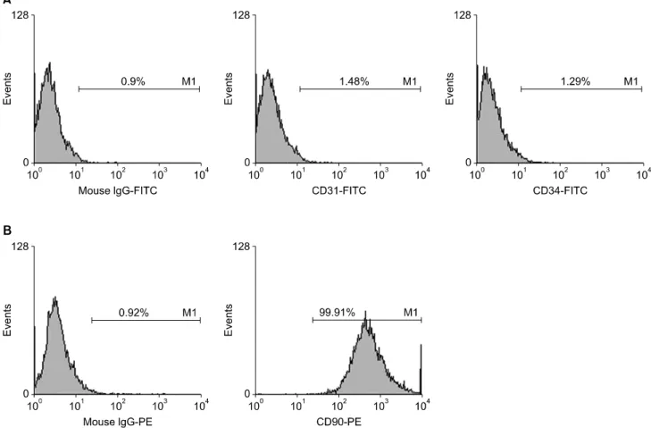
ASC에서는 조혈모세포(hematopoietic stem cell)의 표 적자인 CD34, CD45 및 CD14 등은 발현하지 않지만, MSC의 표적자인 CD29, CD44, CD73, CD90 및 CD105 등은 발현하는 것으로 알려져 있다(24,25). 본 실험에서 이용된 지방유래세포를 3회 계대 배양 후 흐름세포 측정 법으로 세포가 발현하는 표면 표식자를 탐색한 결과 조 혈모세포 표적자인 CD31 및 CD34는 발현하지 않았으며, MSC의 표적자인 CD90를 발현함을 확인할 수 있었다. 이 를 통해 본 실험에 이용된 지방유래세포는 MSC의 범주 에 속하는 ASC임을 확인할 수 있었다(Fig. 1).
2) 효소 면역법을 통한 혈액 내 염증유발 시토카인인 IL-6 정량
IL-6는 T세포 및 대식세포와 같은 면역세포에 의해 분 비되어 면역반응 및 염증반응을 일으키는 시토카인 (proinflammatory cytokine)에 해당한다. C57BL/6 마우 스와 BALB/c 마우스 사이에 피부동종이식을 시행 1일 후, 2일 후, 7일 후 및 20일 후에 혈청 내 IL-6 발현량을 효소 면역법으로 측정하였다. 이식 후 상기 전 기간에 걸 쳐서 ASC 주입군이 PBS 주입군(대조군)에 비해 유의하 게 혈청 내 IL-6 발현량이 감소한 것을 확인할 수 있었다 (P<0.05) (Fig. 2).
3) 이식편 내의 DAPI 양성세포의 관찰
이식 7일째에 ASC 치료군의 피부 동종이식편을 수혜쥐 의 이식편 아래 및 주변 조직(피하조직, 근육조직)과 함께 일괄 절제한 조직으로 4 mm 간격으로 절제한 냉동조직 절편을 만들었다. 이렇게 얻은 냉동조직절편을 형광 현미 경(DM IRE2, Leica Microsystems, Wetzlar, Germany)으로 관찰하여 DAPI 양성세포가 존재하는지 확인하였다. ASC 를 마우스의 꼬리정맥을 통해 이식하기 전 in vitro 상태 에서 ASC의 핵에 DAPI 염색을 시행했으므로 DAPI 양성 세포는 피부 이식편에 도달한 ASC로 간주할 수 있다. 산 발적으로 피부 이식편 및 피부 이식편 아래쪽의 피부 조 직에 산재되어있는 DAPI 양성세포가 관찰되었다(Fig. 3).
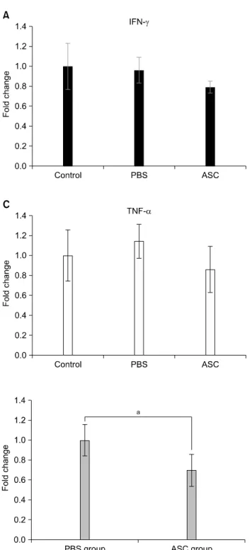
4) 실시간 정량 중합반응을 통한 이식편내 시토카인 발현 분석 IFN-γ, IL-10 및 TNF-α는 염증을 항진시키는 시토카 인(proinflammatory cytokine)으로 알려져 있다. 이식 7
Fig. 1. The expression of cell surface markers in adipose tissue-derived stem cells (ASCs). (A) Representative illustrations of flow cy- tometry demonstrating that ASC did not express hematopoietic stem cell marker (CD31, CD34), and (B) did express mesenchymal stem cell marker (CD90).
Fig. 2. Comparison of the serum levels of proinflammatory cy- tokine interleukin (IL)-6 between phosphate buffered saline (PBS)-infused mice (n=16) and human adipose tissue derived stem cell (ASC)-infused mice (n=16) after skin allograft transfu- sion. Each of four mice in each group were measured at day 1, 2, 7, and 20 posttransplantation, respectively. Overall, ASC-in- fused mice showed statisticalliy significant decrease in serum IL-6 levels than PBS-infused mice did throughout all post- transplantation periods.
aP<0.005.
일째 마우스의 동종 피부 이식편 조직으로 실시간 중합 효소 연쇄반응의 정량분석을 시행하여 동종 피부 이식편 에서의 IFN-γ, IL-10 및 TNF-α 유전자의 mRNA발현을 조사하였다. 대조군과 PBS군 사이에 각각의 유전자 발현 에서 유의한 차이가 나타나지 않았다(Fig. 4). 또한, 대조군 및 PBS 주입군에 비교했을 때, ASC 주입군에서 IFN-γ, IL-10 및 TNF-α 유전자의 mRNA발현이 모두 ASC군에서 가장 낮게 나타났지만 통계학적으로 유의하지는 않았다.
5) 림프구 혼합배양반응
림프구 혼합배양반응을 통해서 ASC 주입이 피부 동종 이식 모델에서 면역학적 관용에 미치는 영향을 이식 30 일째에 알아보았다. 수혜자 BALB/c 마우스의 비장세포 와 mitomycin C로 비활성화시킨 공여자 C57BL/c 마우스의 비장세포로부터 얻은 CD4+세포를 5일간 반응시킨 뒤 CCK assay로 림프구 활성도를 측정하여 비교하였다. ASC 주 입군에서 PBS 주입군보다 림프구 활성도가 유의하게 감 소되었다(1.00±0.15 vs. 0.69±0.16; P=0.002) (Fig. 5).
Fig. 3. Representative photographs which demonstrate a mouse skin allograft specimen containing 4’,6-diamidino-2-phenyl- indole (DAPI)-labeled human adipose-derived stem cells. (A) After obtaining the skin graft at 7-posttransplantation day, (B) the frozen section of the tissue was observed under a fluo- rescent microscope (×100). The staining with DAPI released cy- an fluorescence.
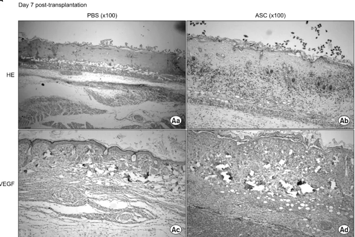
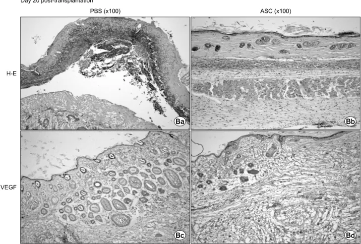
6) 조직학적 검사
이식 7일째 그리고 20일째에 동종 피부 이식편의 병리 조직학적 변화를 관찰하였다. 염증세포의 침윤 및 형태 조직학적 변화를 확인하기 위해 헤마톡실린-에오신 염색 (hematoxylin and eosin, HE stain) 그리고 혈관형성(an- giogenesis) 및 맥관형성(vasulogenesis) 정도에 미치는 영향을 평가하기 위한 VEGF 면역화학염색을 시행했다.
헤마톡실린-에오신 염색에서의 염증 정도는 염증세포(림 프구 및 중성구)의 침윤 정도 및 표피의 굵기 정도에 근 거하여 평가하였다.
먼저 PBS 주입군과 ASC 주입군의 이식 7일째 조직 소 견을 비교하였다(Fig. 6). PBS 주입군에서 HE stain에서 관찰되는 염증 정도가 현저히 나타난 반면, ASC 주입군 에서는 상대적으로 염증 정도가 감소되었다(Fig. 6Aa, Ab). VEGF는 PBS군에서 발현되지 않았지만 ASC군에서 발현이 증가되어 나타났다(Fig. 6Ac, Ad). 이식 20일째 HE 조직 소견에서 PBS 주입군의 피부 이식편 대부분은 수혜자 마우스의 피부로부터 분리되어 나타났다. 하지만 ASC 주입군의 이식편들은 상당수가 분리되지 않고 있었 으며 염증세포의 침윤도 PBS군에 비해 감소되어 있었다 (Fig. 6Ba, Bb). 또한 VEGF 염색에서 PBS 주입군은 VEGF 발현이 7일째와 마찬가지로 뚜렷이 관찰되지 않았 고, ASC 주입군에서는 이식 7일째보다 더 감소되어 나타 났다(Fig. 6Bc, Bd).
7) 피부 이식편의 생존 비교
피부 이식편은 이식 시행 후부터 매일 생존 여부를 확 인하여 이식 후 30일까지 확인하였다. 이식편에서 전체 이식편 면적당 괴사조직 범위가 차지하는 영역이 50% 이 상일 때 거부반응이 발생한 것으로 판정하였다. PBS 주 입군은 이식 18일째까지 모든 피부 이식편에서 거부반응 이 발생하였고, ASC 주입군은 이식 30일째(실험종료시 점) 50%의 이식편 생존 소견을 보였다. 각 군에 따른 이 식편의 생존 곡선을 Fig. 7에 도시하였다. PBS 주입군의 이식편 평균 생존기간은 9.3±1.4일이었다. 하지만 ASC 주입군에서의 이식편 평균 생존기간은 23.9±2.0일로써 PBS군에 비해 유의하게 증가하였다(P<0.001). 또한 ASC 주입군 중 일부(n=2)에서 이식 20일째 이식편의 괴 사소견이 없는 생착소견(graft acceptance)이 관찰되었다 (Fig. 8).
고 찰
MSC는 30여 년 전에 Friedenstein 등(26)에 의해 처음 으로 골수에서 얻어졌지만 그 후 지방 조직, 양수, 뼈 막, 태아 조직 등에서 분리될 수 있음이 밝혀졌다. 이들 조직 중 특히 지방조직의 경우 골수와 비교 시 1) 배양이 쉽고 성장속도가 빠르며, 오랜 계대 배양 후에도 줄기세포의 성상을 잘 유지하며, 2) 높은 paracrine 및 혈관형성 능 력을 지니고, 3) 획득이 용이한 장점으로 인해 MSC원으 로 각광받고 있다.
MSC의 면역억제기능을 나타내는 많은 in vitro 실험 결과에도 불구하고(11,25,27,28), MSC의 면역억제기능은 in vivo 실험에서 충분히 입증되지 않았다(7,13,29). Sbano
Fig. 4. mRNA expressions of proinflammatory (A) interferon (IFN)-γ, (B) interleukin (IL)-10, and (C) tumor necrosis factor (TNF)-α genes in the skin allografts at day 7 posttransplan- tation manifested by real-time polymerase chain reaction.
The mRNA expressions in phosphate buffered saline (PBS) (n=8) and adipose tissue-derived stem cell (ASC) (n=8) were expressed as fold changes in relation to the control group (n=8). The expressions of IFN-γ, IL-2, and TNF-α gene mRNA more decreased in ASC-infused mice than they did in PBS-in- fused mice, though the difference did not reach statistical significance.
Fig. 5. Mixed lymphocyte reaction at day 30 posttransplan- tation. Data are expressed as fold changes related to the phos- phate buffered saline group. The alloreactivity between res- ponder cells (from BALB/c mice of corresponding groups) and stimulating cells (from C57BL/6 mice) was measured using CCK assay. Adipose tissue derived stem cell (ASC) group (n=8) showed significantly reduced alloreactivity compared with PBS group (n=8) (1.00±0.16 vs. 0.70±0.16;
P=0.002).
aP=0.002.
등(22)은 피부 이식편 공여한 동일한 쥐로부터 골수 유래 MSC를 획득하여 수혜쥐에게 이식하였을 때 면역억제요 법만을 단독으로 시행한 쥐보다 피부 이식편의 생존 향 상을 입증하지 못했다. Wang 등(12)은 골수이식과 골수 유래 MSC를 동시에 이식한 마우스 모델에서 공여자 특 이성 면역학적 관용 가능성을 보였다. 2011년 Zografou 등(13)은 처음으로 쥐 모델에서 ASC가 피부 이식편의 생 존율을 높일 수 있음을 보였다. 본 실험은 이상의 실험을 바탕으로 ASC의 이러한 면역억제 능력이 이종이식(xe- nograft)모델을 통해서도 나타남을 처음으로 보이고자 하 였다.
본 실험에서 인간 MSC를 마우스 동종 피부 이식을 시 행받은 마우스에게 이종이식했을 때 이식편의 평균 생존 기간이 증가하였으며, 일부 마우스에서는 이식 후 30일 째 이식편의 안착을 시사하는 소견이 나타났다. 이러한 소견은 MSC가 in vivo 피부동종 이식편 모델에서 면역억 제 및 염증반응 억제기능을 지님을 시사하는 것이다. 따 라서 본 실험은 MSC가 향후 면역학적 관용 유도에 이용 될 수 있는 생물학적 면역억제요법으로 이용될 수 있는 가능성을 열었다고 할 수 있다.
Fig. 6. Representative microphotographs comparing skin allograft specimens between phosphate buffered saline (PBS)-infused mice and adipose tissue-derived stem cell (ASC)-infused mice after skin allograft transplantation. (A) At day 7 posttransplantation, the inflammatory reactions, which had been prominent in PBS-infusion group on day 7 posttransplantation, attenuated after ASC infusion on HE stain (Aa, Ab). Vasoactive endothelial growth factor (VEGF) was minimally or not expressed in PBS group, but its expression was enhanced in ASC-infused group (Ac, Ad). (B) At day 20 posttransplantation, most of full-thickness skin grafts treated with PBS were detached from the recipient’s tissue (Ba). However, the considerable number of grafts treated with ASC was persisted with limited inflammatory cell infiltration (Bb). And, VEGF was lesser expressed on day 20 posttransplantation than it did on day 7 post- transplantation in ASC-infused group (Bc, Bd).
MSC의 면역억제기능은 in vitro 실험에서 충분히 논의 되었다(11,25,27,28). 요약하건대 MSC는 각종 시토카인 등 여러 세크리튬(secretome)을 분비하여 모든 종류의 면역세포, 즉 T세포, B세포, 항원제시세포 각각 면역력, 증식력 및 상호관계에 영향을 미쳐서 면역체계 전반에 걸쳐서 저면역상태를 유도하여 염증반응을 억제한다. 본 실험에서 우리는 두 종류의 이식을 시행하였다. 인간 MSC는 이종이식으로 마우스에게 이식되었으며, 전 층 피 부조직은 동종 이형이식으로 B6 마우스 및 BALB/c 마우 스 간에 시행되었다. 본 실험에서 이식된 MSC는 숙주의 면역감시로부터 벗어나는 면역학적 특권을 지니고 있음 을 이종이식 모델을 통해 입증되었고, MSC의 면역억제 기능이 동종이형 이식편의 생존을 증가시킴을 동종이형 모델을 통해 밝혔다.
본 연구의 모든 결과들은 직접 간접적으로 MSC의 면 역억제기능 및 염증억제기능을 반영한다. IL-6는 T세포 분화에 있어서 중요한 역할을 하는 면역반응 및 염증유 발에 관여하는 시토카인으로 알려져 있다. 본 실험에서 MSC 주입군에서 대조군에 비해 IL-6 분비가 감소된 것을 통해 MSC의 항염증 작용을 in vivo 환경에서 확인할 수 있었다. 우리는 또한 DAPI 염색을 통해 이식 후 7일째에 이식편에 도달한 MSC를 확인할 수 있었다. 이식편에 도 달한 MSC는 이식편에 따라 차이를 보였으나 조밀한 밀 도로 나타나지는 않았고 여러 군데 산재되어 있는 양상 으로 분포되어 있었다. 또한 본 실험에서는 실시간 정량 중합반응으로 이식편 조직에서 이식 7일째 염증항진 시 토카인 IFN-γ, IL-10 및 TNF-α 유전자 발현이 ASC 주 입군에서 감소됨을 확인했으나 sample수가 적어서 통계
Fig. 6. Continued.
Fig. 7. Kaplan-Meier allograft survival curves for mice which had been intravenously administrated phosphate buffered sal- ine (PBS) and adipose tissue-derived stem cells (ASC; 1x10
6).
Each group included 10 mice. ASC infusion markedly increased skin allograft survival in comparison with PBS infusion (
P<0.001).
Fig. 8. Representative photographs of skin allografts showing
skin allograft acceptance. Two adipose tissue-derived stem cells
(ASC)-infused mice (A, B) showed necrosis-free skin graft sug-
gestive of graft acceptance at day 20 posttransplantation, show-
ing the role of ASC inducing immunological tolerance.
학적으로 유의한 수준으로 확인하지는 못했다. 림프구 혼합배양반응은 본 실험들 중에서 면역학적 관용 여부에 대하여 가장 직접적으로 알아보는 실험이다. MSC 주입 군에서 수혜 마우스의 비장세포를 공여 마우스의 CD4+
세포와 반응시켰을 때를 CCK assay를 통해 확인한 림프 구 활성도가 PBS 주입군보다 통계학적으로 유의하게 감 소되었다(P=0.002). 이와 같은 MSC 주입군에서의 저면 역성 및 저염증성은 조직학적 소견에서도 확인할 수 있 었고, 궁극적으로 피부 이식편의 유의한 생존기간 증가로 이어졌다.
본 연구에서는 MSC의 항염증반응과 아울러 MSC의 주 입이 신생혈관 형성에 미치는 영향을 면역조직염색법을 통해 알아보았다. 거부반응이 없는 면역학적 적격한 상 태와 더불어 신생혈관 형성은 피부 이식편의 생존을 결 정하는 중요한 인자이기 때문이다(13,29). 저자들은 MSC 의 주입이 신생혈관 형성인자인 VEGF의 생성을 증가시 킨다는 가정하에서 MSC 주입군에서의 VEGF의 생성과 대조군(PBS 주입군)에서의 VEGF 생성을 비교하였다. 대 조군에서는 VEGF의 발현이 되지 않거나 약하게 발현하 는 데에 비해, MSC 주입군에서는 두드려진 VEGF의 발 현을 확인할 수 있었다.
결 론
결론적으로, 본 실험에서 동종 이형 피부 이식모델에 서 이형 MSC를 주입했을 때 항염증반응, 항면역반응 및 신생혈관 생성 향상을 관찰할 수 있었고 그 결과 이식편 의 생존이 향상된 것으로 추정할 수 있었다. 이러한 실험 결과는 여러 in vitro 선행 연구에서 입증된 MSC의 특성 과 일치한다. 따라서 본 연구를 통해 저자들은 기존의 화 학적 면역억제요법의 한계점을 MSC를 이용한 생물학적 면역억제요법으로 극복할 수 있는 가능성을 열었다고 생 각한다. 더 나아가 본 연구 결과를 바탕으로 향후 MSC의 특성을 이용한 궁극적인 면역학적 관용 유도라는 후속 연구가 이루어질 것을 기대한다.
REFERENCES