ISSN: 2233-601X (Print) ISSN: 2093-6516 (Online)
Received: December 26, 2018, Revised: April 8, 2019, Accepted: April 8, 2019, Published online: October 5, 2019
Corresponding author: Chang Hyun Kang, Department of Thoracic and Cardiovascular Surgery, Seoul National University Hospital, 101 Daehak-ro, Jongno-gu, Seoul 03080, Korea
(Tel) 82-2-2072-3010 (Fax) 82-2-764-3664 (E-mail) chkang@snu.ac.kr
© The Korean Society for Thoracic and Cardiovascular Surgery. 2019. All right reserved.
This is an open access article distributed under the terms of the Creative Commons Attribution Non-Commercial License (http://creativecommons.org/
licenses/by-nc/4.0) which permits unrestricted non-commercial use, distribution, and reproduction in any medium, provided the original work is properly cited.
Lymph Node Status after Neoadjuvant Chemoradiation Therapy for Esophageal Cancer according
to Radiation Field Coverage
Sang Yoon Kim, M.D. 1 , Samina Park, M.D. 2 , In Kyu Park, M.D., Ph.D. 2 , Young Tae Kim, M.D., Ph.D. 2 , Chang Hyun Kang, M.D., Ph.D. 2
1
Department of Thoracic and Cardiovascular Surgery, Daejeon Military Hospital, Armed Forces Medical Command, Daejeon;
2Department of Thoracic and Cardiovascular Surgery, Seoul National University Hospital, Seoul, Korea
Background: To explore the effect of radiation on metastatic lymph nodes (LNs) after neoadjuvant chemo- radiation therapy (nCRT), we examined the metastatic features of LNs according to their inclusion in the ra- diation field. Methods: The patient group included 88 men and 2 women, with a mean age of 61.1±8.1 years, who underwent esophagectomy and lymphadenectomy after nCRT. Dissected LNs were compared in terms of clinical suspicion of metastasis, nodal station, and inclusion in the radiation field. Results: LN pos- itivity did not differ between LNs that were inside (in-field [IF]) and outside (out-field [OF]) of the radiation field (IF: 40 of 465 [9%], OF: 40 of 420 [10%]; p=0.313). In clinical N+ nodal stations, IF stations had a lower incidence of metastasis than OF stations (IF/cN+: 16 of 142 [11%], OF/cN+: 9/30 [30%]; p=0.010).
However, in clinical N- nodal stations, pathological positivity was not affected by whether the nodal stations were included in the radiation field (IF/cN-: 24 of 323 [7%], OF/cN-: 31 of 390 [8%]; p=0.447). Conclusion:
Radiation therapy for nCRT could downstage clinically suspected nodal metastasis. However, such therapy was ineffective when used to treat nodes that were not suspicious for metastasis. Because significant num- bers of residual metastases were identified irrespective of coverage by the radiation field, lymphadenectomy should be performed to ensure complete removal of residual nodal metastases after nCRT.
Key words: 1. Esophageal neoplasms 2. Esophageal surgery 3. Lymph nodes 4. Neoadjuvant therapy 5. Radiotherapy
Introduction
Radical lymphadenectomy has been advocated as an important surgical procedure for curative re- section of esophageal cancer, and it is associated with improved survival after esophagectomy.
However, the importance of radical lymphadenectomy
has been questioned in the present era of tri-modal strategies, including neoadjuvant chemoradiation therapy (nCRT) followed by surgical resection.
Although the results have been interpreted differ- ently, retrospective analyses of data from 2 multi- center randomized controlled studies showed that radical lymphadenectomy did not increase the num-
https://doi.org/10.5090/kjtcs.2019.52.5.353
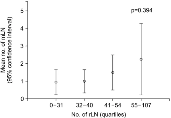
Fig. 1. Change in the number of mLNs according to the number of rLNs in patients who underwent surgery after neoadjuvant che- moradiation therapy. LN, lymph node; mLN, metastatic LN; rLN, resected LN.
ber of metastatic lymph nodes (mLNs) [1,2]. Based on these results, Talsma et al. [1,3] proposed that any therapeutic or diagnostic role for radical lympha- denectomy was limited in patients who underwent nCRT. However, no randomized controlled study has confirmed this suggestion. It also remains unclear how many lymph nodes should be removed and how many nodal stations should be explored during esophagectomy after nCRT. Furthermore, it is not clear how radiation affects the metastatic status of lymph nodes. We thus explored the metastatic status of lymph nodes in terms of inclusion within the radi- ation field. Our aim was to elucidate the role of radi- ation in lymph node metastasis and to explore the utility of radical lymphadenectomy combined with esophagectomy after nCRT.
Methods
1) Study population and specimen collection The Institutional Review Board of Seoul National University Hospital approved this research (1701- 088-824) and waived the requirement for informed consent. A total of 97 patients underwent esophageal resection and lymphadenectomy after nCRT at our institution from February 1998 to May 2016. The in- clusion criteria were: (1) lymphadenectomy after nCRT; (2) availability of detailed information on the radiation field; and (3) availability of information on the metastatic status of specific nodal stations. Seven patients were excluded because they dropped out of nCRT or because preoperative radiation therapy was performed at other hospitals that did not keep ad- equate records of the details of the radiation fields.
In the 90 included patients, 3,904 lymph nodes were resected after lymphadenectomy and grouped by no- dal station. Thoracic, abdominal, and cervical regional nodal stations were defined using the staging classi- fication of the American Joint Cancer Committee [4].
A total of 3,904 lymph nodes were assigned to 885 nodal stations.
2) Neoadjuvant treatment and the surgical approach Pre-treatment clinical staging was performed with the aid of endoscopic ultrasonography, chest and ab- dominal computed tomography, and 18F-fluorodeox- yglucose positron emission tomography. All patients underwent nCRT 4–6 weeks prior to surgical re-
section. The radiation fields were designed by radia- tion oncologists. The nodal stations included in the planning target volumes (PTVs) were considered to be affected by the radiation field. Almost all patients underwent transthoracic esophagectomy and thoracic and abdominal lymphadenectomy. Cervical lymphade- nectomy was added selectively based on clinical judgment. Minimally invasive approaches, such as thoracoscopic or robot-assisted approaches, were chosen for selected patients. In such cases, lympha- denectomy was performed in a manner consistent with open surgery.
3) Statistical analysis
Nodal stations and individual lymph nodes were
divided into in-field (IF) and out-field (OF) groups
according to coverage by the PTV radiation. Categorical
variables, including pretreatment lymph node clinical
and pathological status, are shown as frequencies
with percentages and were compared using the
chi-square test and the Fisher exact test. The num-
bers of resected lymph nodes (rLNs) and mLNs per
patient are given as means±standard deviations. The
mean number of mLNs by the number of rLNs was
investigated using analysis of variance and was dis-
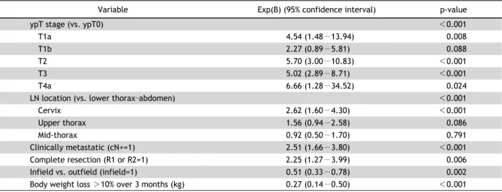
played graphically (Fig. 1). Multiple logistic regression
analysis was used to identify factors influencing
pathological lymph node involvement. A 2-sided
p-value <0.05 was considered to reflect statistical
significance. All statistical analyses were performed
Table 1. Patient characteristics (N=90)
Characteristic Value
Male sex 88 (97)
Age (yr) 61.1±8.1
Body weight (kg) 60.3±9.0
Body mass index (kg/m
2) 21.8±2.9
Body weight loss (kg/3 mo) 2.6±3.7
Smoking status
Never-smoker 8 (9)
Ex-smoker
a)28 (31)
Current smoker 54 (60)
Pack-years 30.1±17.5
Alcohol use (g/day) 44.0±41.2
Eastern Cooperative Oncology Group performance score
0 21 (23)
1 63 (70)
2 6 (7)
Charlson comorbidity index
2 62 (69)
3 20 (22)
4 4 (4)
6 3 (3)
7 1 (1)
Tumor location
Cervix 4 (4)
Upper thorax 21 (23)
Mid-thorax 50 (56)
Lower thorax-gastroesophageal junction 15 (17) cT stage
1a 2 (2)
1b 1 (1)
2 23 (26)
3 64 (71)
cN stage
0 8 (9)
1 53 (59)
2 27 (30)
3 2 (2)
Chemotherapy regimen
5-Fluorouracil/cisplatin 42 (47)
Docetaxel/cisplatin 33 (37)
Cisplatin 8 (9)
Paclitaxel/carboplatin 6 (7)
Paclitaxel/carboplatin/cetuximab 1 (1) Radiation therapy technique
Three-dimensional conformal radiation therapy
86 (96)
Intensity-modulated radiation therapy 4 (4) (Continued to the next page)
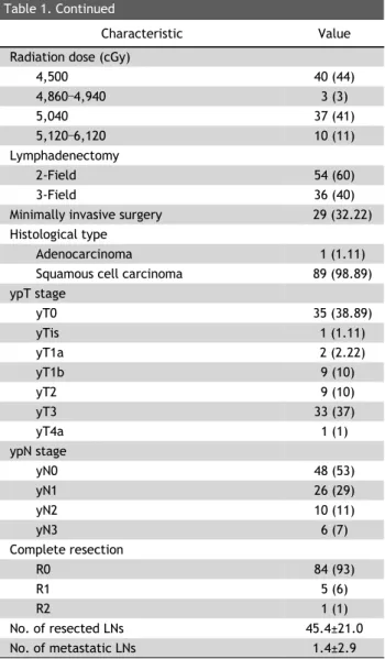
Table 1. Continued
Characteristic Value
Radiation dose (cGy)
4,500 40 (44)
4,860–4,940 3 (3)
5,040 37 (41)
5,120–6,120 10 (11)
Lymphadenectomy
2-Field 54 (60)
3-Field 36 (40)
Minimally invasive surgery 29 (32.22)
Histological type
Adenocarcinoma 1 (1.11)
Squamous cell carcinoma 89 (98.89)
ypT stage
yT0 35 (38.89)
yTis 1 (1.11)
yT1a 2 (2.22)
yT1b 9 (10)
yT2 9 (10)
yT3 33 (37)
yT4a 1 (1)
ypN stage
yN0 48 (53)
yN1 26 (29)
yN2 10 (11)
yN3 6 (7)
Complete resection
R0 84 (93)
R1 5 (6)
R2 1 (1)
No. of resected LNs 45.4±21.0
No. of metastatic LNs 1.4±2.9
Values are presented as frequency (%) or mean±standard deviation.
LN, lymph node; cT, clinical tumor; cN, clinical node; yp, post- therapy pathologic.
a)
An ex-smoker was defined as a smoker who had quit at least 6 months prior.
using PASW SPSS software ver. 18.0.0 (SPSS Inc., Chicago, IL, USA).
Results
1) Patient and primary tumor characteristics
Table 1 shows the clinical characteristics of all pa-
tients and their primary tumor evaluations. All pa-
tients except 2 were men, and more than 90% had
an Eastern Cooperative Oncology Group performance
status ≤1. Squamous cell carcinoma was the pre-
Table 2. Pathological involvement of LN stations: coverage by radiation field (out-field vs. in-field) stratified by pre-treat- ment and clinical metastatic status (cN-/cN+)
Out-field (n=420) In-field (n=465) p-value
cN- 31/390 (8) 24/323 (7) 0.796
cN+ 9/30 (30) 16/142 (11) 0.013
Values are presented as number (%).
LN, lymph node; cN-, clinically non-metastatic LN stations; cN+, clinically metastatic LN stations.
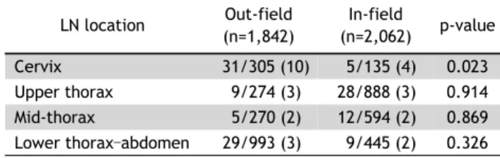
Table 4. Pathological involvement of individual LNs by exposure to radiation field (out-field vs. in-field) stratified by LN location
a)LN location Out-field (n=1,842)
In-field
(n=2,062) p-value
Cervix 31/305 (10) 5/135 (4) 0.023
Upper thorax 9/274 (3) 28/888 (3) 0.914
Mid-thorax 5/270 (2) 12/594 (2) 0.869
Lower thorax–abdomen 29/993 (3) 9/445 (2) 0.326 Values are presented as number (%).
LN, lymph node.
a)
The frequencies of LN involvement are shown as ratios (%).
Table 3. Pathological involvement of individual LNs by exposure to radiation field (out-field vs. in-field) stratified by pre-treat- ment and clinical metastatic status (cN-/cN+)
a)Out-field (n=1,842) In-field (n=2,062) p-value
cN- 54/1,658 (3) 31/1,401 (2) 0.080
cN+ 20/184 (11) 23/661 (3) <0.001
Total 74/1,842 (4) 54/2,062 (3) 0.014
Values are presented as number (%).
LN, lymph node; cN-, clinically non-metastatic LN stations; cN+, clinically metastatic LN stations.
a)