• 검색 결과가 없습니다.

Actinomycotic Osteomyelitis of the Mandible: A Case Report JOMP

N/A
N/A
Protected

Academic year: 2021

Share "Actinomycotic Osteomyelitis of the Mandible: A Case Report JOMP"

Copied!
5
0
0

로드 중.... (전체 텍스트 보기)

전체 글

(1)

pISSN 2288-9272 eISSN 2383-8493 J Oral Med Pain 2019;44(3):140-144 https://doi.org/10.14476/jomp.2019.44.3.140

Actinomycotic Osteomyelitis of the Mandible: A Case Report

Young-Cheol Lee

1

, Lee-Rang Lim

1

, Kyu-Hoon Lee

1

, Dong-Jun Seo

1

, Na-Ra Yun

2

, Ji-Su Oh

1

, Jae-Seek You

1

, Hae-In Choi

1

1

Department of Oral and Maxillofacial Surgery, School of Dentistry, Chosun University, Gwangju, Korea

2

Division of Infectious Disease, Department of Internal Medicine, College of Medicine, Chosun University, Gwangju, Korea

Received August 13, 2019 Revised September 1, 2019 Accepted September 1, 2019

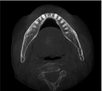
Actinomycosis is rare, chronic, slowly progressive disease caused by gram-positive anaero- bic organisms from the Actinomycosis family that normally colonizes the oral cavity. Acti- nomycotic osteomyelitis is even more rare and refractory disease because diagnosis by bac- terial culture is not easy. In our case, 80-year-old man visited our clinic with a complaint of swelling and severe sinus tracts without teeth evolvement on anterior mandible. Computed tomography (CT) scan demonstrated erosive bone destruction on anterior mandible, clini- cally suspected actinomycotic osteomyelitis. The patient also had thoracic actinomycosis on Lt. lower lung. We could diagnosis actinomycosis by histopathologic examination. He treated by conservative surgery and long term antibiotics. After 2 year, no recurrence was seen in CT scan.

Key Words: Actinomycosis; Actinomycotic osteomyelitis

Correspondence to:

Hae-In Choi

Department of Oral and Maxillofacial Surgery, School of Dentistry, Chosun University , 309 Pilmun-daero, Dong-gu, Gwangju 61452, Korea

Tel: +82-62-220-3810 Fax: +82-62-222-3810 E-mail: [email protected] https://orcid.org/0000-0002-2425-9506 This study was supported by research fund from Chosun University Dental Hospital, 2018.

JOMP

Journal of Oral Medicine and Pain

Copyright

2019 Korean Academy of Orofacial Pain and Oral Medicine. All rights reserved.

CC

This is an open-access article distributed under the terms of the Creative Commons Attribution Non-Commercial License (http://creativecommons.org/licenses/by-nc/4.0/), which permits unrestricted non-commercial use, distribution, and reproduction in any medium, provided the original work is properly cited.

INTRODUCTION

Actinomycosis is rare chronic disease caused by Actino- myces spp. and once mistaken for fungal infection [1].

Actinomyces spp. is Gram-positive anaerobic bacteria, in- cluding Actinomycosis israelii, A. bovis, A. odontolyti- cus, and A. viscus. They can be found in calculus, gingival crevices, infected dentin and commonly live in pharynx and tonsil [2]. Actinomyces has no pathogenic potential when exposed to normal skin or healthy mucosa. Once losing membrane continuity by trauma or dental surgery, Actinomyces can invade into mucous membrane and form localized abscess [3].

Actinomycosis can be divided in three categories: cer- vicofacial (55%), abdominopelvic (20%), thoracic (15%) depending on the region [4]. Cervicofacial actinomycosis is the most common type; it can spread in acute form to

hypopharynx, larynx, lacrimal duct and gland, major sali- vary gland, oral mucous, mandible, paranasal sinus and scalp; it can be chronic condition forming granuloma or multiple sinus tract and fistula [5]. Periapical actinomyco- sis, less invasive subtype of cervicofacial actinomycosis, has been reported, too [6].

Pus culture and bacterial examination is needed for di- agnosis. Culture of Actinomyces spp. require specific oxi- dation-reduction condition associated with microbiome. In clinic, diagnosis of actinomycosis can be difficult even in highly suspected. Several months of antibiotics are required of treating actinomycosis and it can recur if treatment is stopped early. Making early definite diagnosis is important for preventing recurrence of actinomycosis [7].

There are few case reports of actinomycosis and even much

rare case reports of osteomyelitis by Actinomyces except for

medication related osteonecrosis of jaw. This report is about a

(2)

case of 80-year-old man with actinomycotic mandibular os- teomyelitis, which is identified later to co-infection of cervi- cofacial and thoracic type.

CASE REPORT

A 80-year-old man presented with mandibular inci- sor pain and swelling. Pain has been for 20 days and there was no surgery, trauma history and no underlying system- ic disease. In physical examination, right cervical lymph node enlargement and multiple fistula in mandibular lat- eral incisor, canine, first premolar and first molar were ob- served. Elective pulp test on those teeth were all positive and no suspected teeth involvement was observed in pan- orama (Fig. 1) and periapical view. Clinically, actinomyco- sis was suspected and pus culture and bacterial examina- tion were held but failed to culture actinomyces. Computed tomography (CT) and biopsy were perfomed for diagno- sis. Irregular destruction on mandibular incisor labial bone was observed in CT (Fig. 2) and histological examination

confirmed actinomycotic osteomyelits. Patient was admit- ted to the emergency room presenting with severe swelling on right lower face, pain and limited mouth opening during administration of prescribed amoxicillin under cooperating with divsion of infectious diseases. In enhanced neck com- puted tomoraphy, there was abscess pocket showing irregu- alr constract enhancement in right submasseteric space and pterygomandibular space (Fig. 3). drainage on pus pocket and curettage of bone on incisor area were performed under local anesthesia.

Meanwhile, chest compute tomography was performed as routine ward laboratory examination despite of no symp- tom, observing tuberculosis-like lesion in left lower lobe in chest X-ray (Fig. 4A). Considering patient’s history,

Fig. 1. First visit panoramic view.

Fig. 2. First visit computed tomography. Anterior mandibular labial bone resorption was observed.

Fig. 3. Irregular contrast enhancement

was observed in right submassteric and

pterygomandibular space.

(3)

disseminated actinomycosis was suspected, left lower lo- bectomy was performed under general anesthesia in depart- ment of cardiothoracic surgery. It was finally confirmed actinomycosis by histopathologic examination. Patient was prescribed doxycycline over 10 months after discharge.

After 2 years, patient visited for another reason with no sign of recurrence of actinomycosis. CT showed healing state of mandibular bone (Fig. 5).

DISCUSSION

After first reporting of actinomycosis, misunderstood to fungal infection in 1845 by Van Langenbeck, Bollinger et al. assumed that Actinomyces bovis were fungus breaking out ‘lumpy jaw’ in cow. Wolff et al. succeeded in isolation culture from human body in 1891 and Waksman concluded that Actinomyces are Gram-positive anerobic bactera, not fungus in 1960s [8].

Cervicofacial actinomycosis is usually associated with soft tissue of chin and neck area and make multiple fistula on skin with swelling. It is characterized by features that in- fection spreads ignoring anatomical plane. Infected skin has abscess with purplish red and indurated, wood-like area.

Special sulfur granule is observed in fistula drainage. Since another fistula appears when the former fistula disappears, extensive scarring and facial deformity in prolonged dura- tion of untreated patient with actinomycosis.

Actinomycotic osteomyelitis is rare, which accounts for about 12% of total osteomyelitis [9]. Cervicofacial actino- mycotic osteomyelitis usually occurs in mandibular body,

following symphysis, ramus, mandibular angle. Maxilla and temporomandibular joint are comparatively resistant for the disease and prevalence is a ratio mandible:maxilla of 4:1. It is assumed that mandible is more commonly affected than maxilla due to the lesser vascularity and the denser corti- cal bone, similar to osteoradionecrosis [10]. In this case, co- infection of cervicofacial and thoracic type was shown by invasion of symphysis and lung.

Actinomyces in oral flora in patient with poor oral hy- giene could be inhaled to lung through the pharynx. It could be cause of thoracic actinomycosis. Thoracic actino- mycosis forms micro-abscess and focal consolidation in pa- renchyma, fibrosis of local tissue. It was shown like tuber- culosis, infiltration, malignancy in chest PA [11]. Thoracic

A B

Fig. 4. (A) A pathogen similar to tuberculosis is seen in the left and lower lobe of the chest radiography. (B) Mild abscess and sclerosis are seen in chest computed tomography.

Fig. 5. A 2 year follow-up computed tomography view. Healing

state at anterior mandible area.

(4)

actinomycosis cannot be diagnosed without surgical inter- vention, while cervicofacial actinomycosis can be treated by removal of fistula and long-term antibiotics. Biopsy is required to rule out pulmonary malignancy especially [12].

In this case, thoracic type actinomycosis was likely to occur because of long-time untreated condition and in- halation of abscess with poor oral hygiene due to mouth opening limitation because of abscess pocket of masticator space. To prevent thoracic actinomycosis, meticulous oral hygiene is essential in cervicofacial actinomycosis patient.

Pathophysiology of actinomycotic osteomyelitis has not clearly defined yet. Actinomyces can be found in tonsil, sul- cus fluid, mucus membrane, infected dentin and tooth ex- trated area but cannot be infectious by itself. Actinomycotic osteomyelitis may be shown in the circumstance with bro- ken balance of normal flora and pathologic change by chronic inflammation for some reason. Actinomyces itself is less invasive and toxic than other pathogens. Another bacteria help actinomyces to invade tissue with production of toxin and enzyme. These polymicrobial conditions make proper environment for growing anaerobic bacteria hav- ing low oxido-reduction potential [13]. Not forming sulfur granule of actinomyces in laboratory environment, it sug- gests that invasion of host immune system and colonization of associated bacteria are cornerstones to actinomycosis [14].

Once infection established on bone, the host makes inflam- mation response, suppuration, forming granuloma, fibrosis and making fistula.

Actinomycosis is shown clinically like tuberculosis and granuloma mimicking malignant lesion. Actinomycotic os- teomyelitis usually proceeds slowly, but there were severe cases that infection invaded to mid-face, skull base, and cranium [15], and that proceed so rapidly that mandible was fractured, requiring reconstruction in immunosuppres- sive patient [16]. It is very important to diagnose actino- mycosis, but it is reported that usual bacterial examination can confirm actinomycosis in less than 50% and first visit examination in less than 10% [17]. Four case series of acti- nomycotic osteomyelitis of Sezer et al. [18] reported to fail to make definite diagnosis of actinomycosis by usual mi- crobiological examination. If the examination are failed re- peatedly and delayed, it can get more inaccurate by effect of antibiotics. In this case, biopsy is highly recommended

for diagnosis [19]. Actinomyces are strongly stained by H&E and Periodic acid-Schiff, Giemsa (GMS), especially fil- aments are well-shown in GMS [20].

In this case, there was no collection of actinomyces in first test. After several test, penicillin-resistant Streptococcus pneumonia, S. parsanguinis, candida was found but not found actinomyces at all. Definite diagnosis is performed by biopsy of mandible and lung. When actinomycosis is highly suspected clinically, biopsy helps more accurate diagnosis, as well as scrutinizing microbiological test.

It can be treated long-term antibiotic application in case of cervicofacial actinomyces localized in soft tissue, but when osteomyelitis is accompanied as this case, surgical in- tervention is essential. If acute abscess formation and ne- crotic tissue exists, surgical treatment is also required to distribute antibiotics more effectively. Antibiotic treatment for actinomycosis usually consists of 2-6 weeks of penicil- lin antibiotics by intravenous injection and 2-12 months of amoxicillin oral administration after IV [21]. Tetracyclin, minocycline, erythromycin, cephalosporin, sulfonamide, streptomycin can be used If patient has allergy for penicil- lin. In case of penicillin-resistant bacteria, clindamycin can be used by oral administration [22]. In this case, penicillin- resistant S. pneumonia, S. parsanguinis is detected, cef- triaxone and vancomycin was prescribed intravenously.

Doxycycline was also prescribed through oral administra- tion over 10 months after discharge.

In conclusion, early and thorough diagnosis, appropriate surgery, long-term administration of antibiotics is required for actinomycotic osteomyelitis. When actinomycosis is clinically suspected, biopsy is recommended that cannot be diagnosed by typical microbiological culture test.

CONFLICT OF INTEREST

No potential conflict of interest relevant to this article was reported.

ORCID

Young-Cheol Lee

https://orcid.org/0000-0001-6371-7445

Lee-Rang Lim

(5)

https://orcid.org/0000-0001-6751-9091 Kyu-Hoon Lee

https://orcid.org/0000-0002-0236-1788 Dong-Jun Seo

https://orcid.org/0000-0002-4301-5881 Na-Ra Yun

https://orcid.org/0000-0002-4219-0127 Ji-Su Oh

https://orcid.org/0000-0002-8369-5025 Jae-Seek You

https://orcid.org/0000-0001-7638-9583 Hae-In Choi

https://orcid.org/0000-0002-2425-9506

REFERENCES

1. Benoliel R, Asquith J. Actinomycosis of the jaws. 2 Case reports.

Int J Oral Surg 1985;14:195-199.

2. Laskaris G. Oral manifestations of infectious diseases. Dent Clin North Am 1996;40:395-423.

3. Miller M, Haddad AJ. Cervicofacial actinomycosis. Oral Surg Oral Med Oral Pathol Oral Radiol Endod 1998;85:496-508.

4. Brown JR. Human actinomycosis. A study of 181 subjects. Hum Pathol 1973;4:319-330.

5. Nagler R, Peled M, Laufer D. Cervicofacial actinomycosis: a di- agnostic challenge. Oral Surg Oral Med Oral Pathol Oral Radiol Endod 1997;83:652-656.

6. Hirshberg A, Tsesis I, Metzger Z, Kaplan I. Periapical actinomy- cosis: a clinicopathologic study. Oral Surg Oral Med Oral Pathol Oral Radiol Endod 2003;95:614-620.

7. Kaplan I, Anavi K, Anavi Y, et al. The clinical spectrum of Ac- tinomyces-associated lesions of the oral mucosa and jawbones:

correlations with histomorphometric analysis. Oral Surg Oral Med Oral Pathol Oral Radiol Endod 2009;108:738-746.

8. Bala S, Narwal A, Gupta V, Duhan J, Goel P. Actinomycotic os- teomyelitis of mandible masquerading periapical pathology. J Oral Health Community Dent 2011;5:97-99.

9. Acevedo F, Baudrand R, Letelier LM, Gaete P. Actinomycosis: a great pretender. Case reports of unusual presentations and a review of the literature. Int J Infect Dis 2008;12:358-362.

10. Figueiredo LM, Trindade SC, Sarmento VA, de Oliveira TF, Muniz WR, Valente RO. Actinomycotic osteomyelitis of the mandible: an unusual case. Oral Maxillofac Surg 2013;17:299-302.

11. Endo S, Murayama F, Yamaguchi T, et al. Surgical considerations for pulmonary actinomycosis. Ann Thorac Surg 2002;74:185- 190.

12. Taştepe AI, Ulaşan NG, Liman ST, Demircan S, Uzar A. Thoracic actinomycosis. Eur J Cardiothorac Surg 1998;14:578-583.

13. Gannepalli A, Ayinampudi BK, Baghirath PV, Reddy GV. Actino- mycotic osteomyelitis of maxilla presenting as oroantral fistula: a rare case report. Case Rep Dent 2015;2015:689240.

14. Marx RE, Carlson ER, Smith BR, Toraya N. Isolation of Acti- nomyces species and Eikenella corrodens from patients with chronic diffuse sclerosing osteomyelitis. J Oral Maxillofac Surg 1994;52:26-33; discussion 33-34.

15. Shen JY, Futran ND, Sardesai MG. Craniofacial Actinomyces os- teomyelitis evolving from sinusitis. Radiol Case Rep 2017;13:104- 107.

16. Kusuyama Y, Matsumoto K, Okada S, Wakabayashi K, Takeuchi N, Yura Y. Rapidly progressing osteomyelitis of the mandible. Case Rep Dent 2013;2013:249615.

17. Abbaszadeh H, Sheibani MS. Actinomycotic osteomyelitis of mandible. J Craniofac Surg 2016;27:e452-e454.

18. Sezer B, Akdeniz BG, Günbay S, Hilmioğlu-Polat S, Başdemir G.

Actinomycosis osteomyelitis of the jaws: report of four cases and a review of the literature. J Dent Sci 2017;12:301-307.

19. Goldstein BH, Sciubba JJ, Laskin DM. Actinomycosis of the maxilla: review of literature and report of case. J Oral Surg 1972;30:362-366.

20. Chandler FW, Watts JC. Fungal diseases. In: Anderson WAD, Damjanov I, Linder J, eds. Anderson’s pathology. 10th ed. St.

Louis: Mosby; 1996. pp. 951-982.

21. Bennhoff DF. Actinomycosis: diagnostic and therapeutic consid- erations and a review of 32 cases. Laryngoscope 1984;94:1198- 1217.

22. Dubey A, Murthy JG, Banerjee PK. Actinomycosis of the parotid

gland. Indian J Otolaryngol Head Neck Surg 2004;56:306-308.

수치

Fig. 3. Irregular contrast enhancement  was observed in right submassteric and  pterygomandibular space
Fig. 5. A 2 year follow-up computed tomography view. Healing  state at anterior mandible area.

참조

관련 문서

Since every classical or virtual knot is equivalent to the unknot via a sequence of the extended Reidmeister moves together with the forbidden moves, illustrated in Section 2,

Osteomyelitis in the right distal femur with surrounding

The economies of the world will remain highly interdependent through trade, investment and fi nancial system linkages, driving the need for stronger global policy

1 John Owen, Justification by Faith Alone, in The Works of John Owen, ed. John Bolt, trans. Scott Clark, "Do This and Live: Christ's Active Obedience as the

Finally the reason for the cosmetic consumption tendency dependent on the containers used was proposed.. According to a statistical report, a quarter of

Time series of vertical cross section of potential vorticity and wind vector calculated by Case 1 along the A-A' line indicated at Fig.. Same

This report make a description of criminal special code for purification and improvement of terminology and sentence.. All the while, criminal special code make a

 Bent posture, and as a fast check of the measurement major pectoral muscle case, the patient lying immediately behind the head comes along,