유방암 검진에서 초음파의 유용성
전북대학교 의과대학 외과학교실 유방·갑상선외과
윤현조, 안하림, 강상율, 정성후
Efficacy of Ultrasonography in Breast Cancer Screening
Hyun Jo Youn, Ha Rim Ahn, Sang Yull Kang, Sung Hoo Jung
Division of BreastㆍThyroid Surgery, Department of Surgery, Chonbuk National University Medical School, Jeonju, Korea
Received February 11, 2019 Revised April 30, 2019 Accepted May 2, 2019
A good screening test should be widely available, inexpensive, and have high sensitivity and specificity. Mammography is the only screening modality proven to reduce the mortality of breast cancer. On the other hand, with the increasing awareness of mammography limi- tations, particularly in a dense breast, supplemental screening for breast cancer with other imaging modalities has been expanding. Breast ultrasonography has the advantage of detect- ing mammographically occult breast cancers in a dense breast. On the other hand, operator dependence, false positive rates, and rising costs due to increased biopsy rates are a problem.
Thus far, the guidelines for the implementation of screening breast ultrasonography have not been established. Physicians should be able to understand the efficacy of breast ultra- sonography as the screening modality for breast cancer and perform risk-based screening ap- proaches for individual women. This paper reviews the efficacy, limitations and im- plementation of screening breast ultrasonography for breast cancer.
Keywords: Breast neoplasms, Screening, Ultrasonography, Breast density Correspondence to:
Sung Hoo Jung
Division of BreastㆍThyroid Surgery, Department of Surgery, Chonbuk National University Medical School, 20 Geonji-ro, Deokjin-gu, Jeonju 54907, Korea Tel: +82-63-250-2133 Fax: +82-63-271-6197 E-mail: [email protected]
서 론
최근에 발표된 GLOBOCAN 보고에 따르면 전 세계적으 로 2018년 한 해 동안 새롭게 진단받은 약 1,810만 명의 암 환자 중 유방암 환자가 210 만명(11.6%)을 차지해 여성 에서 가장 많은 암 발생률을 보였다.(1) 국내에서도 2016 년 한 해 약 22,000명의 새로운 유방암 환자가 발생해 여 성암 발생률 1위를 차지하였으며, 매년 지속적인 증가 추 세를 보이고 있다.(2) 유방암의 증가 원인으로는 서구화 된 식생활과 에스트로겐(estrogen)에 노출되는 기간의 연 장과 함께 건강에 대한 관심 증가에 따른 검진의 활성화와 영상학적 진단 기술의 발전 등을 요인으로 꼽을 수 있
다.(3)
유방암의 조기 발견은 사망률을 낮출 수 있으며, 유방 촬영술(mammography)은 지금까지 시행된 무작위 대조 군 연구(randomized controlled trial) 결과 유방암 사망 률을 감소시키는 것으로 알려진 유일한 검진 방법 (screening modality)이다.(4) 그러나 유방촬영술의 민 감도(sensitivity)는 유방 밀도가 증가할수록 감소하는 특 징이 있어 치밀 유방(dense breast)의 경우 위음성률 (false negative rate)이 50%에 달하며, 치밀 유방에 의해 발견되지 않은 유방암은 다음 검진 전에 증상을 동반한 간 격암(interval cancer)으로 발견되는 비율이 증가한 다.(5,6) 이는 미국에서 유방 밀도를 고지하는 법률이 시 J Surg Ultrasound 2019;6:1-7
https://doi.org/10.46268/jsu.2019.6.1.1 JSUJournal of Surgical Ultrasound
Fig. 1. Mammographically occult cancer detected by breast ultrasound in a 64-year-old woman with dense breasts. (A) Gray scale breast ultrasound image shows a 1.2 cm invasive ductal carcinoma with irregular shape and angular margin in the left breast. (B) Corresponding same day mammography cranio-caudal and mediolateral-oblique views show dense breast tissue.
행된 배경이 되었고, 유방촬영술 이외의 추가적인 검진 방 법이 필요함을 인식하는 계기가 되었다.(7)
치밀 유방은 유방촬영술의 민감도를 감소시킬 뿐 아니 라 유방암 발생과 관련된 독립적인 위험 인자로 알려져 있 으며, 검진자의 31-43%가 치밀 유방으로 분류될 정도로 매우 흔하다.(8,9) 유방 밀도는 연령이 증가할수록 감소하 는 것으로 알려져 있는데 국내 유방암은 50대에서 가장 많 이 발생하여 나이가 많아질수록 유방암 발생 빈도가 증가 하는 외국과는 차이가 있으며, 따라서 국내 유방암의 역학 적인 특성을 고려한 유방암 검진 권고안의 개발이 필요하 다.(3)
유방 초음파(breast ultrasound)는 유방암의 추가적인 검진 방법 중의 하나로 기존의 보고들을 종합해 보면 검진 받은 여성 1,000명 당 3.2-4.2건의 암 발견율(cancer detection rate; CDR)을 보이며, 특히 치밀 유방으로 유 방촬영술에서 발견하지 못한 병변의 발견에 유용한 것으 로 알려져 있다.(10,11) 하지만 검사자 의존적(operator dependent)이고 위양성률(false-positive rate)이 높으며 추가된 조직 검사로 인한 비용 증가 등의 단점이 있어 유방 암의 검진 방법으로 시행하기 전에 고려해야 할 사항들이 있다.
이에 저자들은 문헌 고찰을 통해 잠재 유방암(occult breast cancer)의 조기 발견을 위한 검진 방법으로서의 유방 초음파, 즉 선별 유방 초음파(screening breast ul- trasound)의 효과와 제한점 그리고 실제 임상 현장에서의 실행에 관하여 살펴보고자 한다.
본 론
1. 선별 유방 초음파의 효과
좋은 선별 검사(screening test)는 이용이 편리하고 저 렴하며 높은 민감도와 특이도를 가져야 한다. 전통적으로 유방암의 선별 검사로 사용된 유방촬영술에 비해 유방 초 음파는 이용이 편리하고 통증이 적으며 방사선 노출이 적 은 장점과 함께 높은 민감도를 가지고 있다. 이러한 특징 으로 과거에 유방촬영술 또는 신체 진찰 후 조직 검사 등의 추가 검사를 위한 진단 방법으로 사용되어 왔던 유방 초음 파는 최근에 유방촬영술의 보조적인 선별 검사(supple- mental screening test)로 사용되고 있다. 선별 유방 초 음파는 유방촬영술 검진의 문제점인 치밀 유방에서 보이
지 않는 유방암을 발견할 수 있는 해결책으로, 특히 크기 가 작고 겨드랑이 림프절 전이가 없는 조기 유방암을 발견 하는데 유용한 것으로 알려져 있다(Fig. 1).
유방 초음파를 이용한 유방암 검진은 1995년에 Gordon 과 Goldenberg(12)에 의해 처음으로 보고되었는데 치밀 유방을 가진 12,706명의 여성에서 예상하지 못한 44개의 침윤암을 발견하여 1,000명 당 3.5 CDR을 보였다. Kolb 등(13)은 11,130명의 여성을 대상으로 27,825건의 유방촬 영술 검진과 13,547건의 유방 초음파 검진을 비교하였을 때 민감도가 77.6%와 75.3%로 유사하였으며, 유방 초음 파만을 시행한 경우 1,000명 당 2.7 CDR로 평균 9.9 mm
의 크기의 유방암을 발견하여 유방 초음파가 유방촬영술 에서 보이지 않는 임상적으로 작은 크기의 유방암 발견에 유용한 것으로 보고하였다. 또한 대규모의 유럽 코호트 연 구 결과에서도 유방촬영술에서 특이 소견을 보이지 않는 치밀 유방 여성에서 선별 유방 초음파를 통해 15.9%의 유 방암 발견율 증가 소견을 보였다.(14)
유방촬영술에 유방 초음파를 추가한 병용 검진에 대한 가장 대표적인 연구는 American College of Radiology Imaging Network (이하 ACRIN) 6666 연구이다. (15) 치 밀 유방을 보인 고위험군 여성 2809명에 대한 다기관 전 향적 연구로 유방촬영술 검진과 유방 초음파 병용 검진을 비교한 결과 유방촬영술에서는 1,000명 당 7.6개의 유방 암을 발견했고 유방 초음파 병용 검진을 했을 때 1,000명 당 11.8개의 유방암을 발견하였다. 유방 초음파에서만 발 견된 유방암은 모두 침윤암이었고 평균 크기는 10 mm로 작았다. 이후 보고된 ACRIN 6666 연구의 추가 분석에 따 르면 유방 초음파 단독 검진은 유방촬영술 검진과 비슷한 정도의 유방암 발견율을 보였으며(52.3% vs.53.2%; P = 0.90), 유방 초음파로 진단된 유방암은 침윤암이 더 많았 다. 하지만 높은 소환율(recall rate)과 조직 검사율, 낮은 조직 검사 양성 예측도를 보였다(유방 초음파 각 10.7%, 5.5%, 11.7%; 유방촬영술 각 9.4%, 2.0%, 38.1%; P < 0.0001). (16)
우리나라를 포함한 동양 여성들은 서양에 비해 치밀 유 방이 많아 유방촬영술의 민감도가 떨어지며 유방암의 발 생 빈도가 낮아 검진 프로그램의 효과에도 제한점이 있 다.(17,18) 13,339명의 여성을 대상으로 유방암 검진 방법 으로 유방촬영술과 유방 초음파의 효과를 비교한 대규모 의 다기관 연구가 중국에서 시행되었으며 유방촬영술, 유 방 초음파 그리고 병용한 경우 1 ,000명 당 각각 0.72, 1.51, 2.02 CDR을 보였다.(19) 유방 초음파만을 시행한 경우 유방촬영술만을 시행한 경우보다 민감도는 더 우수 하였으나(100% vs.57.1%; P = 0.04), 특이도는 두 그룹 간에 차이를 보이지 않았다(100% vs.99.9%; P = 0.51).
2016년에는 Japan Strategic Anti-cancer Randomized Trial (J-START)라는 유방촬영술 검진과 유방 초음파 병 용 검진을 비교한 무작위 대조군 연구 결과가 발표되었다.
(20) 40-49세의 증상이 없는 여성 약 8 만 명이 참여하였 고 유방촬영술과 비교하여 유방 초음파 병용 검진은 1,000명당 1.84개의 유방암을 더 발견하였다. 유방 초음
파 병용 검진을 통해 민감도와 유방암 발견율, 특히 조기 침윤암 발견율이 증가되었고 간격암 발생률은 감소되었 다.
국내에서도 선별 유방 초음파의 유용성에 관한 연구 결 과가 발표되었는데 106,856건의 선별 유방 초음파를 후향 적으로 분석한 Bae 등(21)의 보고에 따르면 유방 초음파 는 1,000명 당 3.4 CDR을 보였으며, 유방 초음파로 발견 된 335개의 유방암 위치를 알고 유방촬영술을 후향적으로 다시 판독해도 81% (272개)에서는 암을 발견할 수 없었 다. 또한 치밀 유방을 가진 무증상의 여성 20,864명에서 유방 초음파를 유방암 검진의 보조 방법으로 사용한 연구 결과 1,000명 당 2.5 CDR을 보였으며 유방촬영술보다 우 수한 민감도를 보여(100% vs.54.55%; P = 0.002), 유방 초음파가 유방암 검진의 보조 방법으로 유용하다고 보고 하였다.(22)
2. 선별 유방 초음파의 제한점
선별 검사로서 유방 초음파는 유방촬영술과 동등한 CDR을 보이고 있지만 유방촬영술에 비해 특이도가 낮은 단점이 있다. 이러한 이유로 유방촬영술의 소환율이 10%
미만인 반면 유방 초음파는 11%-13%의 높은 소환율을 보 이고 있다.(16,23) 낮은 특이도는 높은 위양성률로 표현 할 수 있으며 이로 인한 의료비 상승과 함께 불필요한 조직 검사 또는 단기간의 추적 검사로 인한 환자의 불안감 증가 등의 단점이 있다. 또한 유방 초음파는 석회화 병변의 발 견에 제한이 있어, 최근에 좀 더 높은 주파수의 탐촉자를 사용하면서 석회화 발견율이 증가하기는 했지만 미세석 회화의 선별 검사로는 아직 유용하지 못하다. 이와 같은 선별 유방 초음파의 낮은 양성 예측률(positive pre- dictive value)과 높은 단기간의 추적 관찰 권고율을 극복 하기 위해 전단파 탄성초음파(shear wave elastography) 또는 도플러(doppler) 초음파의 적용이 도움이 될 수 있 다.(24,25)
ACRIN 6666 연구에 따르면 선별 유방 초음파를 시행하 는 경우 판독과 보고 시간을 제외하고 평균 19분의 검사 시간이 추가로 소요되었으며 이는 유방 초음파를 유방암 검진 방법으로 고려할 때 가장 중요한 이슈 중의 하나이 다. 또한 유방 초음파는 검사자 의존적이기에 비록 경험이 많은 방사선사(techonologist)가 유방 초음파를 시행할 때 임상의(physician)와 동등한 검사 결과를 보였지만,
Fig. 2. Three standard images of breast cancer using automated breast ultrasound in a 42-year-old woman.
Cononal view (left), longitudinal (right, upper) and transverse views (right, lower) are synchronously visualized on the screen.
(26) 경험이 적은 기사에 의해 시행될 경우 정확도에 차이 가 있을 것으로 예상된다. 이와 함께 재현성(reproduc- ibility)이 낮은 유방 초음파의 제한점을 극복하기 위해 선 별 유방 초음파로서 자동 유방 초음파(automated breast ultrasound; ABUS)의 시행이 대두하게 되었다.
ABUS는 영상의학과 전문의가 아닌 방사선사가 촬영할 수 있기 때문에 상대적으로 덜 숙련된 인력으로 표준화되 고 일관성 있는 영상을 얻을 수 있는 장점이 있어 수동 유 방 초음파(hand-held ultrasound)의 단점을 보완할 수 있는 새로운 기술로 인정받고 있다(Fig. 2).(27,28) Kelly 등(29)의 보고에 따르면 치밀 유방을 보이는 4,419명의 무 증상 여성을 대상으로 유방촬영술에 ABUS를 추가로 시행 하였을 때 1,000명 당 CDR이 3.6에서 7.2로 상승하였으 며, 민감도도 유방촬영술 단독, ABUS 단독, 병용 사용군 에서 각각 40%, 67%, 81%로 증가하였다. 다른 보고에서 도 유방촬영술 단독 사용에 비해 ABUS를 병용 했을 때 1,000명 당 CDR이 4.2에서 6.6으로 높아졌으며 민감도도 36.4% 증가하였다.(30) 또한 다기관 전향적 연구인 SomoIsight trial에서는 유방촬영술과 ABUS를 함께 시 행하였을 때 1,000명 당 1.9개의 암을 추가로 발견하였으 며, 추가로 발견된 암들은 대부분(93.3%) 낮은 병기의 침 윤암이었다.(31) 이와 같이 ABUS는 기존의 수동 유방 초 음파의 단점을 보완하면서 유방암 선별 검사로서 유방촬
영술과 함께 유용하게 사용될 수 있을 것으로 예상한다.
앞서 기술한 바와 같이 무작위 대조군 연구를 통해 유방 암 사망률을 감소시키는 것으로 알려진 유일한 검진 방법 은 유방촬영술뿐이다. 비록 J-START 연구가 보고 되었 지만 안타깝게도 선별 유방 초음파의 장기간, 대규모 무작 위 대조군 연구 결과는 아직 발표되지 않았다. 환자들이 직접 진료 방향 결정에 적극적으로 참여하고 영상 기술이 빠르게 발전하는 현 의료 상황으로 미루어 볼 때 향후에도 선별 유방 초음파와 관련된 무작위 대조군 연구 시행은 쉽 지 않을 것으로 보인다. 하지만 선별 유방 초음파가 작은 크기의 침윤함을 조기에 발견하고 좋지 않은 예후와 관련 된 간격암의 발생률을 줄일 수 있다면, 유방암 사망률을 낮출 수 있는 합리적인 검진 방법으로 고려될 수 있을 것으 로 생각한다.
3. 선별 유방 초음파의 실행 1) 임상적 적용
검사자 의존적인 초음파의 단점은 ABUS 또는 적절한 교육을 포함한 방사선사의 수련을 통해 극복할 수 있다.
경험이 많은 방사선사는 임상의와 동등한 판독 정확도를 보인다고 알려져 있으며, 일본과 영국에서는 효과적인 수 련 프로그램을 통해 방사선사가 선별 유방 초음파를 적절 하게 시행할 수 있음을 보고하였다.(32,33) 방사선사 수
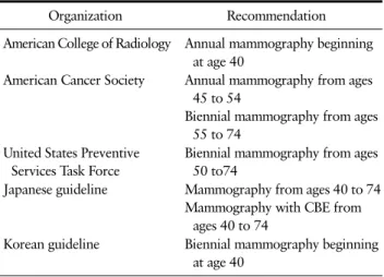
Table 1. Breast Cancer Screening Recommendations
Organization Recommendation
American College of Radiology Annual mammography beginning at age 40
American Cancer Society Annual mammography from ages 45 to 54
Biennial mammography from ages 55 to 74
United States Preventive Services Task Force
Biennial mammography from ages 50 to74
Japanese guideline Mammography from ages 40 to 74 Mammography with CBE from
ages 40 to 74
Korean guideline Biennial mammography beginning at age 40
CBE = clinical breast examination.
련 프로그램의 성공을 위해 건설적인 피드백(feedback) 과 임상의와의 효과적인 소통 또한 필수 요소이다.
검진자의 편의를 위해 유방촬영술과 유방 초음파를 동 일한 날에 시행하면 소환율을 낮출 수 있고 검진의 정확도 도 높일 수 있다. Tohno 등의 보고에 의하면 선별 유방 초 음파를 유방촬영술과 동일한 날에 시행함으로써 유방촬 영술의 소환율을 50% 감소시켰으며, American Roentgen Ray Society 연구에서도 동일한 결과를 보였다.(34,35) 즉각적인 판독(immediate interpretation) 또한 소환율, 환자의 불안감 그리고 의료 비용 감소에 매우 중요한 인자 이다.
American College of Radiology (ACR)에 따르면 유방 초음파는 최소한 5-12 MHz의 탐촉자로 전체 유방을 중첩 해서 최소한 두 단면(planes) 이상 검사해야 하며 유두 뒤 부분의 영상도 포함해야 한다.(36) 복합 낭종(complicated cyst)의 경우 최소한 두 방향에서 길이를 측정하고 기록해 야 하지만, 중년 여성 유방의 약 50%에서 관찰되는 단순 낭종은 선별 유방 초음파에서 별도로 기술하지 않아도 된 다.(37)
2) 위험도 기반 유방암 검진 권고안
다양한 기관에서 보고된 유방암 검진 권고안은 1-2년 주기의 유방 촬영술만 포함되어 있고, 선별 유방 초음파에 관한 검진 권고안은 대규모 무작위 대조군 연구 결과가 충 분하지 않아 현재까지 정립되지 않았다(Table 1). (38-42) 하지만 미국에서는 치밀 유방 여성의 경우 유방 초음파를 추가로 시행할 수 있으며, 프랑스와 독일에서도 유방이 매
우 치밀한(extremely dense) 여성에 대해 추가적인 초음 파 검사가 시행되고 있다.(43) 특히 동양 여성은 서양 여 성보다 치밀 유방의 빈도가 높고 보다 젊은 연령에서 유방 암이 발생하는 특징으로 선별 유방 초음파의 유용성이 높 을 것으로 판단된다.(44)
최근에는 유방암이 발생하는 위험도의 차이를 반영하 여 개인마다 유방암 검진 방법을 다르게 적용하는 맞춤형 유방암 검진을 제시하고 있다. 즉, 유방 밀도와 관계없이 유방암 발생 평균 위험군(life time risk < 15%)에서는 유 방촬영술 검진을 권고하며, 고위험군(life time risk > 20%)에서는 유방 자기공명영상(magnetic resonance imaging; MRI) 검진이 권고된다. 유방암 발생 중간 위험 군(life time risk 15%-20%)이면서 치밀 유방을 가진 군 에서는 선별 유방 초음파를 추가로 시행할 수 있다.(45) 또한 ACR은 유방암 고위험군에서 MRI를 시행할 수 없는 경우 유방 초음파를 고려할 수 있다고 권고하고 있다.(38)
향후 더 많은 연구를 통해 선별 유방 초음파 검진이 필 요한 군의 정의와 위양성률을 줄이고 정확도를 높이기 위 한 국내 실정에 맞는 선별 유방 초음파 검진 권고안의 개발 이 필요하리라 생각한다.
결 론
선별 유방 초음파는 유방촬영술의 민감도가 떨어지고 유방암의 위험도가 증가하는 치밀 유방을 가진 여성의 유 방암 검진 방법으로 유용하며 특히 작은 크기의 림프절 전 이가 없는 초기의 침윤암을 발견하는데 이점이 있다. 낮은 특이도 등의 단점을 보완한다면 ABUS가 좀 더 신속하고 효율적인 유방암 검진 방법으로 활용될 수 있을 것이다.
유방암 검진의 이익은 극대화하고 위해는 최소화하기 위 해 향후 유방 밀도, 유방암 발생 위험률 등의 개개인의 특 성을 고려한 국내 실정에 맞는 맞춤형 유방암 검진을 시행 하는 것이 필요할 것으로 생각한다.
REFERENCES
1. Bray F, Ferlay J, Soerjomataram I, Siegel RL, Torre LA, Jemal A. Global cancer statistics 2018: GLOBOCAN estimates of incidence and mortality worldwide for 36 cancers in 185 countries. CA Cancer J Clin 2018;68:
394-424.
2. National Health Insuarance Service. 2016 cancer reg-
istry statistics in Korea [Internet]. Daejeon: Statistics Korea; 2019 [cited 2019 Feb 25]. Available from:
http://kosis.kr.
3. Korean Breast Cancer Society. Breast Cancer Facts &
Figures 2018. Seoul: Korean Breast Cancer Society;
2018.
4. Myers ER, Moorman P, Gierisch JM, Havrilesky LJ, Grimm LJ, Ghate S, et al. Benefits and harms of breast cancer screening: a systematic review. JAMA 2015;314:1615-34.
5. Mandelson MT, Oestreicher N, Porter PL, White D, Finder CA, Taplin SH, et al. Breast density as a pre- dictor of mammographic detection: comparison of in- terval- and screen-detected cancers. J Natl Cancer Inst 2000;92:1081-7.
6. Freer PE. Mammographic breast density: impact on breast cancer risk and implications for screening.
Radiographics 2015;35:302-15.
7. Boyd NF, Martin LJ, Yaffe MJ, Minkin S. Mammographic density and breast cancer risk: current understanding and future prospects. Breast Cancer Res 2011;13:223.
8. Lee CI, Bassett LW, Lehman CD. Breast density leg- islation and opportunities for patient-centered out- comes research. Radiology 2012;264:632-6.
9. Checka CM, Chun JE, Schnabel FR, Lee J, Toth H.
The relationship of mammographic density and age:
implications for breast cancer screening. AJR Am J Roentgenol 2012;198:W292-5.
10. Berg WA, Zhang Z, Lehrer D, Jong RA, Pisano ED, Barr RG, et al. Detection of breast cancer with addi- tion of annual screening ultrasound or a single screening MRI to mammography in women with ele- vated breast cancer risk. JAMA 2012;307:1394-404.
11. Hooley RJ, Greenberg KL, Stackhouse RM, Geisel JL, Butler RS, Philpotts LE. Screening US in patients with mammographically dense breasts: initial experi- ence with Connecticut Public Act 09-41. Radiology 2012;265:59-69.
12. Gordon PB, Goldenberg SL. Malignant breast masses detected only by ultrasound. A retrospective review.
Cancer 1995;76:626-30.
13. Kolb TM, Lichy J, Newhouse JH. Comparison of the performance of screening mammography, physical examination, and breast US and evaluation of factors that influence them: an analysis of 27,825 patient evaluations. Radiology 2002;225:165-75.
14. Schaefer FK, Waldmann A, Katalinic A, Wefelnberg C, Heller M, Jonat W, et al. Influence of additional breast ultrasound on cancer detection in a cohort study for quality assurance in breast diagnosis-- analysis of 102,577 diagnostic procedures. Eur Radiol 2010;20:1085-92.
15. Berg WA, Blume JD, Cormack JB, Mendelson EB, Lehrer D, Böhm-Vélez M, et al. Combined screening with ultrasound and mammography vs mammography alone in women at elevated risk of breast cancer.
JAMA 2008;299:2151-63.
16. Berg WA, Bandos AI, Mendelson EB, Lehrer D, Jong RA, Pisano ED. Ultrasound as the primary screening test for breast cancer: analysis from ACRIN 6666. J Natl Cancer Inst 2015;108:djv367.
17. El-Bastawissi AY, White E, Mandelson MT, Taplin S.
Variation in mammographic breast density by race.
Ann Epidemiol 2001;11:257-63.
18. Nie K, Su MY, Chau MK, Chan S, Nguyen H, Tseng T, et al. Age- and race-dependence of the fibroglandular breast density analyzed on 3D MRI. Med Phys 2010;
37:2770-6.
19. Shen S, Zhou Y, Xu Y, Zhang B, Duan X, Huang R, et al. A multi-centre randomised trial comparing ultra- sound vs mammography for screening breast cancer in high-risk Chinese women. Br J Cancer 2015;112:
998-1004.
20. Ohuchi N, Suzuki A, Sobue T, Kawai M, Yamamoto S, Zheng YF, et al. Sensitivity and specificity of mam- mography and adjunctive ultrasonography to screen for breast cancer in the Japan Strategic Anti-cancer Randomized Trial (J-START): a randomised con- trolled trial. Lancet 2016;387:341-8.
21. Bae MS, Moon WK, Chang JM, Koo HR, Kim WH, Cho N, et al. Breast cancer detected with screening US:
reasons for nondetection at mammography. Radiology 2014;270:369-77.
22. Chae EY, Kim HH, Cha JH, Shin HJ, Kim H. Evaluation of screening whole-breast sonography as a supple- mental tool in conjunction with mammography in women with dense breasts. J Ultrasound Med 2013;32:1573-8.
23. Rosenberg RD, Yankaskas BC, Abraham LA, Sickles EA, Lehman CD, Geller BM, et al. Performance benchmarks for screening mammography. Radiology 2006;241:55-66.
24. Choi JS, Han BK, Ko EY, Ko ES, Shin JH, Kim GR.
Additional diagnostic value of shear-wave elastog- raphy and color Doppler US for evaluation of breast non-mass lesions detected at B-mode US. Eur Radiol 2016;26:3542-9.
25. Lee SH, Chang JM, Kim WH, Bae MS, Seo M, Koo HR, et al. Added value of shear-wave elastography for evaluation of breast masses detected with screening US imaging. Radiology 2014;273:61-9.
26. Kaplan SS. Clinical utility of bilateral whole-breast US in the evaluation of women with dense breast tissue. Radiology 2001;221:641-9.
27. Chang JM, Moon WK, Cho N, Park JS, Kim SJ.
Radiologists' performance in the detection of benign and malignant masses with 3D automated breast ul- trasound (ABUS). Eur J Radiol 2011;78:99-103.
28. Shin HJ, Kim HH, Cha JH, Park JH, Lee KE, Kim JH.
Automated ultrasound of the breast for diagnosis: in- terobserver agreement on lesion detection and characterization. AJR Am J Roentgenol 2011;197:
747-54.
29. Kelly KM, Dean J, Comulada WS, Lee SJ. Breast can- cer detection using automated whole breast ultra- sound and mammography in radiographically dense breasts. Eur Radiol 2010;20:734-42.
30. Wilczek B, Wilczek HE, Rasouliyan L, Leifland K.
Adding 3D automated breast ultrasound to mammog- raphy screening in women with heterogeneously and extremely dense breasts: report from a hospital- based, high-volume, single-center breast cancer screening program. Eur J Radiol 2016;85:1554-63.
31. Brem RF, Tabár L, Duffy SW, Inciardi MF, Guingrich JA, Hashimoto BE, et al. Assessing improvement in detection of breast cancer with three-dimensional automated breast US in women with dense breast tissue:
the SomoInsight Study. Radiology 2015;274:663-73.
32. Tohno E, Takahashi H, Tamada T, Fujimoto Y, Yasuda H, Ohuchi N. Educational program and testing using images for the standardization of breast cancer screening by ultrasonography. Breast Cancer 2012;19:
138-46.
33. Geisel J, Raghu M, Hooley R. The role of ultrasound in breast cancer screening: the case for and against ultrasound. Semin Ultrasound CT MR 2018;39:25-34.
34. Tohno E, Umemoto T, Sasaki K, Morishima I, Ueno E.
Effect of adding screening ultrasonography to screen- ing mammography on patient recall and cancer de- tection rates: a retrospective study in Japan. Eur J Radiol 2013;82:1227-30.
35. Durand MA, Hooley RJ. Implementation of whole- breast screening ultrasonography. Radiol Clin North Am 2017;55:527-39.
36. American College of Radiology. Breast ultrasound ac- creditation program requirements [Internet]. Reston:
American College of Radiology; 2017 [cited 2019 Mar 1]. Available from: http://www.acraccreditation.org/-/
media/ACRAccreditation/Documents/Breast-Ultrasound/
Requirements.pdf?la=en.
37. Berg WA, Sechtin AG, Marques H, Zhang Z. Cystic breast masses and the ACRIN 6666 experience. Radiol Clin North Am 2010;48:931-87.
38. Monticciolo DL, Newell MS, Hendrick RE, Helvie MA, Moy L, Monsees B, et al. Breast cancer screening for average-risk women: recommendations from the ACR commission on breast imaging. J Am Coll Radiol 2017;
14:1137-43.
39. Oeffinger KC, Fontham ET, Etzioni R, Herzig A, Michaelson JS, Shih YC, et al. Breast cancer screen- ing for women at average risk: 2015 guideline update from the American Cancer Society. JAMA 2015;314:
1599-614.
40. Siu AL. Screening for breast cancer: U.S. Preventive Services Task Force recommendation statement. Ann Intern Med 2016;164:279-96.
41. Hamashima C, Hamashima C C, Hattori M, Honjo S, Kasahara Y, Katayama T, et al. The Japanese guide- lines for breast cancer screening. Jpn J Clin Oncol 2016;46:482-92.
42. Lee EH, Park B, Kim NS, Seo HJ, Ko KL, Min JW, et al. The Korean guideline for breast cancer screening.
J Korean Med Assoc 2015;58:408-19.
43. Vourtsis A, Berg WA. Breast density implications and supplemental screening. Eur Radiol 2019;29:1762-77.
44. Burkett BJ, Hanemann CW. A review of supplemental screening ultrasound for breast cancer: certain pop- ulations of women with dense breast tissue may benefit. Acad Radiol 2016;23:1604-9.
45. Lee CI, Chen LE, Elmore JG. Risk-based breast can- cer screening: implications of breast density. Med Clin North Am 2017;101:725-41.