D I A B E T E S & M E T A B O L I S M J O U R N A L D I A B E T E S & M E T A B O L I S M J O U R N A L
This is an Open Access article distributed under the terms of the Creative Commons Attribution Non-Commercial License (https://creativecommons.org/licenses/by-nc/4.0/) which permits unrestricted non-commercial use, distribution, and reproduction in any medium, provided the original work is properly cited.
Umbilical Cord-Mesenchymal Stem Cell-Conditioned Medium Improves Insulin Resistance in C2C12 Cell
Kyung-Soo Kim1, Yeon Kyung Choi2, Mi Jin Kim2, Jung Wook Hwang2, Kyunghoon Min3, Sang Youn Jung1, Soo-Kyung Kim1, Yong-Soo Choi2, Yong-Wook Cho1
1Department of Internal Medicine, CHA Bundang Medical Center, CHA University School of Medicine, Seongnam,
2Department of Biotechnology, CHA University, Seongnam,
3Department of Rehabilitation Medicine, CHA Bundang Medical Center, CHA University School of Medicine, Seongnam, Korea
Background: Umbilical cord-mesenchymal stem cell-conditioned medium (UC-MSC-CM) has emerged as a promising cell-free therapy. The aim of this study was to explore the therapeutic effects of UC-MSC-CM on insulin resistance in C2C12 cell.
Methods: Insulin resistance was induced by palmitate. Effects of UC-MSC-CM on insulin resistance were evaluated using glu- cose uptake, glucose transporter type 4 (GLUT4) translocation, the insulin-signaling pathway, and mitochondrial contents and functions in C2C12 cell.
Results: Glucose uptake was improved by UC-MSC-CM. UC-MSC-CM treatment increased only in membranous GLUT4 ex- pression, not in cytosolic GLUT4 expression. It restored the insulin-signaling pathway in insulin receptor substrate 1 and protein kinase B. Mitochondrial contents evaluated by mitochondrial transcription factor A, mitochondrial DNA copy number, and per- oxisome proliferator-activated receptor gamma coactivator 1-alpha were increased by UC-MSC-CM. In addition, UC-MSC-CM significantly decreased mitochondrial reactive oxygen species and increased fatty acid oxidation and mitochondrial membrane potential. There was no improvement in adenosine triphosphate (ATP) contents, but ATP synthesis was improved by UC-MSC- CM. Cytokine and active factor analysis of UC-MSC-CM showed that it contained many regulators inhibiting insulin resistance.
Conclusion: UC-MSC-CM improves insulin resistance with multiple mechanisms in C2C12 cell.
Keywords: Culture media, conditioned; Diabetes mellitus; Insulin resistance; Mesenchymal stem cells; Mitochondria; Muscles;
Umbilical cord; Wharton jelly
Corresponding authors: Yong-Wook Cho https://orcid.org/0000-0001-5601-3802 Department of Internal Medicine, CHA Bundang Medical Center, CHA University School of Medicine, 59 Yatap-ro, Bundang-gu, Seongnam 13496, Korea
E-mail: [email protected]
Yong-Soo Choi https://orcid.org/0000-0001-8445-8067
Department of Biotechnology, CHA University, 335 Pankyo-ro, Bundang-gu, Seongnam 13488, Korea
INTRODUCTION
How do we manage type 2 diabetes mellitus (T2DM) more ap- propriately? This is a very difficult problem, because T2DM has a complex pathophysiology [1-3]. Although we have various anti-diabetic drugs to treat patients with T2DM, half of them have not achieved the glycemic target yet [4-6]. Unmet medical needs still exist in this field.
Stem cells are an attractive candidate for treatment of many diseases because of their properties, such as division, renewal,
differentiation, homing, and engraftment [7,8]. Umbilical cord-mesenchymal stem cells (UC-MSC) could be the best choice if one considers their high potential to differentiate into other cells and low immunogenicity [9]. Although decreased insulin secretion and increased insulin resistance are two ma- jor defects in T2DM, most studies using stem cells were fo- cused on increasing insulin secretion [8]. Some studies have shown that UC-MSC treatment improved insulin resistance in patients with T2DM [10,11].
Therapeutic usage of UC-MSC, however, is not easy, because https://doi.org/10.4093/dmj.2019.0191
pISSN 2233-6079 · eISSN 2233-6087
of entrapment in filtering organs and potential tumorigenicity [12]. After it was revealed that the beneficial effects of UC- MSC depend mostly on their paracrine activity, a conditioned medium (CM) that consists of bioactive factors and cytokines has emerged as a promising cell-free therapy [12]. Neverthe- less, UC-MSC-CM has not been studied for the treatment of T2DM so far.
Muscles play a major role in insulin resistance, but there have been no anti-diabetic drugs targeted at reducing insulin resis- tance in muscles [13-16]. If we can improve insulin resistance in the muscles, that will be helpful for patients with T2DM in a way different from already existing anti-diabetic drugs. The aim of this study was to explore the potential therapeutic ef- fects of UC-MSC-CM on insulin resistance in C2C12 cell.
METHODS
Cell culture
Mouse skeletal-muscle cells (CRL-1772, passage #6) were pur- chased from the American Type Culture Collection (ATCC, Manassas, VA, USA) and maintained in Dulbecco’s Modified Eagle’s Medium (DMEM; Hyclone laboratories Inc., Logan, UT, USA) supplemented with 10% fetal bovine serum (FBS;
Gibco, Carlsbad, CA, USA) and 1% penicillin-streptomycin (P/S; Hyclone, Northumberland, PA, USA). To differentiate the myotube, we cultured C2C12 myoblasts in DMEM con- taining 2% horse serum (Gibco, Auckland, New Zealand) for 4 days, with media changed every 2 days. Written informed con- sent was obtained from all patients. This study was approved by the CHA University Institutional Review Board (201803- BR-015-03).
Palmitate solution preparation and induction of insulin resistance
Palmitate (PA; Sigma, St. Louis, MO, USA) was dissolved in 99.9% ethanol by heating at 95°C and adjusted to a concentra- tion of 100 mM. After filtration, 100 mM PA was diluted in DMEM mixed with 2% bovine serum albumin (BSA; Bio Ba- sic, Markham, ON, Canada) at a ratio of 1:100. The final con- centration of PA solution was 1 mM, which was then incubat- ed in a 37°C water bath for 1 hour and stored at –20°C.
Preparation of UC-MSC-CM
UC-MSC was thawed at a density of 1×10⁶ cells in a T-175 flask. When cell density reached 80% to 90%, they were re-
placed with fresh serum-free Minimum Essential Medium Ea- gle Alpha Modification (α-MEM; Hyclone) supplemented with 1% P/S. After 37°C incubation for 48 hours, we harvested them from the medium and centrifuged them at 1,500 rpm for 5 minutes. We filtered them with a 0.2 μM filter and stored the supernatant at –80°C until use.
2-Deoxy glucose uptake
For quantifying glucose uptake in cells, we treated differentiat- ed C2C12 cell with 1 mM PA with or without UC-MSC-CM for 24 hours. Prior to the assay, cells were washed with phos- phate buffered saline twice and starved in Krebs-Ringer-Phos- phate-HEPES buffer for 40 minutes. Cells were then stimulat- ed with 100 nM insulin (Eli Lilly Nederland, Indianapolis, IN, USA) for 30 minutes. We measured glucose uptake by C2C12 cells using the Glucose Uptake Assay kit (ab136955, Abcam, Cambridge, UK) following the manufacturer’s instructions.
Glucose levels were quantified using Microplate reader (BioTek Inc., Winooski, VT, USA).
Membrane protein extraction
Cytosol and membrane were separated by a membrane protein extraction kit (Thermo, Rockford, IL, USA) and differential centrifuge. Additionally, we added a protease inhibitor (PI;
Quartett Immunodiagnostika, Berlin, Germany) to the per- meabilization and solubilization buffer. Briefly, we centrifuged at 300 ×g for 5 minutes and washed the cells twice using wash buffer. After permeabilization buffer treatment, we centrifuged the permeabilized cells for 10 minutes at 4°C with constant mixing and for 15 minutes at 16,000 ×g to transfer the super- natant containing cytosolic proteins to a new tube. To isolate membrane-associated protein, cell pellets were resuspended with solubilization buffer, were incubated at 4°C for 30 minutes with constant mixing, and were centrifuged for 15 minutes at 16,000 ×g.
Western blot analysis
Cell were harvested and centrifuged at 1,500 rpm for 3 minutes and lysed with radioimmunoprecipitation assay buffer includ- ing a PI. The concentration of protein extraction was evaluated by using a bicinchoninic acid protein assay kit (Thermo) at 562 nm with a microplate reader. Western blot for glucose trans- porter type 4 (GLUT4) protein was carried for the membrane fraction and the cytosol fraction. We analyzed other markers using total proteins. Each protein sample were migrated by
12% sodium dodecyl sulfate-polyacrylamide gel electrophore- sis and transferred to a polyvinylidene difluoride membrane (Millipore, Billerica, MA, USA). The membrane was next blocked for an hour with 5% BSA in 1X-Tris buffered saline with Tween 20 (TBST) at room temperature and incubated overnight at 4°C with constant mixing with insulin receptor substrate 1 (IRS-1; Cell Signaling Technology, Danvers, MA, USA; #2386S, #2381S), phosphoinositide 3-kinase (PI3K; Cell Signaling Technology, #4223), protein kinase B (AKT1; Cell Signaling Technology, #9271), GLUT4 (Abcam, ab654), per- oxisome proliferator-activated receptor gamma coactivator 1-alpha (PGC-1α) (Santa Cruz Biotechnology, Santa Cruz, CA, USA; sc-13067), mitochondrial transcription factor A (mtTFA;
Santa Cruz Biotechnology, sc-376672), and β-actin (Santa Cruz Biotechnology, sc-47778). After washing with 1x TBST, we visualized specific secondary antibodies by incubation with 1:2,000 dilution of goat anti-rabbit immunoglobulin G (IgG;
Santa Cruz; sc-2004), goat anti-mouse IgG (Santa Cruz, sc- 2005). We confirmed the band using enhanced chemilumines- cence (ECL component of Pierce Clarity and Western ECL Substrate, Bio-Rad Laboratories, Hercules, CA, USA) with an LAS-4000 imager (Fujifilm Inc., Tokyo, Japan).
Mitochondrial DNA analysis
The relative level of mitochondrial DNA (mtDNA) copy num- ber was calculated using reverse transcription polymerase chain reaction (PCR) by measuring the ratio of Cox2 (mito- chondrial-encoded gene) versus Rsp18 (nuclear-encoded gene). Prepare 1×10⁶ cells and extract RNA using RNA extrac- tion kit (iNTRON, Seongnam, Korea). Then, complementary DNA (cDNA) is synthesized by PCR with a primer (Supple- mentary Table 1) and master mix using a cDNA synthesis kit (iNTRON).
Measurement of reactive oxygen species production and fatty acid oxidation
Intracellular reactive oxygen species (ROS) was measured us- ing chloromethyl-2´,7´-dichlorodihydrofluorescein diacetate (CM-DCFDA, Molecular Probes; Invitrogen, Carlsbad, CA, USA). For positive control, cells were treated with H2O2 (1 mM) for 10 minutes and then incubated with 10 μM of DCF- DA dye concentration for 30 minutes. Intracellular ROS was measured at excitation 485 nm and emission 535 nm. Mito- chondrial ROS was measured using the fluorescent dye Mito- Sox Red, a mitochondrial superoxide indicator (Invitrogen).
Cells were incubated with 5 μM of MitoSox dye concentration at 37°C for 30 minutes. The dye was activated at excitation 510 nm and emission 528 nm. Results from the fluorescence imag- es showed similar trends. Fatty acid oxidation was measured by the non-radioactive fatty acid oxidation assay (Biomedical Research Service Center, E-141).
Analysis of inner mitochondrial membrane potential To confirm the mitochondrial function and viability, cells were stained with JC-1 dye (Invitrogen), which assesses the mito- chondrial membrane potential. For positive control, we used carbonyl cyanide-p-trifluoromethoxyphenylhydrazone (CCCP at 50 μM for 30 minutes; Sigma). We dissolved it in dimethyl sulfoxide to make 1 mM stock, and then diluted the solution in DMEM to a concentration of 50 μM and incubated at 37°C for 30 minutes. After incubation, monomer signal (green) was ac- tivated at excitation 488 nm, emission 528 nm, and the aggre- gate signal (red) was activated at excitation 530 nm, emission 590 nm.
Measurement of ATP contents and ATP synthesis
Intracellular adenosine triphosphate (ATP) was measured with a CellTiter-Glo 2.0 assay kit (Promega, Madison, WI, USA).
We prepared a 1:1 solution containing fresh culture media and CellTier-Glo luminescence test reagent and inserted the mix- ture (200 μL) into each well. After incubation for 30 minutes at room temperature, we read luminescent signal using a lumi- nescence microplate reader. ATP concentration was calculated from a standard curve and normalized by total protein. ATP synthesis was measured after adding adenosine diphosphate (ADP) as a substrate.
Cytokine assay
Our group has already analyzed UC-MSC-CM in a previous study [17]. To summarize briefly, cytokine, growth factor, and active protein contents were analyzed using an antibody-based protein array capable of simultaneously detecting 507 different factors (Ray Biotech Inc., Norcross, GA, USA).
Statistical analysis
All in vitro experiments were independently performed at least three times. Data were analyzed using the SigmaPlot version 11.0 software (Systat Software, San Jose, CA, USA). Quantita- tive results are shown as the mean±standard deviation for each experiment, and P<0.05 was considered statistically significant.
RESULTS
The effect of UC-MSC-CM on glucose uptake in C2C12 myotubes
To investigate the effect of UC-MSC-CM on insulin resistance in C2C12 myotube, we induced insulin resistance by PA and measured 2-deoxy glucose (2-DG) uptake. UC-MSC-CM sig- nificantly improved 2-DG uptake in comparison with the PA- treated group. However, there was no improvement of 2-DG uptake in the experiment using C2C12-CM to verify the inter- ference effect (Fig. 1).
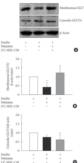
The effect of UC-MSC-CM on protein expression of GLUT4 GLUT4 is a membranous channel that transports glucose into cells. Insulin resistance is influenced by translocation to the cell membrane of GLUT4, not the presence of GLUT4 in cytosol.
PA significantly reduced the expression of membranous GLUT4, and UC-MSC-CM restored it in C2C12 myotube. The level of cytosolic GLUT4 expression was not changed by UC- MSC-CM treatment (Fig. 2).
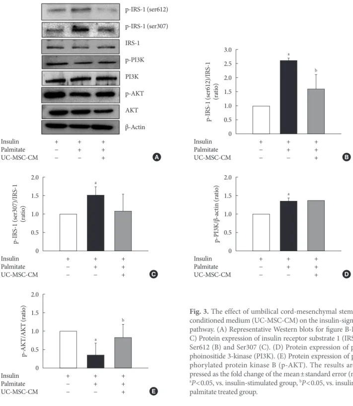
The effect of UC-MSC-CM on the insulin-signaling pathway IRS-1 plays a key role in transmitting signals from the insulin and insulin-like growth factor receptor to intracellular path- ways PI3K/AKT kinase pathway. Phosphorylation of IRS-1 at ser612 results in an inhibition of insulin signaling in the cell and phosphorylation at Ser307 activates dissociation of insulin
receptor [18]. IRS1 contains multiple tyrosine phosphorylation sites, which acts as docking sites for PI3K. PI3K elicit AKT phosphorylation causing translocate GLUT4 onto the cell membrane [19]. Consequently, insulin-dependent transport of
Fig. 2. The effect of umbilical cord-mesenchymal stem cell- conditioned medium (UC-MSC-CM) on protein expression of glucose transporter type 4 (GLUT4). (A) Western blot. (B) Protein expression of membranous GLUT4. (C) Protein ex- pression of cytosolic GLUT4. The results are expressed as the fold change of the mean±standard error (n≥3). aP<0.05, vs.
insulin-stimulated group, bP<0.05, vs. insulin and palmitate treated group.
2.0 1.5 1.0 0.5 0 Membranous GLUT4/ β-actin (ratio)
+ + + − + + − − + Insulin
Palmitate UC-MSC-CM
a
b
B
2.0 1.5 1.0 0.5 Cytosolic GLUT4/β-actin (ratio) 0
+ + + − + + − − + Insulin
Palmitate UC-MSC-CM
b
C + + +
− + + − − + Insulin
Palmitate
UC-MSC-CM A
Membranous GLUT4
Cytosolic GLUT4
β-Actin
Fig. 1. The effect of umbilical cord-mesenchymal stem cell-con- ditioned medium (UC-MSC-CM) on glucose uptake in C2C12 myotubes. The results are expressed as the fold change of the mean±standard error (n≥3). 2-DG, 2-deoxy glucose. aP<0.05, vs. insulin-unstimulated group, bP<0.05, vs. insulin-stimulated group, cP<0.05, vs. insulin and palmitate treated group.
− + + + + − − + + + − − − + − − − − − + Insulin
Palmitate UC-MSC-CM C2C12-CM
2.5 2.0 1.5 1.0 0.5 0
2-DG uptake (ratio) a
b c
glucose is initiated. UC-MSC-CM decreased the phosphoryla- tion of IRS1 at Ser612 and Ser307 (Fig. 3A-C). However, al- though the phosphorylation of PI3K was increased by PA treatment, there was no change by UC-MSC-CM treatment (Fig. 3D). The expression of phosphorylated AKT1 at Ser473 was up-regulated by UC-MSC-CM (Fig. 3E).
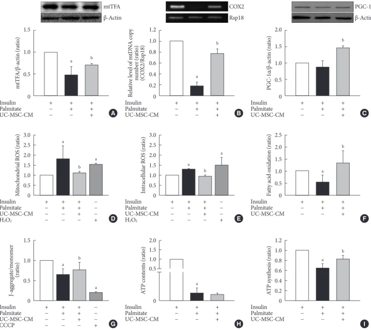
The effect of UC-MSC-CM on mitochondria Contents
mtTFA plays a key role for the regulation of mtDNA replica- tion and its level is proportional to mtDNA [20]. The protein level of mtTFA was restored by UC-MSC-CM treatment (P<0.05) (Fig. 4A). In addition, UC-MSC-CM treatment in-
Fig. 3. The effect of umbilical cord-mesenchymal stem cell- conditioned medium (UC-MSC-CM) on the insulin-signaling pathway. (A) Representative Western blots for figure B-E. (B, C) Protein expression of insulin receptor substrate 1 (IRS1) at Ser612 (B) and Ser307 (C). (D) Protein expression of phos- phoinositide 3-kinase (PI3K). (E) Protein expression of phos- phorylated protein kinase B (p-AKT). The results are ex- pressed as the fold change of the mean±standard error (n≥3).
aP<0.05, vs. insulin-stimulated group, bP<0.05, vs. insulin and palmitate treated group.
2.0 1.5 1.0 0.5 0 3.0 2.5 2.0 1.5 1.0 0.5 0
2.0 1.5 1.0 0.5 0 2.0 1.5 1.0 0.5 0
p-PI3K/β-actin (ratio)p-IRS-1 (ser612)/IRS-1 (ratio)
p-AKT/AKT (ratio)p-IRS-1 (ser307)/IRS-1 (ratio)
+ + + − + + − − + + + + − + + − − + + + +
− + + − − +
+ + + − + + − − + + + + − + + − − +
Insulin Palmitate UC-MSC-CM Insulin Palmitate UC-MSC-CM Insulin
Palmitate UC-MSC-CM
Insulin Palmitate UC-MSC-CM Insulin Palmitate UC-MSC-CM
a a
b
b a
D B A
E C p-IRS-1 (ser612) p-IRS-1 (ser307) IRS-1
p-PI3K PI3K p-AKT AKT β-Actin
a
creased the relative level of mtDNA copy number (Fig. 4B).
PGC-1α is widely regarded as the master regulator of mito- chondrial biogenesis [21,22]. There was no difference in con- tent of PGC-1α between the control and PA-treated C2C12 myotubes. Treatment with UC-MSC-CM increased the con- tent of PGC-1α (Fig. 4C).
Function
Oxidative stress plays an important role in glucotoxicity and insulin resistance [23]. In this study, we found that UC-MSC- CM significantly decreased the level of mitochondrial ROS as well as intracellular ROS, in contrast to PA-treated C2C12 myotubes (Fig. 4D and E). Fatty acid oxidation increases glu-
Fig. 4. The effect of umbilical cord-mesenchymal stem cell-conditioned medium (UC-MSC-CM) on mitochondrial contents and functions. (A) Protein level of mitochondrial transcription factor A (mtTFA). (B) The relative level of mitochondrial DNA (mtD- NA) copy number. (C) Protein level of peroxisome proliferator-activated receptor gamma coactivator 1-alpha (PGC-1α). (D) Mi- tochondrial reactive oxygen species (ROS). (E) Intracellular ROS. (F) Fatty acid oxidation. (G) Mitochondrial membrane poten- tial. (H) Adenosine triphosphate (ATP) contents. (I) ATP synthesis. The results are expressed as the fold change of the mean±standard error (n≥3). CCCP, carbonyl cyanide-p-trifluoromethoxyphenylhydrazone. aP<0.05, vs. insulin-stimulated group, bP<0.05, vs. insulin and palmitate treated group.
1.5 1.0 0.5 0
3.0 2.5 2.0 1.5 1.0 0.5 0
1.5 1.0 0.5
0 0
2.0 1.5 1.0 0.5 3.0 2.5 2.0 1.5 1.0 0.5 0
2.5 2.0 1.5 1.0 0.5 0
1.2 1.0 0.8 0.6 0.4 0.2 0 1.2
1.0 0.8 0.6 0.4 0.2 0
2.0 1.5 1.0 0.5 0
mtTFA/β-actin (ratio)Mitochondrial ROS (ratio)J-aggregate/monomer (ratio) ATP contents (ratio)Intracellular ROS (ratio) Fatty acid oxidation (ratio)ATP synthesis (ratio)
Relative level of mtDNA copy number (ratio) (COX2/Rsp18) PGC-1α/β-actin (ratio)
+ + + − + + − − +
+ + + − − + + − − − + − − − − +
+ + + − − + + − − − + − − − − +
+ + + − + + − − + + + + − − + + − − − + − − − − +
+ + + − + + − − +
+ + + − + + − − + + + +
− + + − − +
+ + + − + + − − + Insulin
Palmitate UC-MSC-CM
Insulin Palmitate UC-MSC-CM H2O2
Insulin Palmitate UC-MSC-CM CCCP
Insulin Palmitate UC-MSC-CM Insulin Palmitate UC-MSC-CM H2O2
Insulin Palmitate UC-MSC-CM
Insulin Palmitate UC-MSC-CM Insulin
Palmitate UC-MSC-CM
Insulin Palmitate UC-MSC-CM
a
a
a
a a
a b
a
a
a b
b
b
b b
b
b
A
D
G H
E F
I
B C
mtTFA β-Actin
COX2 Rsp18
PGC-1α β-Actin
a
a
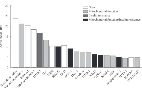
Fig. 5. Analysis of secreted factors in umbilical cord mesenchymal stem cell-conditioned medium. EDA-A2, ectodysplasin-A2;
IGFBP, insulin-like growth factor binding protein; TIMP, tissue inhibitor of metalloproteinases; IL-6, interleukin-6; MIP2, mac- rophage inflammatory protein 2; SPARC, secreted protein acidic and rich in cysteine; GRO, growth related oncogene; MCP, monocyte chemoattractant protein; Dkk, Dickkopf-related protein; VEGF, vascular endothelial growth factor; GDF, growth dif- ferentiation factor; HGF, hepatocyte growth factor; MMP-1, matrix metalloproteinase-1; FGF, fibroblast growth factor; KGF, ke- ratinocyte growth factor.
30
25
20
15
10
5
0 Active factor (104)
Thrombospondin
Thrombospondin-1EDA-A2 IGFB
P-rp1/IGFBP-7
TIMP-2 IL-6MIP2SPARC GROMCP -1Dkk-1
Activin A TIMP
-1VEGFGDF-15
Smad 4 HGF Angiopoietin-1
MMP -1 IGFB
P-6 FGF-7/KGF None
Mitochondrial function Insulin resistance
Mitochondrial function/Insulin resistance
cose extraction and glycogen synthesis, so incomplete fatty acid oxidation contributes to muscle insulin resistance. UC- MSC-CM increased fatty acid oxidation (Fig. 4F). Mitochon- drial function is usually monitored by mitochondria mem- brane potential (MMP) as well as intracellular ATP contents [24]. The MMP was not as low as CCCP, but it was decreased by PA. Treatment with UC-MSC-CM increased MMP (Fig.
4G). After ATP contents were diminished by PA, there was no improvement by UC-MSC-CM treatment (Fig. 4H). However, ATP synthesis was increased by UC-MSC-CM treatment when ATP was measured after adding ADP as a substrate (Fig. 4I).
Analysis of secreted factors in UC-MSC-CM
There were many cytokines, growth factors, and active protein contents affecting insulin resistance and mitochondrial func- tion in UC-MSC-CM. Among them, tissue inhibitor of metal- loproteinases-2 (TIMP-2), secreted protein acidic and rich in cysteine (SPARC), monocyte chemoattractant protein-1 (MCP-1), vascular endothelial growth factor (VEGF), growth differentiation factor-15 (GDF-15), and angiopoietin-1 are known regulators inhibiting insulin resistance (Fig. 5).
DISCUSSION
T2DM has become one of the greatest challenges to human health nowadays. More and more people are suffering from T2DM, and many drugs are being used to treat T2DM, which is not easy to treat, because of its complex pathophysiology, and we still need other treatment options [1-3]. UC-MSC-CM is an attractive candidate for treatment of T2DM and can over- come the problem of cell therapy [8-12]. However, there were few studies to investigate whether UC-MSC-CM could im- prove insulin resistance in muscles. In this study, we demon- strated that UC-MSC-CM improved insulin resistance in C2C12 cells. Possible mechanisms we have identified were im- proving GLUT4 translocation, the insulin-signaling pathway, and mitochondrial contents and functions.
MSCs exert beneficial effects on T2DM through differentia- tion into insulin-producing cells, promotion of islet cell regen- eration, protection of endogenous islet cells, and ameliorating insulin resistance [8]. In fact, many studies have focused on improving insulin secretory function by MSCs [8,25]. Some studies investigated whether UC-MSC improved insulin resis-
tance in T2DM [10,11]. Xie et al. [26] showed that UC-MSC alleviated insulin resistance by directing macrophages into an alternatively activated phenotype (anti-inflammatory, M2) in adipose tissues of T2DM rats. A study in 22 patients with T2DM demonstrated that treatment with UC-MSC can im- prove glucose profiles and beta-cell function by improving sys- temic inflammation and/or immunological regulation [10].
However, direct use of MSC has several problems, such as po- tential tumorigenicity, entrapment in filtering organs, scarce overall availability, and low survival rate [12]. UC-MSC-CM is a charming candidate for treating T2DM, because it is not a cell therapy and its beneficial effects depend mostly on their paracrine activity [12]. Shree and Bhonde [27] demonstrated that adipose-derived MSC-CM restored insulin resistance in C2C12 cells by improving inflammation and insulin signaling.
UC-MSC-CM suppressed ROS generation and improved muscle atrophy in atrophied muscles [17,28]. There is no study about the effects of UC-MSC-CM on insulin resistance in skel- etal muscles yet. In this study, UC-MSC-CM improved glucose uptake in PA-treated C2C12 myotubes. The expression of membranous GLUT4 was increased and insulin signaling, such as IRS1 and AKT1, was improved when UC-MSC-CM was used. In addition, mitochondrial contents and functions were also improved by UC-MSC-CM treatment. To the best of our knowledge, this is the first study to show that UC-MSC- CM improved insulin resistance via multiple mechanisms in C2C12 cell.
Mitochondria are organelles that are a critical contributor to cellular and organismal homeostasis [29]. Mitochondrial bio- genesis might be associated with insulin resistance through GLUT4 translocation, the insulin-signaling pathway, and in- flammation [30-32]. However, whether mitochondrial dys- function is a cause or a consequence of insulin resistance is not clear. There were many efforts to improve insulin resistance by restoring mitochondrial dysfunction [33], but there has been no study investigating the effects of UC-MSC-CM on mito- chondrial dysfunction. In this study, mitochondrial contents assessed by mtTFA, the relative level of mtDNA copy number, and PGC-1α were increased by UC-MSC-CM treatment. In terms of mitochondrial function, UC-MSC-CM treatment de- creased mitochondrial ROS and increased MMP in C2C12 cells. ATP contents were not increased by UC-MSC-CM treat- ment, but ATP synthesis was increased. Because ATP genera- tion is a complex process and is associated with various factors, UC-MSC-CM treatment could not restore it completely. How-
ever, because ATP synthesis was increased after adding ADP as a substrate, so we could assume that UC-MSC-CM influenced the improvement of ATP synthesis. Based on previous studies, these improvements for mitochondrial dysfunction might be associated with improving insulin resistance.
UC-MSC-CM consists of various cytokines, growth factors, and active proteins [17]. Among them, TIMP-2, SPARC, MCP-1, VEGF, GDF-15, and angiopoietin-1 are known regu- lators that improve insulin resistance [34-36]. Thrombospon- din-1, interleukin-6 (IL-6), SPARC, MCP-1, Dickkopf-related protein-1 (Dkk-1), activin A, TIMP-1, VEGF, GDF-15, smad- 4, hepatocyte growth factor (HGF), angiopoietin-1, matrix metalloproteinase-1 (MMP-1), and fibroblast growth factor-7 (FGF-7) were factors that influenced mitochondria [37-39].
There would also be active factors that aggravated insulin resis- tance and mitochondrial dysfunction. We could not find out which factors have mainly beneficial effects on insulin resis- tance. However, we could assume that UC-MSC-CM is a mix- ture that has beneficial effects on insulin resistance, although we do not know the exact ratio of active factors. One possible explanation might be that UC-MSC-CM is made when UC- MSCs are cultured, so it should have beneficial effects in all as- pects. Furthermore, many of these were known factors, but an unknown one that could have beneficial effects on insulin re- sistance and mitochondrial dysfunction still might exist.
In conclusion, our findings demonstrated that UC-MSC- CM improves insulin resistance in C2C12 cells. GLUT4 trans- location and the insulin-signaling pathway were improved by UC-MSC-CM treatment. Mitochondrial contents and func- tions were also improved, which is a novel finding, never re- ported previously. All these effects might be beneficial for in- sulin resistance in C2C12 cell. Further study is warranted to fully elucidate the effects of UC-MSC-CM on insulin resis- tance in animals or humans.
SUPPLEMENTARY MATERIALS
Supplementary materials related to this article can be found online at https://doi.org/10.4093/dmj.2019.0191.
CONFLICTS OF INTEREST
No potential conflict of interest relevant to this article was re- ported.
AUTHOR CONTRIBUTIONS
Conception or design: K.S.K., Y.S.C., Y.W.C.
Acquisition, analysis, or interpretation of data: K.S.K., Y.K.C., M.J.K., J.W.H., K.M., S.Y.J., S.K.K., Y.S.C., Y.W.C.
Drafting the work or revising: K.S.K., Y.K.C., M.J.K., Y.S.C., Y.W.C.
Final approval of the manuscript: K.S.K, Y.S.C., Y.W.C.
ORCID
Kyung-Soo Kim https://orcid.org/0000-0002-7738-2284 Yong-Wook Cho https://orcid.org/0000-0001-5601-3802 Yong-Soo Choi https://orcid.org/0000-0001-8445-8067
FUNDING
This work was supported by a grant (Kyung-Soo Kim, 2016F- 2) from the Korean Diabetes Association and the National Re- search Foundation of Korea (NRF) grant funded by the Korea government (MSIT) (NRF-2018R1C1B5042633).
ACKNOWLEDGMENTS
None
REFERENCES
1. Ahn YH. A journey to understand glucose homeostasis: start- ing from rat glucose transporter type 2 promoter cloning to hyperglycemia. Diabetes Metab J 2018;42:465-71.
2. Kwak SH, Park KS. Pathophysiology of type 2 diabetes in Kore- ans. Endocrinol Metab (Seoul) 2018;33:9-16.
3. Kim KS, Lee BW, Kim YJ, Lee DH, Cha BS, Park CY. Nonalco- holic fatty liver disease and diabetes. Part II: treatment. Diabe- tes Metab J 2019;43:127-43.
4. American Diabetes Association. 6. Glycemic targets: standards of medical care in diabetes. 2019. Diabetes Care 2019;42(Suppl 1):S61-70.
5. Ko SH, Han K, Lee YH, Noh J, Park CY, Kim DJ, et al. Past and current status of adult type 2 diabetes mellitus management in Korea: a National Health Insurance Service database analysis.
Diabetes Metab J 2018;42:93-100.
6. Won JC, Lee JH, Kim JH, Kang ES, Won KC, Kim DJ, et al. Di- abetes fact sheet in Korea, 2016: an appraisal of current status.
Diabetes Metab J 2018;42:415-24.
7. Diecke S, Jung SM, Lee J, Ju JH. Recent technological updates and clinical applications of induced pluripotent stem cells. Ko- rean J Intern Med 2014;29:547-57.
8. Zang L, Hao H, Liu J, Li Y, Han W, Mu Y. Mesenchymal stem cell therapy in type 2 diabetes mellitus. Diabetol Metab Syndr 2017;9:36.
9. Nagamura-Inoue T, He H. Umbilical cord-derived mesenchy- mal stem cells: their advantages and potential clinical utility.
World J Stem Cells 2014;6:195-202.
10. Liu X, Zheng P, Wang X, Dai G, Cheng H, Zhang Z, et al. A preliminary evaluation of efficacy and safety of Wharton’s jelly mesenchymal stem cell transplantation in patients with type 2 diabetes mellitus. Stem Cell Res Ther 2014;5:57.
11. Sun X, Hao H, Han Q, Song X, Liu J, Dong L, et al. Human umbilical cord-derived mesenchymal stem cells ameliorate in- sulin resistance by suppressing NLRP3 inflammasome-medi- ated inflammation in type 2 diabetes rats. Stem Cell Res Ther 2017;8:241.
12. Ranganath SH, Levy O, Inamdar MS, Karp JM. Harnessing the mesenchymal stem cell secretome for the treatment of cardio- vascular disease. Cell Stem Cell 2012;10:244-58.
13. Periasamy M, Herrera JL, Reis FCG. Skeletal muscle thermo- genesis and its role in whole body energy metabolism. Diabetes Metab J 2017;41:327-36.
14. American Diabetes Association. 9. Pharmacologic approaches to glycemic treatment: standards of medical care in diabetes.
2019. Diabetes Care 2019;42(Suppl 1):S90-102.
15. Ko SH, Hur KY, Rhee SY, Kim NH, Moon MK, Park SO, et al.
Antihyperglycemic agent therapy for adult patients with type 2 diabetes mellitus 2017: a position statement of the Korean Dia- betes Association. Diabetes Metab J 2017;41:337-48.
16. Raveendran AV, Deshpandae A, Joshi SR. Therapeutic role of yoga in type 2 diabetes. Endocrinol Metab (Seoul) 2018;33:
307-17.
17. Kim MJ, Kim ZH, Kim SM, Choi YS. Conditioned medium derived from umbilical cord mesenchymal stem cells regener- ates atrophied muscles. Tissue Cell 2016;48:533-43.
18. Li HB, Yang YR, Mo ZJ, Ding Y, Jiang WJ. Silibinin improves palmitate-induced insulin resistance in C2C12 myotubes by attenuating IRS-1/PI3K/Akt pathway inhibition. Braz J Med Biol Res 2015;48:440-6.
19. Huang X, Liu G, Guo J, Su Z. The PI3K/AKT pathway in obesi- ty and type 2 diabetes. Int J Biol Sci 2018;14:1483-96.
20. Choi YS, Kim S, Pak YK. Mitochondrial transcription factor A
(mtTFA) and diabetes. Diabetes Res Clin Pract 2001;54 Suppl 2:S3-9.
21. Handschin C, Spiegelman BM. The role of exercise and PG- C1alpha in inflammation and chronic disease. Nature 2008;
454:463-9.
22. Samjoo IA, Safdar A, Hamadeh MJ, Glover AW, Mocellin NJ, Santana J, et al. Markers of skeletal muscle mitochondrial func- tion and lipid accumulation are moderately associated with the homeostasis model assessment index of insulin resistance in obese men. PLoS One 2013;8:e66322.
23. Yuan Y, Shi M, Li L, Liu J, Chen B, Chen Y, et al. Mesenchymal stem cell-conditioned media ameliorate diabetic endothelial dysfunction by improving mitochondrial bioenergetics via the Sirt1/AMPK/PGC-1α pathway. Clin Sci (Lond) 2016;130:2181- 98.
24. Kim MJ, Hwang JW, Yun CK, Lee Y, Choi YS. Delivery of exog- enous mitochondria via centrifugation enhances cellular meta- bolic function. Sci Rep 2018;8:3330.
25. Kadam S, Muthyala S, Nair P, Bhonde R. Human placenta-de- rived mesenchymal stem cells and islet-like cell clusters gener- ated from these cells as a novel source for stem cell therapy in diabetes. Rev Diabet Stud 2010;7:168-82.
26. Xie Z, Hao H, Tong C, Cheng Y, Liu J, Pang Y, et al. Human umbilical cord-derived mesenchymal stem cells elicit macro- phages into an anti-inflammatory phenotype to alleviate insu- lin resistance in type 2 diabetic rats. Stem Cells 2016;34:627-39.
27. Shree N, Bhonde RR. Conditioned media from adipose tissue derived mesenchymal stem cells reverse insulin resistance in cellular models. J Cell Biochem 2017;118:2037-43.
28. Park CM, Kim MJ, Kim SM, Park JH, Kim ZH, Choi YS. Um- bilical cord mesenchymal stem cell-conditioned media prevent muscle atrophy by suppressing muscle atrophy-related proteins and ROS generation. In Vitro Cell Dev Biol Anim 2016;52:68- 76.
29. Yi HS. Implications of mitochondrial unfolded protein re- sponse and mitokines: a perspective on fatty liver diseases. En- docrinol Metab (Seoul) 2019;34:39-46.
30. Morino K, Petersen KF, Shulman GI. Molecular mechanisms of insulin resistance in humans and their potential links with mitochondrial dysfunction. Diabetes 2006;55 Suppl 2(Suppl 2):S9-15.
31. Pagel-Langenickel I, Bao J, Pang L, Sack MN. The role of mito- chondria in the pathophysiology of skeletal muscle insulin re- sistance. Endocr Rev 2010;31:25-51.
32. Szendroedi J, Phielix E, Roden M. The role of mitochondria in insulin resistance and type 2 diabetes mellitus. Nat Rev Endo- crinol 2011;8:92-103.
33. Fealy CE, Mulya A, Axelrod CL, Kirwan JP. Mitochondrial dy- namics in skeletal muscle insulin resistance and type 2 diabe- tes. Transl Res 2018;202:69-82.
34. Jaworski DM, Sideleva O, Stradecki HM, Langlois GD, Habi- bovic A, Satish B, et al. Sexually dimorphic diet-induced insu- lin resistance in obese tissue inhibitor of metalloproteinase-2 (TIMP-2)-deficient mice. Endocrinology 2011;152:1300-13.
35. Xu L, Ping F, Yin J, Xiao X, Xiang H, Ballantyne CM, et al. Ele- vated plasma SPARC levels are associated with insulin resis- tance, dyslipidemia, and inflammation in gestational diabetes mellitus. PLoS One 2013;8:e81615.
36. Kempf T, Guba-Quint A, Torgerson J, Magnone MC, Haefliger C, Bobadilla M, et al. Growth differentiation factor 15 predicts future insulin resistance and impaired glucose control in obese nondiabetic individuals: results from the XENDOS trial. Eur J Endocrinol 2012;167:671-8.
37. Frazier EP, Isenberg JS, Shiva S, Zhao L, Schlesinger P, Dimitry J, et al. Age-dependent regulation of skeletal muscle mitochondria by the thrombospondin-1 receptor CD47. Matrix Biol 2011;
30:154-61.
38. Sandhir R, Halder A, Sunkaria A. Mitochondria as a centrally positioned hub in the innate immune response. Biochim Bio- phys Acta Mol Basis Dis 2017;1863:1090-7.
39. Wu L, Tan X, Liang L, Yu H, Wang C, Zhang D, et al. The role of mitochondria-associated reactive oxygen species in the am- yloid β induced production of angiogenic factors by ARPE-19 cells. Curr Mol Med 2017;17:140-8.
Supplementary Table 1. Primer sequences used in this study
Gene name Forward primer (5ʹ-3ʹ) Reverse primer (3ʹ-5ʹ)
Rsp18 TGTGTTAGGGGACTGGTGGACA CATCACCCACTTACCCCCAAAA
COX2 ATAACCGAGTCGTTCTGCCAAT TTTCAGAGCATTGGCCATAGAA