척추에 염증성 병변이 있는 경우에 화농성 척추염과 결핵 성 척추염의 감별을 위해,또는 척추의 여러 부분에 결절성 병 소가 있을 때 전이암과 다발성 골수암의 감별을 위해, 방사선 학적 검사로만은 어려움이 있으며 조직학적 확진이 필요한 경우가 있다. 특히 원발암을 진단 받은 환자에서 척추에 압박 골절이 있을 때 전이성 암에 의한 것인지, 또는 외상에 의한 것인지 감별하는 것은 매우 중요하다. 전산화단층촬영(이하 C T )의 발달로 척추와 척추 주변부의 병변을 CT 유도하 경피 적 생검술을 이용하여 보다 용이하고 안전하게 생검을 시행 할 수 있게 되었다 (1-3). 이에 저자들은 척추와 주변 연부 조 직에 시행한 경피적 생검의 진단적 가치 및 효율을 알아보고 자 하였다.
대상 및 방법
1 9 9 6년 8월부터 1 9 9 8년 1 1월까지 척추와 주변 연부 조직의 CT 유도하 경피적 생검을 시행한 2 3명을 대상으로 하였으며 남자가 1 1명, 여자가 1 2명이었고, 나이는 1 0세에서 7 5세로 평
균 5 1 . 5세였다.
생검을 통해 조직학적 진단이 내려지기 전 대상군의 임상진 단은 결핵성 척추염(n=5), 화농성 척추염(n=4), 나비형 척추 체(n=1), 압박 골절(n=3), 추간판염(n=1), 혈관종(n=1), 전 이성 척추암(n=7) 및 다발성 골수암( n = 1 )이었다 (Table 1).
C T는 HiSpeed Advantage Scanner(GE medical system, Milwaukee, U.S.A.) 기종을 사용하였고, 생검용 바늘은 O s t y c u t bone-biopsy needle(16G, Angiomed, Germany) 을이용하였다.
환자의 자세는 복와위로 하고, 먼저 생검할 병소를 3 -5m m 두께로 스캔한 후, 생검 바늘이 들어갈 위치를 결정한 다음, 피부로부터 병변까지의 거리를 측정하였다. 2% lidocaine hy- drochloride(Kwang Myung Pharmacy, Seoul, Korea)로 생검 부 위까지 진행시켜 가며 국소 마취를 하였다. 생검을 위한 정확 한 위치를 알기 위해 마취 바늘을 제 위치에 둔 채 피부를 약 3mm 가량 절개한 후, 생검 바늘을 국소 마취한 바늘과 평행 하게 점진적으로 생검 부위를 향해 진행시켜 가며 스캔을 반 복하였다(5mm 두께, 3cuts).
생검을 시행한 부위는 경추 주변 연부 조직(n=1), 흉추체 와 주변 연부 조직(n=9) 및 요추체와 주변 연부 조직( n = 1 3 ) 이었으며, 흉추의 경우 상흉추에 2예, 하흉추에 7예였다 (Table 1). 병변의 주 병소와 접근 용이성 등을 고려하여 2예 에서는 가장 근접한 피부에서 후면 접근을 하였으며, 11예에
목적 : 척추에 발생한 염증과 종괴성 병변을 대상으로 시행한 전산화단층촬영 유도하 경피
적 생검의 진단적 가치를 알아보고자 하였다.
대상 및 방법 : 전산화단층촬영 유도하에 척추와 주변 연부 조직의 경피적 생검을 시행한 2 3
명의 환자를 대상으로 하였다. 대상군의 생검 전 임상 진단은 결핵성 척추염(n=5), 화농성 척추염(n=4), 나비형 척추체(n=1), 압박 골절(n=3), 추간판염(n=1), 혈관종(n=1), 전이성 척추암(n=7) 및 다발성 골수암( n = 1 )이었고, 생검을 시행한 부위는 경추(n=1), 흉추( n = 9 ) 및 요추( n = 1 3 )이었다. 병소로의 접근은 척추 주위 조직의 병변은 후측면(n=11) 또는 후면 (n=2) 접근법을, 척추체의 병변은 척추경을 통한 접근법( n = 1 0 )을 사용하였다.
결과 : 생검을 시행한 총 2 3예 중 2 1예( 9 1 % )에서 조직학적으로 질환의 특성을 나타내기에
충분한 조직 표본을 얻었으며, 21예 중 1 9예에서는 임상 진단을 뒷받침하는 병리 조직학적 소견을 보였고, 나머지 2예에서는 상이한 결과가 나왔다. 시술 도중 또는 후에 심한 동통, 출 혈, 감염 및 신경학적 후유증, 내부 장기 손상 등과 같은 합병증은 없었다.
결론 : 전산화단층촬영 유도하 경피적 생검은 척추체 또는 척추주위 병변의 조직학적 진단
에 있어서 안전하고 유용한 검사이며 치료 방침 결정에 유용하였다.
1인하대학교 의과대학 방사선과학교실
2인하대학교 의과대학 정형외과학교실
3인하대학교 의과대학 일반외과학교실
본 연구는 인하대학교 1 9 9 8년도 연구비 지원에 의하여 수행되었음.
이 논문은 1 9 9 9년 2월 1 9일 접수하여 1 9 9 9년 8월 1 7일에 채택되었음.
척추 질환에서 전산화단층촬영 유도하 경피적 생검의 진단적 유용성
1강경진・김원홍・변준수・조영국・조규정2・문경호2・석을혜・신석환3・한 헌
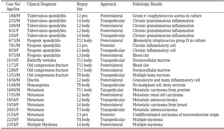
Table 1.Summary of CT-guided Percutaneous Biopsy of Spine and Paravertebral Lesions in 23 Patients Case No/ Clinical Diagnosis B i o p s y A p p r o a c h Pathologic Results
A g e / S e x S i t e
01 / 6 8 / M Tuberculous spondylitis L3 pvs P o s t e r o l a t e r a l Gram(+) staphylococcus aureus in culture 02 / 3 5 / M Tuberculous spondylitis L4 body T r a n s p e d i c u l a r Chronic granulomatous inflammation 03 / 4 0 / M Tuberculous spondylitis L2 body P o s t e r o l a t e r a l Chronic granulomatous inflammation 04 / 3 1 / F Tuberculous spondylitis L2 body P o s t e r o l a t e r a l Chronic granulomatous inflammation 05 / 5 0 / F Tuberculous spondylitis L4 body T r a n s p e d i c u l a r Chronic granulomatous inflammation 06 / 7 3 / M Pyogenic spondylitis T12 body P o s t e r o l a t e r a l β-hemolytic streptococcus group D in culture 07 / 6 1 / M Pyogenic spondylitis L3 pvs P o s t e r i o r Chronic inflammatory cell
08 / 5 9 / F Pyogenic spondylitis L5 body T r a n s p e d i c u l a r Chronic inflammatory cell 09 / 7 5 / M Pyogenic spondylitis L3 body P o s t e r o l a t e r a l Blood clot
1 0 / 1 0 / F Butterfly vertebra T12 body T r a n s p e d i c u l a r Normocellular marrow 1 1 / 7 2 / F Old compression fracture T12 body P o s t e r o l a t e r a l Blood clot
1 2 / 6 8 / F Old compression fracture T11 body P o s t e r o l a t e r a l Normocellular marrow 1 3 / 5 3 / M Old compression fracture T8 body T r a n s p e d i c u l a r Multiple bony necrosis
1 4 / 5 6 / M D i s c i t i s L2 body P o s t e r o l a t e r a l Granulocyte and many inflammatory cell 1 5 / 4 8 / M H e m a n g i o m a T8 body T r a n s p e d i c u l a r No malignant cell, bony necrosis 1 6 / 6 0 / M M e t a s t a s i s T11 body T a n s p e d i c u l a r Metastatic carcinoma from prostate 1 7 / 5 5 / M M e t a s t a s i s L2 body P o s t e r o l a t e r a l Metastatic renal cell carcinoma 1 8 / 5 4 / F M e t a s t a s i s L2 body T r a n s p e d i c u l a r Metastatic adenocarcinoma 1 9 / 3 9 / F M e t a s t a s i s L4 body P o s t e r o l a t e r a l Metastatic carcinoma from breast 2 0 / 4 1 / F M e t a s t a s i s T3 body T r a n s p e d i c u l a r Metastatic adenocarcinoma
2 1 / 5 4 / F M e t a s t a s i s C3 pvs P o s t e r i o r Undifferentiated carcinoma of neuroendocrine origin 2 2 / 2 9 / F M e t a s t a s i s T4 body T r a n s p e d i c u l a r Multiple myeloma
2 3 / 5 4 / F Multiple Myeloma L4 body P o s t e r o l a t e r a l Multiple myeloma pvs: paravertebral soft tissue
1 2 3
Fig. 1. Posterolateral approach to L2 vertebral body in the patient with suspected discitis. The needle traversed obliquely in the vertebral body, passing lateral to the right transverse process. Pathologic result was some granulocytes and many inflammatory c e l l s .
Fig. 2. Transpedicular approach to L1 vertebral body in the patient with suspected hemangioma. The needle passed through the right pedicle and the needle tip was located within the vertebral body.
Fig. 3. Ten-year-old girl. CT scan shows midsagittal cleft and soft tissue interposition in T7 vertebral body suggestive of butterfly vertebra. Pathologic result was normal marrow cells.
서는 후측면 접근법 (Fig. 1)을, 10예에서는 척추경을 통한 접 근법 (Fig. 2)을 통해 시행하였다. 생검은 조직의 육안적 상태 및 병변의 성격에 따라 1 4예에서 2회, 5예에서 3회, 3예에서 1 회 시행하였다. 생검이 끝난 후에는 다시 CT 스캔을 하여 출 혈, 기흉, 골절 등의 합병증 유무를 확인하였다.
생검한 조직은 병리조직학적 결과를 얻었으며, 염증성 병변 이 의심된 예에서는 조직 배양을 함께 시행하였다.
결 과
총 2 3예 중 2 1예( 91% )에서는 생검을 통해 얻은 조직이 종 양, 감염성 질환, 정상 골조직 등과 같은 병리학적 진단이 가 능하였다 (Table 1).
병리 조직 검사에서 나비형 척추체는 정상 골조직 (Fig. 3), 압박 골절 3예 중 2예는 가골 형성과 골괴사, 추간판염은 염 증세포와 과립구를 보여 임상 진단을 뒷받침하였고, 다발성 골수염 1예도 생검 전 임상 진단과 일치하였다. 화농성 척추 염으로 진단한 4예 중 1예는 β-hemolytic streptococcus group D 가 배양되었고 2예에서 만성 염증 세포를 보였으며, 결핵성 척추염은 5예 중 4예에서 만성 육아종성 염증 소견을 보였다.
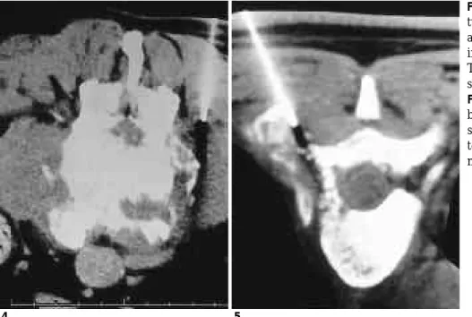
생검 전 결핵성 척추염으로 생각했던 1예는 그람 양성 구균이 배양되어 화농성 척추염으로 진단되었다 (Fig. 4). 전이성 척 추암은 선암종이 2예, 유방의 도관세포암, 신세포암과 신경내 분비 기원의 미분화 암종 및 전립선암이 각각 1예였으며, 다 발성 골수암이 1예였다 (Fig. 5). 혈관종은 조직학적 확진은 할 수 없었으나 악성을 배제할 수 있었다. 화농성 척추염의 1 예와 압박 골절의 1예에서는 혈종만이 나와 진단을 내리기에 부적절하였다.
시술 도중 또는 시술 후에 환자는 심한 동통은 호소하지 않 았고, 출혈, 기흉, 감염과 신경학적 후유증 및 내부 장기 손상 등의 합병증은 없었다.
고 찰
척추 병변의 정확한 진단은 치료 방향의 결정에 필수적이 다. 예를 들면 원발암이 있는 환자에서 병기 결정을 위한 검 사 중 척추에 병변이 발견되었을 때, 이의 정확한 진단은 수 술로 치료 할 것인지, 척추 전이로 판단하여 방사선 치료 또 는 화학적 치료를 할 것인지 결정하는데 매우 중요하다. 또한 척추에 감염성 질환이 있은 경우에도 병원체에 따른 약물의 선택을 위해 병변의 조직학적 진단이 요구된다. 최근 CT 및 자기공명영상술 등의 영상 매체의 발달로 병변의 조기 발견 이 가능해 짐에 따라 이러한 요구는 더욱 증가되고 있다. 그 러나 수술을 통한 생검은 그 자체로 많은 위험과 합병증을 수 반하기 때문에 덜 침습적인 경피적 생검을 위한 방법들이 모 색되었다.
척추의 경피적 생검술은 1 9 3 5년 R o b e r t s o n과 Ball (4)에 의 해 처음 보고되었으며, 이들은 방사선학적 위치 선정 없이 종 양부위로 생검침을 삽입하였다. 1949년 S i f f e r t와 Arkin (5)이 방사선 유도하의 생검을 처음 시도하였으나, 생검침의 위치 선정을 위해 시술 도중 방사선 사진을 얻는 정도에 불과하였 다. 그러나 영상 매체의 발전으로 보다 안전한 경피적 생검을 위한 방법들이 모색되었고 투시하 경피적 생검술의 다양한 기법들이 보고되었다 (6-8). 또한 CT 유도하의 생검은 안전 성과 정확한 위치 선정 등의 이유로 1 9 8 1년 Adapson 등 ( 9 )에 의해 처음 시도된 이후 척추 생검을 위한 일반적인 시술이 되 었으며 높은 진단률을 보이고 있다 (10-12).
과거에는 경추와 흉추 및 주변 연부 조직의 생검은 주변 중 요 장기에 손상을 입히거나 기흉 발생의 가능성 때문에 두려 움과 어려움이 많았다. 그러나 CT 유도하에서의 생검은 병변 과 주변 장기의 해부학적 관계를 정확히 알 수 있고 생검침의 끝과 경로를 볼 수 있게 되어 합병증을 최소화할 수 있게 되 Fig. 4. In 67 year-old-man diagnosed as tuberculous spondylitis in the initial di- agnosis, staphylococcus aureus grew in specimen obtained through biopsy.
Tissue diagnosis confirmed pyogenic s p o n d y l i t i s .
Fig. 5. In 30 year-old woman with back pain and lower leg weakness, CT scan shows soft tissue mass in left pos- terior compartment of T4. Tissue diag- nosis confirmed multiple myeloma.
4 5
었다 (13-15). 이 연구에서도 접근이 어려운 부위로 여겨져 왔 던 상흉추의 2예와 경부의 1예에서도 별 어려움 없이 시술할 수 있었으며 동반된 합병증이 없었다. 또한 요통이 있어 시행 한 검사상 나비형 척추체를 보인 소아에서 다른 병인을 배제 하기 위해 시행한 생검에서도 성인에서와 마찬가지로 어려움 없이 시술이 가능하여 협력 가능한 소아에서 나이가 시술의 금기사항은 되지 않는 것으로 생각된다.
병변으로의 접근 방법에 있어 후측면 접근법은 Craig (7)가 생검침을 이용한 수술적 생검에서 처음 시도한 이래 가장 보 편화되었으며, 이 접근 방법을 통한 척추 및 주위조직의 생검 의 높은 진단률이 보고되었다 (9, 12, 16). 그러나 병변이 척추 체의 중앙이나 전면에 있을 때, 척추 주위 혈종, 기흉 및 신경 학적 합병증 등의 위험이 높아 척추경을 통한 접근법이 새로 이 시도되었고, 그 장점과 유용성이 보고되고 있다 ( 1 7 - 1 9 ) . 이 연구에서는 병변의 위치에 따라 접근 방법을 변화시켰는 데, 즉 척추체 주변의 연부 조직에 있는 병소는 후측면 또는 후면 접근법을, 척추체의 병소는 척추경을 통한 접근법을 시 행하였다. 병소로의 접근 방법은 특별한 기준이 있는 것으로 생각되지 않으며 생검 전 CT 소견을 확인하고 병변의 위치에 따라 결정하는 것이 바람직한 것으로 보인다.
이 연구에서 폐결핵을 앓은 병력과 완만한 증상 발현 등의 임상적 소견을 종합하여 결핵성 척추염으로 진단하고 항결핵 성 약을 투여한 환자에서 생검하여 얻은 검체에서 포도상 구 균이 배양되어 균에 잘 반응하는 항생제로 바꿨으며, 전이성 암으로 진단한 1예에서 병리소견상 다발성 골수암으로 나와 향후 검사 방향과 치료 방침이 변경되었다. 일차성 암이 있었 던 한 예에서는 척추의 다발성 병소가 외상성 골절에 의한 것 인지 또는 전이에 의한 것인지 확진을 요하였고 병리 소견상 골괴사를 보여 골전이를 배제할 수 있었다. 이상에서와 같이 생검은 환자의 향후 치료 방침의 결정에 많은 도움을 줄 수 있다.
결론적으로, CT 유도하 생검은 경부, 흉부 및 요추체와 주 변 연부 조직의 조직학적 진단에 있어 안전하고 간단한 시술 이며 임상적으로 유용한 검사로 사료된다.
참 고 문 헌
1 . Bender CE, Berquist TH. Imaging assisted percutaneous biopsy of the thoracic spine. Mayo Clin Proc 1 9 8 6 ; 6 1 : 9 4 2 - 9 5 0
2 . Bernardino ME. Percutaneous biopsy. AJR 1 9 8 4 ; 1 4 2 : 4 1 - 4 5 3 . Kattapuram SV, Rosenthal DI. Percutaneous biopsy of the cervical
spine using CT guidance. A J R 1987;149:539-541
4 . Robertson RC, Ball RP. Destructive spine lesions: diagnosis by needle biopsy. J Bone Joint Surg[Am] 1935;57:749-758
5 . Siffert RS, Arkin AM. Trephine bone biopsy with special reference to the lumbar vertebral bodies. J Bone Joint Surg[Am] 1 9 4 9 ; 3 1 : 1 4 6 - 149
6 . Mazet R, Cozen L. The diagnostic value of vertebral body needle biopsy. Ann Surg 1952;135:245-252
7 . Craig FS. Vertebral body biopsy. J Bone Joint Surg[Am] 1 9 5 6 ; 3 8 : 9 3 - 1 0 2
8 . Deeley TJ. The drill biopsy of bone lesions Clin Radiol 1 9 7 2 ; 2 3 : 536-540
9 . Adapon BD, Legada BD, Lim EVA, Silao JV, Dalmacio-Cruz A. CT- guided closed biopsy of the spine. J Comput Assist Tomogr 1 9 8 1 ; 5:73-78
1 0 . de Santos LA, Lukeman JM, Wallace S, Murray JA, Ayala AG.
Percutaneous needle biopsy of bone in cancer patient. A J R 1 9 7 8 ; 130:641-649
1 1 . Kattapuram SV, Khurana JS, Rosenthal DI. Percutaneous needle biopsy of the spine. S p i n e 1992;17:561-564
1 2 . Stoker DJ, Kissin CM. Percutaneous vertebral biopsy: a review of 135 cases. Clin Radiol 1 9 8 5 ; 3 6 : 5 6 9 - 5 7 7
1 3 . Valls J, Ottolenghi CE, Schajowicz F. Aspiration biopsy in diagno- sis of lesions of vertebral bodies. J A M A 1 9 4 8 ; 1 3 6 : 3 7 6 - 3 8 2 1 4 . Franker CJ. Aspiration biopsy of the spine. J Bone Joint Surg [Am]
1954; 36:69-74
1 5 . Mclaughlin RE, Miller WR, Miller CW. Quadriparesis after needle aspiration of the cervical spine: report of a case. J Bone Joint Surg [ A m ] 1976; 58:1167-1168
1 6 . Babu NV, Chittaranjan S, Prem H. Computed tomographically guided biopsy of the spine. S p i n e 1994; 19(21):2436-2442
1 7 . Renfrew DL, Whitten CG, Wiese JA, El-Khouri GY, Harris KG.
CT-guided percutanous transpedicular biopsy of the spine.
R a d i o l o g y 1991; 180:574-576
1 8 . 김용훈, 윤정희, 조우호, 차순주, 허감. CT 유도하의경피적척추체 생검: 척추경을통한접근법. 대한방사선의학회지1998; 39:395-398 1 9 . James SJ, Mark JK, Richard G, et al. Percutaneous transpedicular
biopsy of vertebral body lesions. S p i n e 1996; 21:2035-2040
J Korean Radiol Soc 1999;41:1 1 95-1 1 9 9
Address reprint requests to : Won-Hong Kim, M.D., Department of Radiology, Inha University College of Medicine
#7-206, 3rd street, Shinheung-dong, Choong-gu, Inchon, 400-103, Korea.
Tel. 82-32-890-2767 Fax. 82-32-890-2743
Diagnostic Usefulness of CT-guided Pe rcutaneous Biopsy of the Spine
1Kyung Jin Kang, M.D., Won-Hong Kim, M.D., Joon Soo Byun, M.D., Young Kook Cho, M.D., Kyu Jung Cho, M.D.2, Kyung Ho Moon, M.D.2, Eul Hye Seok, M.D.,
Seok Hwan Shin, M.D.3, Heon Han, M.D.
1Department of Radiology, Inha University College of Medicine
2Department of Orthopedics, Inha University College of Medicine
3Department of General Surgery, Inha University College of Medicine
Purpose : To evaluate the diagnostic value of CT-guided percutaneous biopsy of inflammatory and tumorous lesions of the spine and paraspinal soft tissue.
Materials and Methods : Twenty-three patients underwent CT-guided percutaneous biopsy of the spine and paraspinal soft tissue. Tentative clinical diagnoses determined before biopsy were tuberculous spondylitis (n=5), pyogenic spondylitis (n=4), butterfly vertebra (n=1), old compression fracture (n=3), discitis (n=1), hemangioma (n=1), metastasis (n=7) and multiple myeloma (n=1). Biopsy was performed at the following levels: cervical-(n=1), thoracic-(n=9), and lumbar-spine(n=13). The approach to biopsy of the spine and paraspinal soft tissue lesions was posterolateral (n=11), posterior (n=2), or transpedicular (n=10).
Results : Tissue considered adequate by the pathologist involved was obtained in 21 (91%) of the 23 cases. In 19 cases, pathologic findings supported the clinical diagnoses determined before biopsy. In two cases, patho- logic and clinical diagnoses differed. Complications such as severe pain, bleeding, infection, neurologic deficit or damage to internal organs were detected neither during or after the procedure.
Conclusion : CT-guided percutaneous biopsy is a safe and reliable method of obtaining a diagnosis in many cases involving different spinal and paraspinal lesions.
Index words :Spine, biopsy
Computed tomography (CT), guidance Spine, diseases