Coronary artery bypass graft surgery (CABG) has been widely used for patients with multivessel coronary artery disease. The post-operative patency of the bypass
graft is important to predict the clinical outcome (1-3).
Postoperative myocardial infarction is a rare, but poten- tially severe complication after CABG. So, the early recognition of the failure of a bypass graft on the early postoperative imaging is necessary to evaluate the quali- ty of the surgical anastomosis and the graft patency (4, 5).
Conventional coronary angiography (CCA) is current- ly the standard diagnostic modality that’s used to assess the status of bypass grafts. However, it is an invasive
Evaluation of Coronary Artery Bypass Grafts in the Early Postoperative Period Using 64-Slice MDCT
1Yu Mi Jeong, M.D., Jeong Ho Kim, M.D., Chul Hyun Park, M.D.2, Kook Yang Park, M.D.2, Sung Su Byun, M.D., Hee Young Hwang, M.D., Chul Hi Park, M.D., Hyung Sik Kim, M.D.
Departments of Radiology1 and Thoracic and Cardiovascular Surgery2, Gachon University, Gil Hospital
Received July 3, 2008 ; Accepted October 14, 2008
Address reprint requests to : Jeong Ho Kim, M.D., Department of Radiology, Gachon University, Gil Hospital, 1198 Guwol-dong, Namdong- gu, Incheon 405-760, Korea.
Tel. 82-32-460-3063 Fax. 82-32-460-3065 E-mail: [email protected]
Purpose: The aim of this study was to evaluate the accuracy of 64-slice multidetector row CT (MDCT), as compared with conventional coronary angiography (CCA), for as- sessing coronary artery bypass grafts (CABG) in the early (< 1 month) postoperative period.
Materials and Methods: Twenty-four patients underwent both 64-slice MDCT (0.6 mm collimation, 0.37s gantry rotation) and CCA within 15 days after CABG. Sixty-five by- pass grafts (20 venous grafts and 45 arterial grafts) and 67 distal runoff arteries in 24 patients were included in the analysis. The bypass grafts and distal runoffs were evalu- ated for the presence of significant stenosis, including obstruction. The CT findings were compared with the CCA findings.
Results: CCA confirmed that 62 of the 65 grafts were patent and that 3 had significant stenosis or obstruction. Sixty of the 62 patent grafts were correctly identified by MD- CT. One of the three grafts that had significant stenosis was misinterpreted on the MD- CT images. The sensitivity, specificity and positive and negative predictive values for detecting stenosis were 67%, 97%, 50% and 98%, respectively, with an overall diag- nostic accuracy of 95%. MDCT also correctly detected all the stenotic distal runoff ar- teries (4 of 67).
Conclusion: 64-slice MDCT is a promising alternative diagnostic modality for evaluat- ing a CABG in the early postoperative period.
Index words :Tomography, X-ray computed Coronary angiography Coronary artery bypass graft Graft occlusion, vascular
procedure requiring a long procedure time, it can cause graft injury or spasm, and it has the risk of complica- tions (6-10), and especially in the early postoperative pe- riod. Therefore, a less invasive imaging modality would be desirable for evaluating the patency of a graft. MDCT has recently gained increasing acceptance for noninva- sive cardiac imaging. Several recent studies have shown that 64-slice MDCT angiography achieved excellent im- age quality and high diagnostic accuracy for evaluating coronary arterial bypass grafts, as compared with CCA (11-17). However, to the best our knowledge, there have been no studies that have reported on the value of 64-slice MDCT for evaluating bypass grafts in the early postoperative period.
The purpose of this study was to investigate the accu- racy of 64-slice MDCT, as compared with CCA, for the assessment of the patency of a CABG in the early (< 1 month) postoperative period. In addition to the assess- ment of a CABG,we also assessed the distal coronary runoff arteries in this study.
Materials and Methods
Patient Population
From March 2005 to February 2006, eighty one pa- tients underwent CABG at our hospital due to triple ves- sel coronary artery disease. Overall, seventy patients underwent postoperative imaging within 15 days after CABG. Forty six patients (46/70, 65.7%) underwent CCA to assess their graft’s patency. The patients who had a regular heart beat without difficulty when holding their breath participated in this study, and they under- went additional 64-slice coronary MDCT within 7 days (mean: 2 days, range: 1-7 days) after CCA. The patients with a history of renal insufficiency, contrast allergy or atrial fibrillation were excluded. Overall, twenty four patients (13 men and 11 women, mean age: 61.8 years, range: 42 to 74 years) were included in this comparative study. This study was approved by the hospital institu- tional review board, and informed consent was ob- tained from all the patients for the CCA and MDCT.
MDCT Angiography
The scans were performed on a 64-slice multidetector system (Somatom Sensation 64; Siemens, Erlangen, Germany). The patients were positioned in the gantry in the supine position with electrocardiographic leads placed on the anterior thorax to enable a retrospectively gated scan. The scan parameters were 800-900 effective
mA, 120 kV, a gantry rotation time of 0.37s, a detector collimation of 64 × 0.6 mm and a pitch of 0.24. Scans were performed in the craniocaudal direction, with a scan range from the thoracic inlet through the lung bases. For the cases with a gastroepiploic arterial graft, the scan range was expanded into the upper abdomen.
All the scans could be performed within one single breath-hold. A total of 70-100 mL of contrast media (Visipaque 320; Amersham Health, Cork, Ireland) was injected at a rate of 5.0 ml/second, and this was followed by an injection of 40 ml saline at a rate of 5.0 ml/second.
A test bolus technique was used to optimize graft visual- ization. A small amount of contrast material (20 mL) was intravenously injected during the acquisition of a series of dynamic low-dose monitoring scans at the level of the ascending aorta. The scan delay time was determined by calculating the peak time plus 2 seconds. The pa- tients with a heart rate exceeding 70 beats per minute (bpm) received 40 mg-80 mg of oral �- blocker (propra- nolol), unless underlying contraindications were pre- sent. In addition, 0.6 mg oral nitroglycerin was given to all the patients immediately before MDCT scanning.
In order to optimally visualize the coronary arteries, image reconstruction was performed between 30% and 70% relative to the RR interval by using 5% increments.
The best phases with the least cardiac motion for the im- age analysis were determined on the basis of the pre- view series of scans that were reconstructed at 9 differ- ent R-R interval positions. A slice thickness of 0.6 mm with increments of 0.4 mm each and a B25f smooth++
Kernel were used for the reconstruction. The recon- structed data of the MDCT was transferred to a work- station (Wizard; Siemens, Erlangen, Germany) for fur- ther processing.
Table 1. Graft Types
Graft Type n = 65
Arterial Left IMA to the LAD or a diagonal branch 22
(n=45) Left IMA to the OM 01
Right IMA to the LAD or a diagonal branch 09
Right IMA to the OM 07
Right IMA to the ramus intermedius 01
Right GEA to the RCA 02
RA to the OM or the ramus intermedimus 03
Venous SV to the RCA or the PDA 17
(n=20) SV to the LAD 01
SV to the OM 02
IMA = internal mammary artery; LAD = left anterior descending artery; OM = obtuse marginal branch; GEA = gastroepiploic artery; RCA = right coronary artery; RA = radial artery; SV = saphenous vein; PDA = posterior descending artery
Conventional Coronary Angiography
Arterial catheterization and selective conventional an- giography of the coronary arteries and the bypass grafts were performed according to standard techniques.
Quantitative coronary angiography was performed on two orthogonal projections of the coronary arteries (Coroskop, Siemens, Erlangen, Germany) to identify the segments larger than 2.0 mm in diameter. An experi- enced interventional cardiologist and a thoracic surgeon reviewed the angiographic images for stenotic lesions or occlusions in the bypass grafts and in the runoff arteries that were distal to the grafts.
Analysis
Two radiologists working in consensus reviewed the conventional contrast enhanced axial slices, as well as the three-dimensional images, including the volume rendered (VR), curved multiplanar reformatted (MPR) and maximum intensity projection (MIP) images, to evaluate graft patency and the runoff arteries distal to the grafts on the MDCT images. The image quality of an exam was grade as ‘good’, ‘adequate’ or ‘poor’. Each by- pass graft was visually classified as either ‘evaluable’ or
‘unevaluable’, according to the vessel size (>2 mm) or the presence of motion artifacts. The evaluable bypass grafts were assessed for the presence of significant stenosis or occlusion. Significant stenosis was defined as a reduction in the luminal diameter of more than 50%.
All the coronary branches that were supplied by a
patent bypass graft were also assessed for significant lu- minal narrowing. The CT findings were compared with those findings of the CCA. Based on the findings of the CCA, the sensitivity, specificity, positive and negative predictive values and the accuracy of 64-slice MDCT were calculated. The 95% confidence intervals (CIs) were calculated using binomial proportions.
Results
Among the eighty one postoperative patients, fifty sev- en patients were referred to undergo MDCT, but nine patients (9/57, 15.8%) failed to undergo MDCT because of arrhythmia. There were twenty four patients who didn’t undergo CCA, but they did undergo MDCT be- cause they refused to receive invasive procedures. All 24 patients enrolled in this study completed their MD- CT examination successfully without serious complica- tions. The mean heart rate during the scan was 66 bpm (range: 57-86). In 3 of the 24 patients, the cardiac rhythm was regular, but the heart rate was over 70 bpm after the administration of oral β-blockers.
The image quality of each exam was graded as ‘good’
for 12 (50%) examinations and ‘adequate’ for 12 (50%) examinations. No patient was excluded from the analy- sis because of poor image quality.
There were five sequential grafts and eighteen com- posite grafts. Each consecutive anastomosis in the cases of composite grafts was regarded as a separate graft seg-
A B C
Fig. 1. Representative images from a 70-year-old male with a patent left internal mammary artery (LIMA) graft to the left anterior descending artery (LAD).
A, B. The anastomotic site (arrow) is well visualized on a volume rendering image and on a MPR image.
C. The corresponding CCA shows a patent LIMA graft (arrow).
ment. Therefore, the 24 patients had a total of 68 bypass grafts (21 venous grafts and 47 arterial grafts). One of the 68 grafts was classified as ‘unevaluable’ and this was excluded from the analysis due to motion artifact on MDCT. Two of the 68 grafts were excluded because of catheterization failure during CCA, although both of them were well visualized on MDCT; 1 graft was a right gastroepiploic artery grafted to a posterolateral branch, and the other graft was a saphenous vein graft connect- ed to a diagonal branch. So, 65 grafts in 24 patients were finally included in the comparative analysis, of which 45 were arterial grafts and 20 were venous grafts (Table 1).
CCA confirmed that 62 of the 65 grafts were patent and that 3 had significant stenoses or obstruction. Sixty of the 62 patent grafts were correctly identified by MD-
CT (Fig. 1, 2). Two cases of graft stenoses or occlusions were erroneously suspected on MDCT, but this was not present on CCA. The reviewers correctly identified two of the three grafts that had significant stenoses or ob- structions on the CCA (Fig. 3, 4); one was a radial artery graft to an obtuse marginal branch, and the other was saphenous vein graft to the right coronary artery. Only one case of significant stenosis (a right internal mamma- ry artery (RIMA) to the left anterior descending artery (LAD)) was missed on 64-slice MDCT (Fig. 5).
According to the CCA, there was a total of 69 distal coronary runoff arteries supplied by 62 patent grafts. Of these, 68 distal runoffs could be assessed with MDCT, except for one due to motion artifact. Four of them had significant stenosis on the CCA, and CT detected all these lesions (Fig. 6). Therefore, with regard to the as-
A B C
A B
Fig. 3. A 62-year-old male with a radial artery (RA) graft from the LIMA to an obtuse marginal branch (OM).
A. The arterial graft shows total occlu- sion (arrows) from the anastomotic site with the LIMA.
B. The corresponding CCA confirmed the RA graft occlusion (arrows).
Fig. 2. Representative images from a 64-year-old male with a patent saphenous vein graft (SVG).
A. The SVG originated from the ascending aorta.
B, C. The SVG is first anastomosed (black short arrows) to the distal segment of the right coronary artery (RCA) and then it consec- utively jumps to the posterolateral branch (secondary anastomosis) (white long arrow). The findings of the MDCT angiograms con- curred with those of the CCA.
A B C
A B C
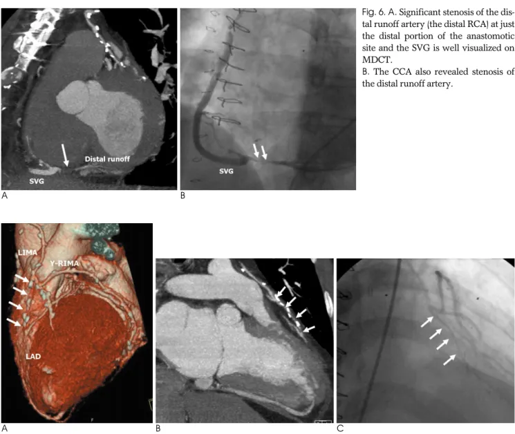
Fig. 5. Mismatch between the MDCT and CCA in a 61-year-old male with a LIMA connected to the LAD via a fragment of the right internal mammary artery (I-RIMA).
A, B. The volume rendered and MIP images show focal discontinuity (arrow) just proximal to a clip. The reviewers considered that it was due to beam-hardening artifact that developed due to a metallic clip and the graft was considered as patent.
C. The CCA shows about 70% stenosis (arrow) at the same site that is the anastomosis between the LIMA and the I-RIMA.
Fig. 4. An example of graft body stenosis in a SVG connected to the distal RCA (d-RCA) in a 59-year-old female. On the MIP and 3- D volume rendering images (A, B), there seems to be significant stenosis of the distal SVG graft with luminal irregularity.
Intraluminal thrombus (white arrow) is suggested by these CT images.
C. The CCA shows sluggish blood flow and a filling defect (black arrows) at the distal graft body, and this is also suggestive of intra- luminal graft thrombus. However, it was initially missed by a cardiologist.
sessment of the distal runoff coronary arteries, our study demonstrated 100% diagnostic accuracy without any cases of false interpretation.
Evaluation of the competitive flow was limited on MDCT. One of the two patent bypass grafts that was er- roneously interpreted as occluded by MDCT was a left internal mammary artery (LIMA) attached to the LAD (Fig. 7). The LIMA was faintly visualized on MDCT, and so it was considered to be a diffusely stenotic graft. The graft was patent with competitive flow between the LI- MA and the LAD on the CCA, although 70% stenosis was present in the LAD.
Using the CCA findings as the standard of reference, the sensitivity and specificity of MDCT for the detection
of significant stenosis or obstruction of bypass grafts were 67% (95% CI: 9.43-99.16%) and 97% (88.83- 99.61%), respectively. The positive and negative predic- tive values were 50% (6.76-93.24%) and 98% (91.20- 99.96%), respectively, with an overall diagnostic accura- cy of 95% (Table 2).
Discussion
The patients with a CABG require follow-up evalua- tions to monitor the patency of their grafts because it is known from previous studies that the clinical outcome of a CABG primarily depends on the graft’s integrity (1- 3). The patency of the bypass graft in the early postoper-
A B
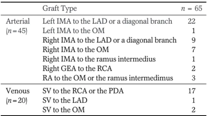
Fig. 6. A. Significant stenosis of the dis- tal runoff artery (the distal RCA) at just the distal portion of the anastomotic site and the SVG is well visualized on MDCT.
B. The CCA also revealed stenosis of the distal runoff artery.
A B C
Fig. 7. A 53-year-old male with a LIMA connected to the LAD.
A. On a volume rendered image, the distal segment of the LIMA is poorly visualized (arrows).
B. An MIP image shows diffuse narrowing (arrows) of the graft from just distal to the junction between the LIMA and the RIMA.
Therefore, we thought that the graft was diffusely stenotic.
C. The CCA shows a patent graft with a relatively small diameter and competitive blood flow.
ative period is also associated with the long-term out- come after CABG (18). Early stenosis or occlusion of the bypass graft is rare, but this may cause significant post- operative complications, including myocardial is- chemia. A study by Berger et al. reported that early stenosis or occlusion was reported in 9.1% of the pa- tients in whom a LIMA was anastomosed to the LAD with a CABG (19). Another study revealed that the im- mediate-term postoperative coronary angiography de- tected graft failure or occluded native vessels in 13 of 40 patients (32.5%), although that study was performed on patients who had clinically suspected myocardial is- chemia after CABG (20).
During the early postoperative period, the primary mechanism of graft failure is thrombosis. Early graft thrombosis is related to technical issues, including fail- ure to reverse the vein graft, a tight suture at the anasto- mosis, poor targets with poor runoff, injury to the graft during harvesting and the occasional grafting of vessels for which the proximal stenosis is less than 70% (2, 21).
Graft closure from thrombosis at one month post- surgery has been reported in 10-15% of cases (2).
However, many patients with occluded grafts have no signs of trouble during the early postoperative period (6). The signs (e.g., electrocardiography alteration and el- evation of the cardiac enzymes) and symptoms (e.g., chest pain and dyspnea) are nonspecific in the postoper- ative condition. Thus, early complications such as graft closure from thrombosis, graft malposition and kinking must be detected by postoperative imaging to plan the
appropriate management such as interventional coro- nary procedures or additional surgery (22-24). So, this current investigation was carried out to assess the diag- nostic accuracy of 64-slice MDCT for the assessment of patients in the early postoperative period after CABG.
Although some non-invasive diagnostic tools such as MDCT and MRI have been reported on, CCA is current- ly considered the gold standard for postoperative imag- ing (23, 25, 26). However, selective catheterization of the coronary artery bypass graft is invasive and it may be difficult; complications can occur during the early postoperative period and these are due to an irregular heart beat, a small vessel size, graft spasm or graft injury (6-8). Coronary artery spasm or graft spasm may severe- ly complicate the immediate postoperative period of pa- tients who underwent CABG, and this can result in cir- culatory collapse or cardiac arrest. The catheterization of the small grafts may initiate or aggravate the graft spasm. On rare ocassions, serious complications of CCA can occur such as conduit dissection, embolism, my- ocardial infarction, stroke and death (7, 9, 10).
Therefore, the use of highly sensitive and non-invasive diagnostic modalities for evaluating bypass grafts would be of great clinical benefit to decrease graft injury and the complications of CCA during the early postoperative period.
MDCT has recently been used for noninvasive imag- ing of coronary artery disease and for determining the patency of bypass grafts. Several studies have reported that 64-slice MDCT achieved good imaging quality and high diagnostic accuracy in assessing coronary artery bypass grafts, as compared with CCA (11-17). However, those studies were performed in the late postoperative setting. In contrast, our study was performed in the ear- ly postoperative setting in which the patients complain of respiratory difficulty or they have irregular heart beats. Therefore, 64-slice MDCT is a convenient tool to evaluate bypass grafts in postoperative patients because it allows more rapid image acquisition than CCA and even 4- or 16-slice MDCT (27).
For the detection of bypass graft stenosis or occlusion after CABG, as compared with CCA, the sensitivity with using 64-slice MDCT has been reported to range from 85% to 100% and the specificity is between 91.4% and 100% (11-17). In this current study, 64-slice MDCT also achieved favorable overall diagnostic accuracy (95%), high specificity (97%) and a high negative predictive val- ue (98%), although the sensitivity (67%) and positive predictive value (50%) were relatively low. This is be- Table 2. Diagnostic Results of MDCT Angiography for the
Bypass Graft Patency, as Compared with Catheter Angiography, in 65 Coronary Artery Bypass Grafts
Graft No. MDCT Angiography* Accuracy
TP TN FP FN (%)
Arterial IMA 40 0 38 1 1 095
GEA 02 0 02 0 0 100
RA 03 1 02 0 0 100
Venous SV 20 1 18 1 0 095
Total** 65 2 60 2 1 095
TP = true positive; TN = true negative; FP = false positive; FN = false negative; IMA = internal mammary artery; GEA = gas- troepiploic artery; RA = radial artery; SV = saphenous vein
* The positive result was defined as significant stenosis or occlu- sion. Significant stenosis means a reduction in the luminal diame- ter of more than 50%.
** Based on these results, the sensitivity, specificity and the posi- tive and negative predictive values are 67% (95% CI: 9.43- 99.16%), 97% (88.83-99.61%), 50% (6.76-93.24%), and 98%
(91.20-99.96%), respectively.
cause of the paucity of true positive cases (only three grafts) in this study. Yet the high negative predictive val- ue suggests that MDCT can rule out the presence of sig- nificant bypass graft failure in postoperative patients.
The 64-slice MDCT is superior to conventional angiog- raphy in that it shows the cardiac structure, the global function of the left ventricle and the coronary vascula- ture, as well as non-cardiac structures (28-30). In addi- tion, not only luminal evaluation, but also demonstra- tion of neointimal plaque or thrombus is possible with the increased spatial resolution of 64-slice MDCT.
During conventional angiography, the direction of blood flow may be changed and it may even be reversed by catheter engagement or by the rate of injecting the contrast material. So CCA may not be a standard of ref- erence in some cases. Therefore, MDCT and CCA should be complementary diagnostic tools for the evalu- ation of postoperative graft patency.
Despite the marked technical improvements, the pre- vious studies have reported that there are still some limi- tations of 64-slice MDCT. First of all, the major draw- back of retrospective ECG-gated MDCT is its high radia- tion dose. A previous study revealed that the mean ef- fective dose for coronary MDCT was significantly high- er than that for CCA (31). Dill et al reported that the mean effective dose for 16-slice coronary MDCT is 2.1 times higher than that for CCA in the patients with by- pass grafts (32). They described the cause of MDCT’s higher radiation dose in bypass patients as the increased length to the through-plane (z-axis), which is particularly needed in patients with IMA grafts. Therefore, for re- ducing the radiation dose, the ECG-correlated tube cur- rent modulation and a reduced tube voltage can be ap- plied (33). Otherwise, image acquisition by prospective ECG-triggering can reduce the radiation exposure while maintaining the diagnostic performance of retrospective ECG-gated MDCT (34).
Evaluating the blood flow is limited with MDCT, which is unlike that for coronary angiography, and the MDCT results for this can be misleading. After CABG, there may be competitive blood flow when an arterial grafts is used to bypass a coronary artery that has only moderate proximal stenosis (35). Alterations in the flow patterns in the graft occur during competitive flow and the total graft flows are notably decreased during com- petitive flow (36). According to these phenomena, the patent graft can be rated as stenosed or occluded on MDCT. Actually, in one of the false positive case in this study, there was competitive blood flow between the LI-
MA graft and the LAD. So, the LIMA graft was pre- sumed to be stenosed on MDCT. Because an arterial graft’s patency decreases as the coronary competitive flow increases (37), another limitation of coronary MD- CT is that it cannot detect the competitive flow. Further, beam hardening artifacts caused by surgical clips or pre- viously inserted stents in the neighboring native vessels can distort the images of 64-slice MDCT.
The limitations of our study need to be addressed.
First, the number of patients included in this study was quite small. The lack of hemodynamically significant stenosis in this series is responsible for the relatively low sensitivity and the low positive predictive value.
Second, there may be a selection bias in this study.
Because the patients with arrhythmia and severe respi- ratory difficulty were not included in this study, good or adequate image quality was achieved in all the examina- tions and MDCT showed results that were comparable to CCA. However, our current study is worthy of note because it was performed in the early postoperative set- ting with using 64-slice MDCT.
Many problems such as respiratory difficulty and a fast, irregular cardiac rhythm are present during the ear- ly recovery period after CABG. Although a small num- ber of patients were studied, 64-slice MDCT achieved favorable diagnostic imaging quality and it showed high diagnostic accuracy and a high negative predictive value as compared the standard invasive CCA. In conclusion, coronary 64-slice MDCT allows physicians to reliable evaluate bypass grafts and the distal arteries, and it can exclude the presence of significant bypass graft failure in the postoperative patients. A future study with a larg- er population of CABG patients will be needed to fur- ther evaluate the value of performing postoperative MDCT angiography for assessing graft patency.
References
1. Zellweger MJ, Lewin HC, Lai S, Dubois EA, Friedman JD, Germano G, et al. When to stress patients after coronary artery by- pass surgery? Risk stratification in patients early and late post- CABG using stress myocardial perfusion SPECT; implications of appropriate clinical strategies. J Am Coll Cardiol 2001;37:144-152 2. Fitzgibbon GM, Kafka HP, Leach AJ, Keon WJ, Hooper GD,
Burton JR. Coronary bypass graft fate and patient outcome: angio- graphic follow-up of 5,065 grafts related to survival and reopera- tion in 1,388 patients during 25 years. J Am Coll Cardiol 1996;28:
616-626
3. Engelmann MG, Knez A, von Smekal A, Wintersperger BJ, Huehns TY, Ho¨fling B, et al. Non-invasive coronary bypass graft imaging after multivessel revascularization. Int J Cardiol 2000;76:
65-74
4. Diegeler A, Matin M, Kayser S, Binner Ch, Autschbach R, Battellini R, et al. Angiographic results after minimally invasive coronary bypass grafting using the minimally invasive direct coro- nary bypass grafting (MIDCAB) approach. Eur J Cardiothoracic Surg 1999;15:680-684
5. Wiklund L, Johansson M, Brandrup-Wognsen G, Bugge M, Rådberg G, Berglin E. Difficulties in the interpretation of coronary angiogram early after coronary artery bypass surgery on the beat- ing heart. Eur J Cardiothorac Surg 2000;17:46-51
6. Zehr JK, Handa N, Bonilla LF, Abel MD, Holmes DR Jr. Pitfalls and results of immediate angiography after off-pump coronary artery bypass grafting. Heart Surg Forum 2000;3:293-299
7. Solodky A, Birnbaum Y, Assali A, Ben Gal T, Strasberg B, Herz I.
Coronary air embolism treated by bubble aspiration. Catheter Cardivasc Interv 2000;49:452-454
8. Suda Y, Otsuka K, Niinami H, Ichikawa S, Ban T, Higashita R, et al. Changes in ultra-low and very low frequency heart rate vari- ability after coronary artery bypass grafting. Biomed Pharmacother 2001;55 Suppl 1:S110-S114
9. Wyman RM, Safian RD, Portway V, Skillman JJ, McKay RG, Baim DS. Current complications of diagnostic and therapeutic cardiac catheterization. J Am Coll Cardiol 1988;12:1400-1406
10. Hung MJ, Kuo LT, Wang CH, Cherng WJ. Irreversible myocardial damage after coronary air embolism - a case report. Angiology 2002;53:213-216
11. Ropers D, Pohle FK, Kuettner A, Pflederer T, Anders K, Daniel WG, et al. Diagnostic accuracy of noninvasive coronary angiogra- phy in patients after bypass surgery using 64-slice spiral computed tomography with 330-ms gantry rotation. Circulation 2006;114:
2334-2341
12. Onuma Y, Tanabe K, Chihara R, Yamamoto H, Miura Y, Kigawa I, et al. Evaluation of coronary artery bypass grafts and native coronary arteries using 64-slice multidetector computed tomogra- phy. Am Heart J 2007;154:519-526
13. Malagutti P, Nieman K, Meijboom WB, van Mieghem CA, Pugliese F, Cademartiri F, et al. Use of 64-slice CT in symptomatic patients after coronary bypass surgery: evalution of grafts and coronary arteries. Eur Heart J 2007;28:1879-1885
14. Feuchtner GM, Schachner T, Bonatti J, Friedrich GJ, Soegner P, Klauser A, et al. Diagnostic performance of 64-slice computed to- mography in evaluation of coronary artery bypass grafts. AJR Am J Roentgenol 2007;189:574-580
15. Jabara R, Chronos N, Klein L, Eisenberg S, Allen R, Bradford S, et al. Comparison of multidetector 64-slice computed tomographic angiography to coronary angiography to assess the patency of coronary artery bypass grafts. Am J Cardiol 2007;99:1529-1534 16. Meyer TS, Martinoff S, Hadamitzky M, Will A, Kastrati A,
Schomig A, et al. Improved noninvasive assessment of coronary artery bypass grafts with 64-slice computed tomographic angiogra- phy in an unselected patient population. J Am Coll Cardiol 2007;49:946-950
17. Dikkers R, Willems TP, Tio RA, Anthonio RL, Zijlstra F, Oudkerk M. The benefit of 64-MDCT prior to invasive coronary angiogra- phy in symptomatic post-CABG patients. Int J Cardiovasc Imaging 2007;23:369-377
18. Halabi A, Alexander J, Shaw L, Lorenz T, Liao L, Kong D, et al.
Relation of early saphenous vein graft failure to outcomes follow- ing coronary artery bypass surgery. Am J Cardiol 2005;96:1254- 1259
19. Berger PB, Alderman EL, Nadel A, Schaff H. Frequency of early occlusion and stenosis in a left internal mammary artery to left an- terior descending artery bypass graft after surgery through a medi-
an sternotomy on conventional bypass. Circulation 1999;100:2353- 2358
20. Alter P, Vogt S, Herzum M, Irqsusi M, Rupp H, Maisch B, et al.
Indications for angiography subsequent to coronary artery bypass grafting. Am Heart J 2005;149:1082-1090
21. Motwani JG, Topol EJ. Aortocoronary saphenous vein graft dis- ease. pathogenesis, predisposition, and prevention. Circulation 1998;97:916-931
22. Vernhet-Kovacsik H, Battistella P, Demaria R, Pasquie JL, Bousquet C, Dogas G, et al. Early postoperative assessment of coronary artery bypass graft patency and anatomy: value of con- trast-enhanced 16-MDCT with restrospectively ECG-gated re- constructions. AJR Am J Roentgenol 2006;186:S395-S400
23. Rasmussen C, Thiis JJ, Clemmensen P, Efsen F, Arendrup HC, Saunama¨ki K, et al. Significance and management of early graft failure after coronary artery bypass grafting: feasibility and results of acute angiography and re-re-vascularization. Eur J Cardiothorac Surg 1997;12:847-852
24. Frazier AA, Qureshi F, Read KM, Gilkeson RC, Poston RS, White CS. Coronary artery bypass grafts: assessment with multidetector CT in the early and late postoperative settings. Radiographics 2005;25:881-896
25. Stanford W, Galvin JR, Skorton DJ, Marcus ML. The evaluation of coronary bypass graft patency: direct and indirect technique other than coronary arteriography. AJR Am J Roentgenol 1991;156:15-22 26. Bartnes K, Sildnes T, Iqbal A, Dahl-Eriksen O, Trovik T, Steigen
TK, et al. Coronary bypass graft patency cannot be determined by multidetector spiral computed tomography. Scand Cardiovasc J 2006;40:83-86
27. Nikolaou K, Knez A, Rist C, Wintersperger BJ, Leber A, Johnson T, et al. Accuracy of 64-MDCT in the diagnosis ischemic heart dis- ease. AJR Am J Roentgenol 2006;187:111-117
28. Salem R, Remy-Jardin M, Delhaye D, Khalil C, Teisseire A, Delannoy-Deken V, et al. Integrated cardio-thoracic imaging with ECG-gated 64-slice multidetector-row CT: initial findings in 133 patients. Eur Radiol 2006;16:1973-1981
29. Pannu HK, Jacobs JE, Lai S, Fishman EK. Coronary CT angiogra- phy with 64-slice MDCT: assessment of vessel visibility. AJR Am J Roentgenol 2006;187:119-126
30. Onuma Y, Tanabe K, Nakazawa G, Aoki J, Nakajima H, Ibukuro K, et al. Noncardiac findings in cardiac imaging with multidetector computed tomography. J Am Coll Cardiol 2006;48:402-406 31. Coles DR, Smail MA, Negus IS, Wilde P, Oberhoff M, Karsch KR,
et al. Comparison of radiation dose from multislice computed to- mography coronary angiography and conventional diagnostic an- giography. J Am Coll Cardiol 2006;47:1840-1845
32. Dill T, Deetjen A, Ekinci O, Mollmann S, Conradi G, Kluge A, et al. Radiation dose exposure in multislice computed tomography of the coronaries in comparison with conventional coronary angiog- raphy. Int J Cardiol 2008;124:307-311
33. Hausleiter J, Meyer T, Hadamitzky M, Huber E, Zankl M, Martinoff S, et al. Radiation dose estimates from cardiac multislice computed tomography in daily practice: impact of different scan- ning protocols on effective dose estimates. Circulation 2006;113:
1305-1310
34. Horiguchi J, Kiguchi M, Fujioka C, Shen Y, Arie R, Sunasaka K, et al. Radiation dose, image quality, stenosis measurement, and CT densitometry using ECG-triggered coronary 64-MDCT angiogra- phy: a phantom study. AJR Am J Roentgenol 2008;190:315-320 35. Sabik JF, Blackstone EH. Coronary artery bypass graft patency
and competitive flow. J Am Coll Cardiol 2008;51:126-128
36. Pagni S, Storey J, Ballen J, Montgomery W, Qaqish NK, Etoch S, et
al. Factors affecting internal mammary artery graft survival: how is competitive flow from a patent native coronary vessel a risk fac- tor? J Surg Res 1997;71:172-178
37. Sabik JF, Lytle BW, Blackstone EH, Khan M, Houghtaling PL, Cosgrove DM. Does competitive flow reduce internal thoracic artery graft patency? Ann Thorac Surg 2003;76:1490-1497
대한영상의학회지 2009;60:303-312
64절편 MDCT를 이용한 관상동맥우회이식편의 수술 후 초기 평가1
1가천의과학대학교 길병원 영상의학과
2가천의과학대학교 길병원 흉부외과
정유미∙김정호∙박철현2∙박국양2∙변성수∙황희영∙박철희∙김형식
목적: 본 연구의 목적은 관상동맥우회술(coronary artery bypass graft, CABG) 후 한 달 이내의 초기 관상동맥 이식편을 평가하는 방법으로 고식적 관상동맥 조영술과 64-다절편 전산화 단층촬영의 정확도를 비교하고자 하였다.
대상과 방법: 24명의 환자가 이식편을 평가하기 위하여 수술 후 15일 이내에 고식적 관상동맥 조영술과 64-다절편 전산화단층촬영을 모두 시행 받았다. 총 65개(정맥이식편 20개, 동맥이식편 45개)의 이식편과 67개의 문합원위부 혈관이 본 연구의 분석에 포함되었다. 64-다절편 전산화 단층촬영으로 이식편과 문합원위부혈관에 의미 있는 협착 이나 폐쇄 소견이 있는지를 평가하고, 고식적 관상동맥 조영술과 그 소견을 비교하였다.
결과: 고식적 관상동맥 조영술 상, 분석에 포함된 총 65개의 이식편 중, 62개의 이식편은 개통성이 좋았으나, 3개의 이식편이 의미있는 협착 또는 폐쇄를 보였다. 64-다절편 전산화 단층촬영상, 62개의 개통된 이식편 중 60개는 고식 적 관상동맥 조영술과 그 소견이 일치하였다. 또한, 의미 있는 협착 또는 폐쇄를 보인 3개의 이식편 중 한 개의 이식 편은 전산화단층촬영에서 협착이 보이지 않았다. 민감도, 특이도, 양성예측도와 음성예측도는 각각 67%, 97%, 50%, 98%였으며, 진단적 정확도는 97%였다. 전산화단층촬영상, 문합원위부혈관의 협착은 모두 발견되었다.
결론: 관상동맥우회술 후 초기 이식혈관을 평가할 때 64-다절편 전산화단층촬영이 유용한 대체 검사방법이 될 것으 로 기대된다.