서론
단일치 및 다수치 상실의 경우에 임플란트는 훌륭한 치료법 으로 자리잡았다. 하지만 기능을 위한 부하를 위해서는 골융 합이 이뤄지기까지 걸리는 수개월의 시간이 걸림돌이 될 때가
많다. 따라서 임플란트의 표면처리를 통해 골융합이 이뤄지기 까지 걸리는 시간을 단축시키려는 많은 연구가 진행되어 왔 다. 그 중에는 임플란트 표면의 물리/화학적인 성질을 변화시 키려는 연구와, 표면에 특정 성장인자를 처리하여 골융합을 앞당기려는 연구가 있다. 전자의 경우에는 거친 표면의 임플 http://dx.doi.org/10.4047/jkap.2014.52.4.298 ORIGINAL ARTICLE
rhTGF-β2/PLGA 복합체를 electrospray법으로 코팅한
타이타늄 임플란트 골 유착의 microCT 계측: a preliminary rabbit study
이우성1∙김성균1*∙허성주1∙곽재영1∙이주희2∙박지만3∙박윤경4
1서울대학교 치의학대학원 치과보철학교실, 2울산대학교 서울아산병원 치과보철과,
3이화여자대학교 이대목동병원 치과보철과, 4서울대학교 치의학대학원 치학연구소
Osseointegration of the titanium implant coated with rhTGF-β β2/PLGA particles by electrospray: a preliminary microCT analyzing rabbit study
Woo-Sung Lee1, Seong-Kyun Kim1*, Seong-Joo Heo1, Jai-Young Koak1, Joo-Hee Lee2, Ji-Man Park3, Yoon-Kyung Park4
1Department of Prosthodontics, Seoul National University Dental Hospital, School of Dentistry, Seoul National University, Seoul, Republic of Korea
2Department of Prosthodontics, Asan Medical Center, College of Medicine, University of Ulsan, Seoul, Republic of Korea
3Department of Prosthodontics, Ewha Womans University, Seoul, Republic of Korea
4Department of Dental Research Institute, Brain Korea 21, Seoul National University, Seoul, Republic of Korea
Purpose: This preliminary rabbit study was conducted to evaluate the effect of recombinant human transforming growth factor-β2 (rhTGF-β2)/poly lactic-co-glycolic acid (PLGA) coating on osseointegration of the titanium (Ti) implant. Materials and methods: Eight Ti implants were anodized with 300 voltages for three minutes. Four of those were coated with rhTGF-β2/PLGA by an electrospray method as the experimental group. The implants were placed into tibiae of four New Zealand rabbits, two implants per a tib- ia, one implant per each group. After 3 and 6 weeks, every two rabbits were sacrificed and micro-computed tomography (microCT) was taken for histomorphometric analy- sis. Results: In scanning electron microscope (SEM) image, the surface of rhTGF-β2/PLGA coated Ti implant showed well distributed particles. Although statistically insignif- icant, microCT analysis showed that experimental group has higher bone volume / total volume (BV/TV) and trabecular thickness (Tb.Th) values relatively. Cross sectional view also showed more newly formed bone in the experimental group. Conclusion: In the limitation of this study, rhTGF-β2/PLGA particles coating on the Ti implant show the possibility of more favorable quantity of newly formed bone after implant installation. (J Korean Acad Prosthodont 2014;52:298-304)
Key words: TGF-beta2; PLGA; Titanium; Implant; Osseointegration
c cc
2014 The Korean Academy of Prosthodontics
This is an Open Access article distributed under the terms of the Creative Commons Attribution Non-Commercial License (http://creativecommons.org/licens- es/by-nc/3.0) which permits unrestricted non-commercial use, distribution, and reproduction in any medium, provided the original work is properly cited.
*Corresponding Author: Seong-Kyun Kim
Department of Prosthodontics, Seoul National University Dental Hospital, School of Dentistry, Seoul National University, 101 Daehak-ro, Jongno-gu, Seoul, 110-744, Republic of Korea +82 2 2072 2661: e-mail, [email protected]
Article history: Received 5 August, 2014 / Last Revision 5 September, 2014 / Accepted 16 September, 2014
* This study was supported by a grant (2011-434) from the Asan Institute for Life Sciences, Seoul, Korea and National Research Foundation of Korea grant funded by the Korea government (No. 2011-0028067), and by a grant of the Korean Health Technology R&D Project, Ministry of Health & Welfare, Republic of Korea (A120304) .
란트,1양극산화 임플란트 표면 처리,2수산화인회석 코팅,3샌드 블라스팅 표면,4샌드블라스팅 후 산부식 처리5등이 있으며 이 중 다수가 이미 시판되어 있다. 후자의 경우는 생물학적인 관 점에서 치유기간을 앞당기려는 노력이며 이것은 임플란트 표 면과 주변 조골세포 사이의 상호작용을 포함한다. Huang 등6은 기능적 부하를 견디기 위해서 임플란트 표면과 조골세포가 강 한 세포 부착을 일으켜야 한다고 발표한 바 있다. 이를 위해 bone morphogenetic protein (BMP), growth hormone (GH), platelet-derived growth factor (PDGF), insulin-like growth factor (IGF), platelet released growth factors (PRGFs), transforming growth factor-beta (TGF-β) 등 여 러 성장인자들이 연구되어 왔다.7
Bosetti 등8은 성장인자들에 대한 in vitro 실험에서 TGF-βisoform 이 인간 조골세포의 증식을 촉진한다고 하였으며, Kim과 Valentini9는 PDGF와 TGF-β2에 대한 인간 조골세포 연구에서 TGF-β2가 단백질 합성을 증가한다고 보고하였다. De Ranieri 등10 은 쥐 실험에서 TGF-β2의 국소적 적용이 임플란트 주변의 골 부 피와bone-implant contact (BIC)의증가를보여준다고보고하였다.
어떤 성장인자를 사용하느냐의 문제와 함께 성장인자를 어 떻게 효과적으로 적용하는지도 중요한 문제로, 단순히 pipet- ting하여 임플란트 표면에 적용시키는 것뿐만 아니라 다양한 전달 시스템이 개발되어왔다. 그중 poly lactic-co-glycolic acid (PLGA)는 생체친화성을 갖고 조직 내에서 서서히 분해되는 성 질을 가진 고분자 화합물로서 성장인자의 유출 속도를 조절할 수 있는 운반체로서 사용되어왔다. Lee11은 PLGA를 운반체로 이용하여 rhBMP를 타이타늄 디스크에 코팅하여 실험한 결과 세포증식효과와 분화속도가 증가함을 발표하였으며, Fan 등12 은 TGF-β3를 PLGA에 고정하여 연골 재생에 대한 실험을 한 결 과 TGF-β3가 연골 재생에 효과적임을 보고하였다.
이에 근거하여 본 실험에서는 rhTGF-β2와 PLGA 복합체를 submicron 크기로 타이타늄 임플란트에 코팅하였을 때 골융합 에 어떤 영향을 미치는지 조사하였다. 본 실험은 선행 연구로, rhTGF-β2와 PLGA 복합체가 동물 실험에서 골융합을 촉진시 킬 수 있다는 가능성을 알아보는데 의의가 있다.
재료 및 방법
1. 타이타늄 임플란트 제작과 rhTGF-β2/PLGA 복합체 표 면 처리
직경 3.75 mm, 길이 7 mm, 나사산 간격 0.6 mm의 grade 4 com- mercially pure titanium 임플란트 8개를 제조하였다(Warantec Co., Seoul, Korea). 임플란트는 0.15 M calcium acetate monohydrate (Ca(CH3COO)2H2O)와 0.02 M calcium glycerophosphate (CaC3H7O6P) 수용액 환경에서 300 V 전압으로 상온에서 3분간 산화과정을 거쳤다. 증류수 세척 후 ethylene oxide (EO) gas로 소독한 후 다음 과정에 따라 각각 4개씩 실험군과 대조군으로 분류하였다.
- 대조군(Control group, C): 표면 처리하지 않은 그룹
- 실험군(Experimental group, E): electrospray법으로 rhTGF-β2/
PLGA 복합체 (3 μg/임플란트)를 코팅한 그룹
실험군 표면처리는, 0.4% w/v 아세톤에 용해되어 있는 PLGA (PURAC Biochem BV, Gorinchem, Holland) 4 mL와 증류수에 25 μg/mL 농도로 용해되어 있는 rhTGF-β2 (Prospec-TechnoGene Ltd., East Brunswick, NJ, USA) 4 mL을 혼합하여 electrospray법으로 코팅하 였다. Electrospray 처리 방법은 기존의 연구에서 시행했던 대로 진행하였다.13,14전압은 17kV, 바늘 끝에서 임플란트까지의 거 리는 6.0 cm, 유출 속도는 24 μL/min이었으며, 임플란트당 10분씩 표면처리하였다. 실험군과 대조군의 임플란트 표면을 scanning electron microscopy (FE-SEM, Hitachi S-4700, Tokyo, Japan)으로 관 찰하였다.
2. 동물 실험과 임플란트 식립
무게 3 - 3.5kg인 4마리의 New Zealand white mature rabbit이 실험 에 사용되었다. 실험은 동물윤리위원회의 승인을 거쳐 진행되 었다(SNU-140129-3, Institutional Animal Care and Use Committee, School of Dentistry, Seoul National University). 수술 시에는 10 mg/kg Zoletil (Vibac, Carros, France)와 0.15 mL/kg Rompun (Bayer Korea, Ansan, Korea) 을 근주(intramuscular)하여 전신마취하였고, mesial proximal tibiae 를 면도하고 소독하였다. 2% lidocaine solution (1 : 100,000 Epinephrine) (Yu-han Co., Seoul, Korea)으로 국소마취 후 절개하여 proximal tibiae를 노출시키고 제조회사의 설명대로 2개의 임플란트를 식립하였다. 식립 후 근육과 피부를 나누어 봉합하여 primary closure를 얻었다.
3. MicroCT 촬영과 계측
MicroCT 촬영 및 계측을 위해 2마리는 3주, 2마리는 6주 후 희 생하였다.15희생은 전신마취하 KCl 정주로 시행하였다. 임플 란트와 주변 골을 en bloc으로 얻은 후 formalin으로 고정하고 microCT (SkyScan 1173, Kontich, Belgium)를 촬영하였다. 촬영의 해 상도는 8.17 μm이며 2240 × 2240 pixel 이미지를 얻은 후 분석하 였다. 계측시 새로 형성된 골은 임플란트 표면 주변 0.5 mm로 제한하였으며,16기존 피질골에 닿아 있는 부분은 제외하였다.
또한 양측 cortical bone 중 더 낮은 지점의 경계로부터 2번째 thread에서 cross section하여 주변 골의 양을 관찰하였다(Fig. 1). 새 로 형성된 해면골을 분석하기 위한 parameter로는 bone volume per total volume (BV/TV), trabecular thickness (Tb.Th), trabecular number (Tb.N), structure model index (SMI)를 사용하였다. MicroCT scanning 에서 위의 측정값들에 대한 정의 및 분석방법은 Bouxsein 등17이 발표한 최근의 가이드라인을 이용하였다. 통계 처리는 비모수 적 방법인 Mann-Whitney test를 사용하여 3주와 6주로 분리하여 진행하였다. 통계 프로그램은 SPSS를 사용하였다.
결과
대조군과 실험군의 임플란트 표면은 SEM 사진으로 확인하 였다(Fig. 2). 대조군의 임플란트에서 양극 산화된 임플란트 표 면 위로 고르게 분산된 rhTGF-β2/PLGA submicron 입자들을 관찰
할 수 있다.
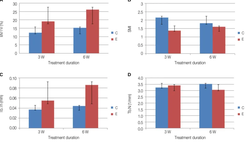
Table 1은 microCT 계측을 시행한 결과이다. 분석시 새로 형성 된 해면골은 임플란트 표면에서부터 0.5 mm 거리까지로 제한 하였다. Sample size가 작아서 통계적으로 유의한 차이를 보일 수 는 없었지만, BV/TV, Tb.Th에서 실험군이 대조군보다 높은 값을 나타내는 경향성을 확인할 수 있다(Fig. 3). 반면 SMI값은 실험군 이 대조군보다 작았으며, Tb.N은 비슷하였다. SMI는 해면골의 구조를 나타내는 수치로, 판상골(plate-like bone)이 막대형골(rod- like bone)보다 작은 값으로 나타나며, 수치는 약간 음수의 값 (plate-like)에서 약 3(rod-like)까지 나타날 수 있다.16
Fig. 4는 각각의 군에 대한 microCT의 cross sectional view이다. 3 주 대조군에서는 약하게 형성된 해면골이 관찰되는 반면, 3주 실험군에서는 더 많은 양의 해면골이 형성되어있음을 관찰할 수 있다. 6주의 경우 대조군, 실험군 모두 3주에 비해 더 많은 양 의 골이 형성되었음을 볼 수 있으며, 실험군에서 대조군보다 성숙한 골이 임플란트 표면 주변으로 더 많이 형성되었음이 확인되었다.
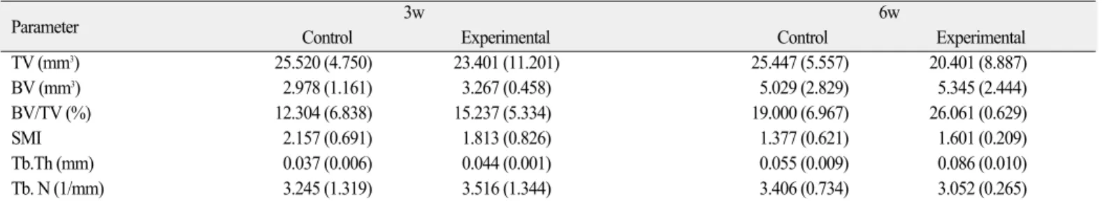
Table 1. MicroCT analysis, mean (SD)
Parameter 3w 6w
Control Experimental Control Experimental
TV (mm3) 25.520 (4.750) 23.401 (11.201) 25.447 (5.557) 20.401 (8.887)
BV (mm3) 2.978 (1.161) 3.267 (0.458) 5.029 (2.829) 5.345 (2.444)
BV/TV (%) 12.304 (6.838) 15.237 (5.334) 19.000 (6.967) 26.061 (0.629)
SMI 2.157 (0.691) 1.813 (0.826) 1.377 (0.621) 1.601 (0.209)
Tb.Th (mm) 0.037 (0.006) 0.044 (0.001) 0.055 (0.009) 0.086 (0.010)
Tb. N (1/mm) 3.245 (1.319) 3.516 (1.344) 3.406 (0.734) 3.052 (0.265)
Fig. 1. Schematic image of implant. (A) Region of interest (ROI) for microCT, (B) reference for cross sectional view.
A B
Fig. 2. SEM image of implant surface (×5,000). (A) Control group (implant without surface coating), (B) Experimental group (implant with rhTGF-β2/PLGA coating via electrospray method).
A B
Fig. 3. MicroCT analysis. (A) BV/TV (bone volume per total volume), (B) SMI (structure model index), (C) Tb.Th (trabecular thickness), (D) Tb.N (trabecular number), C (control), E (experimental) (P>.05).
30 25 20 15 10 5 0
3 W 6 W
BV/TV (%) C
E
Treatment duration
3 2.5 2 1.5 1 0.5 0
3 W 6 W
SMI C
E
Treatment duration
0.10 0.08 0.06 0.04 0.02 0.00
3 W 6 W
Tb.Th (mm) C
E
Treatment duration
4.0 3.5 3.0 2.5 2.0 1.5 1.0 0.5
0.0 3 W 6 W
Tb.N (1/mm)
C E
Treatment duration
A B
C D
Fig. 4. Cross sectional view of microCT. (A) 3 weeks control, (B) 3 weeks experimental (rhTGF-β2/ PLGA coated implant), (C) 6 weeks control, (D) 6 weeks experimental (rhTGF-β2/ PLGA coated implant).
A B
C D
고찰
임플란트 식립 후에 나타나는 골 치유 반응은 여러 단계 과 정에 의해 일어나는 복잡한 일련의 현상으로, 각 단계마다 여 러 유전자의 발현 및 단백질의 기능에 의해 좌우된다.18그 중 TGF-β는 bone matrix component의 합성 및 분해 과정에 개입하여 골이 형성되는 것을 조절하는 중요한 역할을 담당한다.19TGF-β 는 세포 성장을 조절하며,20-23골막(periosteal) 및 골내막(endosteal) 골-유래 세포 환경(bone-derived cell culture)에서 조골세포를 분화 하게끔 하는데 영향을 미친다.19,24,25TGF-β2의 in vivo 실험에 대한 보고를 보면, De Ranieri 등10은 쥐에서 TGF-β2의 국소적 적용이 임플란트 주변의 골 부피와 BIC의 증가를 보여준다고 보고한 바 있다. 한편 Sena 등26은 쥐에서 TGF-β2의 국소적 적용이 임플 란트 주변의 골 부피는 증가시키지만, 고농도의 TGF-β2의 경우 (20 μg)에는 오히려 BIC가 감소하고 strength of fixation 역시 TGF- β2를 처리한 그룹에서 감소한다고 보고하였다. 언급된 논문에 서는 TGF-β2를 처리할 때, 단순히 임플란트 표면에 TGF-β2 용액 을 pipetting하여 처리한 것으로, 임플란트 식립 후에 혈류에 의 해서 빠르게 씻겨나갈 것이라는 추측을 해볼 수 있다. 따라서 본 실험에서는 생체 내에서 독성 없이 서서히 분해되면서 rhTGF-β2를 유출시킬 수 있는 운반체로서 PLGA를 선택하였 으며, rhTGF-β2의 농도 역시 기존 논문에서 언급된 농도보다 작은 농도를 선택하였다. 이는 rhTGF-β2/PLGA 복합체가 생체 내에서 서서히 유리된다면, 더 적은 농도의 rhTGF-β2로도 같은 효과를 낼 수 있을 것이라는 가정 때문이다.
PLGA는 친수성인 poly glycolic acid (PGA)와 소수성인 poly lactic acid (PLA)의 합성체로 PGA와 PLA의 비율을 조절하여 방출 속도를 결정할 수 있다.27PGA와 PLA의 비율을 1 : 1로 할 경우 가수분해에 의하여 약 5 - 20일 사이에 합성체가 분해되어 성장 인자를 모두 방출시킬 수 있게 된다.28결과적으로 PLGA가 분해 되면서 함께 방출되는 rhTGF-β2가 임플란트 표면에서 작용하 여 초기 치유 과정을 앞당기고, 골 성장 반응 속도를 증가시킬 수 있는 환경을 만들게 된다.
본 실험에서 사용한 코팅 방법인 electrospray법을 통해, 코팅되 는 rhTGF-β2/PLGA 복합체의 입자를 submicron 크기로 조절하여 임플란트 식립시 입자가 골 계면에서 떨어져 나가는 것을 감 소시킬 수 있다. Catledge 등29은 줄기간엽세포의 부착을 위해서 작용하는 세포 물질의 크기가 나노 수준이며, 따라서 임플란 트 표면 역시 나노 수준에서 조절해야 효과적이라고 보고하였 다. 신체에서 가장 작은 모세혈관 크기인 5 μm 보다 작은 submi- cron 입자 코팅을 통해 세포내 확산 및 대사가 쉽고,30임플란트 에 적용하였을 때 주위 세포에 빠르게 영향을 미칠 수 있을 것 으로 사료된다.
본 실험은 선행 연구로, 최소한의 동물 희생을 통해 rhTGF-β 2/PLGA 복합체가 골 반응에 미치는 영향을 알아보고자 하였다.
따라서 경향성을 위주로 본 실험의 결과를 해석하였으며 이는 연구의 표본 조사를 통하여 이루어졌다. Table 1 및 Fig. 3에서 확
인할 수 있듯이, rhTGF-β2/PLGA 복합체로 코팅한 실험군의 경 우 BV/TV 및 Tb.Th가 대조군에 비해서 우수한 결과를 보였다.
이는 5 - 20일 사이에 분해되도록 한 rhTGF-β2/PLGA 복합체에 의 해서 유리된 rhTGF-β2가 작용하여 초기 치유 과정을 앞당긴 것 으로 추측해볼 수 있다. 형성된 골의 양적인 측면은 BV/TV를 통해서, 형성된 골의 성상은 Tb.Th를 통해서 예상할 수 있다.
양쪽 parameter를 종합해 볼 때 rhTGF-β2에 의해서 더 많은 양의 골이, 그리고 더 두꺼운 양상의 해면골이 형성될 수 있음을 짐 작할 수 있다. Tb.N은 1 mm 안에 얼마나 많은 수의 해면골의 곁 가지가 존재하는 지를 나타내는 수치로, 본 실험에서는 실험 군과 대조군의 큰 차이를 발견할 수 없었다. 비슷한 Tb.N을 가 지면서 더 높은 Tb.Th를 갖는 실험군의 경우에는, 해면골의 곁 가지의 수가 비슷하지만 각각의 가지의 두께는 더 두꺼운 것 으로 해석할 수 있다. SMI가 실험군에서 더 작은 수치를 보인 것은 실험군의 해면골 대조군에 비해 좀 더 판상(plate-like)이라 는 것을 의미한다.
본 실험을 통해 제한적이지만 rhTGF-β2/PLGA 복합체가 초기 골 유착을 앞당기고 골의 양적 성장을 야기할 수 있는 예비결 과를 확인하였다. 초기 골 유착을 앞당길 수 있는 최소한의 rhTGF-β2 농도 및 성숙된 골을 만드는 정확한 메커니즘은 추후 의 연구가 필요할 것으로 사료된다. 또한 통계적으로 유의미 한 차이를 증명하기 위한 추후의 실험이 필요하다.
결론
본 실험은 rhTGF-β2/PLGA 복합체를 electrospray법으로 코팅한 임플란트가 골 유착에 미치는 영향을 알아보기 위해 토끼를 모델로 실험하였다. SEM 사진으로부터 rhTGF-β2/PLGA 복합체 가 임플란트 표면에 균일하게 분포함을 확인하였으며, microCT 계측 결과 통계적으로 유의하지는 않으나 실험군이 대조군보 다 더 높은 BV/TV, Tb.Th 값을 갖는 경향성을 보였다. Cross sec- tional view에서도 실험군 임플란트 주변으로 더 많은 신생골이 형성되었음을 확인하였다. 본 선행 연구를 통해 electrospray법으 로 rhTGF-β2/PLGA 복합체를 코팅한 임플란트가 초기 골 유착을 앞당기고 골의 양적 성장을 촉진시키는 경향성을 보임을 확인 하였다.
References
1. Hermann JS, Cochran DL, Nummikoski PV, Buser D. Crestal bone changes around titanium implants. A radiographic evaluation of unloaded nonsubmerged and submerged implants in the ca- nine mandible. J Periodontol 1997;68:1117-30.
2. Sul YT, Johansson CB, Petronis S, Krozer A, Jeong Y, Wennerberg A, Albrektsson T. Characteristics of the surface oxides on turned and electrochemically oxidized pure titanium implants up to dielectric breakdown: the oxide thickness, micropore con- figurations, surface roughness, crystal structure and chemical com-
position. Biomaterials 2002;23:491-501.
3. Xie J, Baumann MJ, McCabe LR. Osteoblasts respond to hy- droxyapatite surfaces with immediate changes in gene expres- sion. J Biomed Mater Res A 2004;71:108-17.
4. Ivanoff CJ, Hallgren C, Widmark G, Sennerby L, Wennerberg A. Histologic evaluation of the bone integration of TiO(2) blasted and turned titanium microimplants in humans. Clin Oral Implants Res 2001;12:128-34.
5. Orsini G, Assenza B, Scarano A, Piattelli M, Piattelli A. Surface analysis of machined versus sandblasted and acid-etched titanium implants. Int J Oral Maxillofac Implants 2000;15:779-84.
6. Huang HH, Ho CT, Lee TH, Lee TL, Liao KK, Chen FL.
Effect of surface roughness of ground titanium on initial cell ad- hesion. Biomol Eng 2004;21:93-7.
7. Junker R, Dimakis A, Thoneick M, Jansen JA. Effects of implant surface coatings and composition on bone integration: a systematic review. Clin Oral Implants Res 2009;20:185-206.
8. Bosetti M, Boccafoschi F, Leigheb M, Cannas MF. Effect of dif- ferent growth factors on human osteoblasts activities: a possible application in bone regeneration for tissue engineering. Biomol Eng 2007;24:613-8.
9. Kim HD, Valentini RF. Human osteoblast response in vitro to platelet-derived growth factor and transforming growth fac- tor-beta delivered from controlled-release polymer rods.
Biomaterials 1997;18:1175-84.
10. De Ranieri A, Virdi AS, Kuroda S, Shott S, Leven RM, Hallab NJ, Sumner DR. Local application of rhTGF-beta2 enhances peri- implant bone volume and bone-implant contact in a rat model.
Bone 2005;37:55-62.
11. Lee SY. Effect of poly(D,L-lactide-co-glycolide)/bone mor- phogenic protein-2 coating of anodized titanium surface on osteoblast-like cells. MS Thesis. In: Korea, Seoul University, 2010.
12. Fan H, Tao H, Wu Y, Hu Y, Yan Y, Luo Z. TGF-β3 immobilized PLGA-gelatin/chondroitin sulfate/hyaluronic acid hybrid scaf- fold for cartilage regeneration. J Biomed Mater Res A 2010;95:982- 92.
13. Woosley JP, Turnbull RJ, Kim K. Field injection electrostatic spray- ing of liquid hydrogen. J Appl Phys 1988;64:4278-84.
14. Kim K, Turnbull RJ. Generation of charged drops of insulating liquids by electrostatic spraying. J Appl Phys 1976;47:1964-9.
15. Park IP, Kim SK, Lee SJ, Lee JH. The relationship between ini- tial implant stability quotient values and bone-to-implant contact ratio in the rabbit tibia. J Adv Prosthodont 2011;3:76-80.
16. Xu B, Zhang J, Brewer E, Tu Q, Yu L, Tang J, Krebsbach P, Wieland M, Chen J. Osterix enhances BMSC-associated os- seointegration of implants. J Dent Res 2009;88:1003-7.
17. Bouxsein ML, Boyd SK, Christiansen BA, Guldberg RE, Jepsen KJ, Mu¨ller R. Guidelines for assessment of bone mi-
crostructure in rodents using micro-computed tomography. J Bone Miner Res 2010;25:1468-86.
18. Sims NA, Gooi JH. Bone remodeling: Multiple cellular interactions required for coupling of bone formation and resorption. Semin Cell Dev Biol 2008;19:444-51.
19. Centrella M, McCarthy TL, Canalis E. Current concepts review : transforming growth factor-beta and remodeling of bone. J Bone Joint Surg 1991;73A:1418-28.
20. Centrella M, McCarthy TL, Canalis E. Transforming growth fac- tor beta is a bifunctional regulator of replication and collagen syn- thesis in osteoblast-enriched cell cultures from fetal rat bone. J Biol Chem 1987;262:2869-74.
21. Lomri A, Marie PJ. Bone cell responsiveness to transforming growth factor beta, parathyroid hormone, and prostaglandin E2 in nor- mal and postmenopausal osteoporotic women. J Bone Miner Res 1990;5:1149-55.
22. Lomri A, Marie PJ. Effects of transforming growth factor type beta on expression of cytoskeletal proteins in endosteal mouse osteoblastic cells. Bone 1990;11:445-51.
23. Machwate M, Jullienne A, Moukhtar M, Lomri A, Marie PJ. c- fos protooncogene is involved in the mitogenic effect of trans- forming growth factor-beta in osteoblastic cells. Mol Endocrinol 1995;9:187-98.
24. Robey PG, Young MF, Flanders KC, Roche NS, Kondaiah P, Reddi AH, Termine JD, Sporn MB, Roberts AB. Osteoblasts syn- thesize and respond to transforming growth factor-type beta (TGF- beta) in vitro. J Cell Biol 1987;105:457-63.
25. Rosen DM, Stempien SA, Thompson AY, Seyedin SM.
Transforming growth factor-beta modulates the expression of os- teoblast and chondroblast phenotypes in vitro. J Cell Physiol 1988;
134:337-46.
26. Sena K, Sumner DR, Virdi AS. Effect of recombinant human trans- forming growth factor-beta2 dose on bone formation in rat femur titanium implant model. J Biomed Mater Res A 2010;92:1210- 7.
27. Agrawal CM, Niederauer GG, Athanasiou KA. Fabrication and Characterization of PLA-PGA Orthopedic Implants. Tissue Eng 1995;1:241-52.
28. Yoo SY, Kim SK, Heo SJ, Koak JY, Lee JH, Park YK, Kim E.
A study of mesenchymal stem cell proliferation and surface char- acteristics of the titanium discs coated with MS275/PLGA by an electrospray. J Korean Acad Prosthodont 2012;50:285-91.
29. Catledge SA, Vohra YK, Bellis SL, Sawyer AA. Mesenchymal stem cell adhesion and spreading on nanostructured biomateri- als. J Nanosci Nanotechnol 2004;4:986-9.
30. Hans ML, Lowman AM. Biodegradable nanoparticles for drug delivery and targeting. Curr Opin Solid State Mater Sci 2002;6:
319-27.
ORIGINAL ARTICLE
rhTGF-β2/PLGA 복합체를 electrospray법으로 코팅한
타이타늄 임플란트 골 유착의 microCT 계측: a preliminary rabbit study
이우성1∙김성균1*∙허성주1∙곽재영1∙이주희2∙박지만3∙박윤경4
1서울대학교 치의학대학원 치과보철학교실, 2울산대학교 서울아산병원 치과보철과,
3이화여자대학교 이대목동병원 치과보철과, 4서울대학교 치의학대학원 치학연구소
목적: 본 선행 연구는 recombinant human transforming growth factor-β2 (rhTGF-β2)/ poly lactic-co-glycolic acid (PLGA) 복합체를 타이타늄 임플란트에 처 리하였을 때 골 유착에 미치는 영향을 알아보기 위해 시행된 것으로 토끼 모델을 사용하였다.
재료 및 방법: 8개의 임플란트를 300V에서 3분 동안 양극 산화하였다. 그 중 4개는 electrospray법으로 rhTGF-β2/PLGA를 코팅하여 실험군으로 설정하 였다. 4마리의 New Zealand rabbit의 tibiae에 1개씩의 실험군과 대조군 임플란트를 식립하였으며, 3주와 6주에 2마리씩 희생하여 micro-computed tomog- raphy (microCT) 촬영 후 분석하였다.
결과: Scanning electron microscope (SEM) 사진에서 rhTGF-β2/PLGA 입자가 임플란트 표면에 균일하게 분산되어 있음을 확인하였다. MicroCT 분석 결 과 통계적으로 유의하지는 않지만 rhTGF-β2/PLGA를 처리한 임플란트가 bone volume/total volume (BV/TV)와 trabecular thickness (Tb.Th) 값이 더 높은 경향성을 보였으며, cross sectional view에서 더 많은 골이 형성되었음을 확인하였다.
결론: rhTGF-β2/PLGA 표면처리된 임플란트가 주변 골의 양적 성장을 촉진시킬 수 있으며 임플란트 초기 골 유착을 증진시킬 수 있는 가능성을 보였 다. (대한치과보철학회지 2014;52:298-304)
주요단어: TGF-beta2; PLGA; 타이타늄; 임플란트
*교신저자: 김성균
110-744 서울시 종로구 대학로 101 서울대학교 치과병원 치과보철과 02-2072-2661: e-mail, [email protected]
원고접수일: 2014년 8월 5일 / 원고최종수정일: 2014년 9월 5일 / 원고채택일: 2014년 9월 16일
2014 대한치과보철학회
이 글은 크리에이티브 커먼즈 코리아 저작자표시-비영리 3.0 대한민국 라이선스에 따라 이용하실 수 있습니다.
c cc
* 이 논문은 아산 생명과학재단의 연구기금 (2011-434), 보건복지부의 Korean Health Technology R&D Project (A120304) 및 한국연구재단 (NRF)의 지원 을 받아 연구되었음 (No. 2011-0028067).