서론
골유착성 치과 임플란트는 1965년 스웨덴의 Bra�nemark에 의해 처음 개발된 이후 임상적, 실험적 연구를 통해 성공적인 치료 법으로 인정받아1-3오늘날 치과 분야에서 단일 치아 결손이나 부분 무치악 환자, 완전 무치악 환자의 치료에 널리 사용되고 있다.
임플란트의 성공과 실패는 임플란트 식립 시의 초기 고정과 치유과정 중에 얻어지는 골유착에 의해 결정된다. Bra�nemark 등4,5은 골유착을 광학현미경의 확대 수준에서 임플란트의 표 면과 살아있는 골의 직접적인 접촉이라고 정의하였다.
Albrektsson 등6은 임상적인 의미에서 골유착을 이종물질의 증상 이 없는 견고한 고정을 얻는 것과 기능교합 하에서 골이 유지 되는 것이라고 정의하였다. 골유착은 임플란트의 안정성을 위 한 필수 요소이며, 이를 통해 임플란트가 기능 하중을 성공적 으로 받을 수 있게 한다.7
임플란트가 기능 하중을 받기 전에 골유착에 실패한 것을 초 기 실패(early failure)라 하며, 이는 임플란트 시술 후 수 주 또는 수 개월 내에 나타나고, 임플란트가 기능 하중을 받은 후에 골유 착이 실패한 경우를 후기 실패(late failure)라 한다.8,9임플란트의 초기 실패와 후기 실패를 감별해 내기 위해서는 임플란트의
골유착 상태를 확인해야 한다. 골유착을 정확하게 평가하는 방법으로는 광학현미경이나 전자현미경을 이용하여 조직학 적으로 임플란트 표면과 인접한 골조직이 직접 접촉하는 것을 관찰하는 것이나,10,11임상에서 이러한 방법을 실제로 적용할 수는 없으므로 임상적으로 골유착 상태를 평가하기 위한 진단 방법들이 소개되었다. Adell 등12은 임상에서 골유착을 평가하 는 가장 신뢰할 만한 방법은 방사선학적으로 임플란트 주위의 변연골의 흡수량을 평가하는 방법과 임플란트 동요도를 검사 하는 방법이라고 하였다. 그러나 방사선학적 방법은 진단의 정확성이 떨어지며,13방사선학적 검사상 정상이더라도 여러 가지 임상조건들과 함께 임플란트의 골유착을 신중히 진단해 야 한다.14이에 대한 보완책으로 임플란트 안정성을 객관적인 수치로 측정하기 위해 자기공명장치를 이용한 Osstell� (Integration Diagnostics AB, Gothenburg, Sweden)을 이용하여 초 기 고정과 골유착 정도를 평가하는 것이 이용되고 있다.15
임플란트 성공률에 영향을 끼칠 수 있는 요소에는 임플란트 의 가공소재나 디자인, 표면 처리 등과 같은 임플란트 자체요 소들이 있다.16전 세계적으로 약 95 - 96% 이상 임플란트 시술 성 공률의 평준화가 이루어지고 있으며, 생물학적 적합성과 숙주 의 골유도 능력을 증가시키기 위하여 많은 종류의 표면 처리 방법들이 제안되고 있다.17-22보다 높은 성공률과 장기적인 예후
*교신저자: 김김진진욱욱
700-705 대구광역시 중구 달구벌대로 2175 경북대학교 치의학전문대학원 구강악안면외과학교실 053-600-7575: e-mail, [email protected] 원고접수일: 2012년 7월 12일 / 원고최종수정일: 2012년 7월 18일 / 원고채택일: 2012년 7월 19일
부분 무치악 환자에서 마그네슘 이온주입 임플란트의 성공률에 대한 전향적 임상연구
최수정1∙유정호1∙이규복1∙김진욱2*
경북대학교 치의학전문대학원1치과보철학교실, 2구강악안면외과학교실
연구 목적: 부분 무치악 환자에서 Mg titanate implant (M Implant system, Shinhung, Korea)의 보철 후 1년간의 방사선사진을 이용한 변연골 흡수량과 Osstell�을 이용한 임플란트 안정성 평가 결과를 분석하여 임상적인 성공률에 대해 알아보고자 한다.
연구 재료 및 방법: 38명의 환자에 79개의 임플란트를 식립하여 보철 후 1년 동안 변연골 흡수량, 임플란트의 동요도, 임플란트 식립 위치에 따른 분포 및 성공률, 보철 전후 에 따른 성공률을 분석했다.
결과:변연골의 흡수량은 보철물 장착 후 1년간의 평가에서 상악에서는 평균 1.537 mm, 하악에서는 평균 1.172 mm의 변연골 흡수가 관찰되었으며, 전체적인 상하악의 평균 변연골 흡수량은 1.255 mm로 관찰되었다. 수술 후 시간이 경과함에 따라 ISQ값은 미약하게 감소하였다가 증가하는 경향을 보였으나, 상악과 하악 모두에서 식립 당일 이후 예비 인상일, 최종 보철물의 장착일과 주기적인 평가 기간 동안 ISQ값의 큰 증가나 감소를 나타내지는 않았다. 성공률은 상악에서의 94.12%였고, 하악에서의 98.39%였다.
결론: 본 실험의 결과로 미루어 Mg titanate implant는 임상적으로 양호한 결과를 보여주었다. (대한치과보철학회지 2012;50:176-83) 주요단어: 마그네슘 타이타늄산 임플란트; 변연골 소실; 치아동요도; 임플란트 성골률; 전향적 연구
를 위한 새로운 패러다임의 접근 항목으로 임플란트 표면 처 리, 즉 표면 활성화의 기술개발에 주목하고 있는데, 표면 처리 는 초창기 machined surface에서 rough surface, 이후 다양한 표면 코 팅 및 처리 기법이 등장했다. 예를 들어 전기 산화를 이용해 티 타늄 표면에 산화층 및 다공성의 표면을 형성시켜 그 속으로 골이 성장하여 견고한 고정을 얻을 수 있게 하는 양극산화법 이 많이 연구되고 있으며, 표면의 화학성분을 변경하여 골과 의 화학적 결합을 유도하는 방식으로서 HA (Hydroxyapatite) 코팅 법, CMP (Calcium metaphosphate) 코팅법, HF (Hydrofluoric acid) 코 팅법 및 Mg 코팅법 등 다양한 방법들이 연구되고 있다. 이 가운 데 본 연구에 사용하고자 하는 임플란트는 Mg를 표면 처리한 Mg titanate implant로 그 특징은 다음과 같다.
① 표면처리는 Mg이온을 첨가한 신물질로 빠른 골유착을 유도한다.
② 3.5mm의 microthread 패턴부위가 골흡수를 최소화하고 초기 고정력을 증가시킨다.
③ Conical seal design (11�)이 screw loosening을 방지하여 안정적 인 보철물의 유지가 가능하다.
④ One connection, three platform이 고정체 직경과 상관없이 보 철물의 호환이 가능 하게 함으로써 편의성이 증대되었다.
⑤ Vertical, conical, tapered등 종류별로 다양한 고정체 직경과 길 이를 구비하여 다양한 임플란트 식립 체계를 갖추었다.
⑥ 3개의 cutting edge설계로 self-tapping으로 인해 임플란트 식 립이 용이하고, 골 저항 성을 최소화하였다.
본 연구에서는 부분 무치악 환자에서 Mg titanate implant (M Implant system, Shinhung, Korea)의 2년 6개월간의 방사선사진을 이 용한 변연골 흡수량과 Ostell�을 이용한 동요도 평가 결과를 분 석하여 임상적인 성공률에 대해 알아보고자 한다.
연구 재료 및 방법 1. 연구 대상
경북대학교 치과병원 임상시험 센터에서 2007년 9월부터 모 집한 피험자 가운데 당뇨나 갑상선 질환 등의 내분비 질환이 없는 비흡연자 중 파노라마와 CT상 별도의 골이식없이 임플 란트 식립이 가능한(잔존 치조골 높이 10 mm 이상, 치조골 폭 6 mm 이상) 환자를 임상시험 대상자로 선정하였다. 모두 44명이 선정되었으며 임플란트 식립 후 2년 6개월간 조사하였다. 그 중에서 One stage surgery, Screw type prosthesis에 한정시킨 임상시 험 protocol과 다르게 Two stage surgery, Cement type prosthesis로 치 료되었거나 재소환에 응하지 않은 환자를 제외한 총 38명의 환 자가 이번 연구의 대상이 되었다. 그 중 16명이 남자, 22명이 여 자였고, 이들의 평균 나이는 49.1세였으며, 총 식립한 임플란트 의 개수는 79개였다. 본 연구는 임상시험심사위원회(Institutional Review Board)의 승인을 받았으며(승인번호: 의연 74005-1008), 피 험자들의 서면 동의하에 진행하였다.
2. 연구 방법
1) 임상시험 protocol
① 임상시험 대상자가 선정되면 임플란트 식립 시 임플란트 의 유지력이 없어 고정되지 않는 경우를 제외하면 모든 임플란트는 one stage surgery를 원칙으로 한다.
② 임플란트 식립 후 상악은 6개월, 하악은 3개월 후 치근단 방사선 사진 촬영, ISQ 측정, 보철물 제작을 위한 인상을 채득한다.
③ 임플란트 상부구조는 screw type으로 제작하고 보철물 제 작 후 1, 3, 6, 12개월 마다 치근단 방사선 사진 촬영, ISQ 측 정(상부구조물을 제거하고 transducer를 연결 후 측정)을 시행한다.
2) 변연골의 흡수량
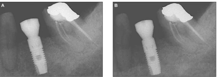
환자 개개인마다 customized XCP bite를 제작한 후 방사선 촬영 을 하여 변연골 흡수량을 측정하였다(Figs. 1 - 3).
방사선 사진의 판독은 동일한 악안면 방사선 전문의가 시행 하였으며, 사용한 임플란트의 smooth surface의 길이는 3.5 mm로 동일하다는 것을 기준으로 하여 계산하였다. 먼저 방사선 사
Fig. 2. Image of installing customized resin bite in XCP.
Fig. 1. Manufacture of customized resin bite.
진 상의 smooth surface의 길이를 측정한 후, smooth surface 최상방 점을 기준으로 변연골의 높이를 정한 다음, 수학적 방정식을 이용하여 실제 변연골의 높이를 구하였다. 이후 촬영된 일련 의 방사선 사진은 동일한 방법으로 실제 변연골의 높이를 구 하였으며, 그 차이를 계산하여 소실량을 정하였다.
Adobe Photoshop CS (Adobe Systems Incorporated, San Jose, California, USA) 프로그램 상에서 계측 도구를 이용하여 소실량 을 측정하였고, 포토샵 이용 시 사진의 어그러짐이 없는 한도 내에서 최대한 확대를 하여 분석하였으며, 포토샵을 이용한 계측 후 수학적 방정식을 이용함으로써 사진 촬영 시 방사선 사진의 확대와 관구의 수직적 오차를 최대한 보정하려고 노력 하였다.
보철 후 1, 3, 6, 12개월 이후의 치근단 방사선 사진을 촬영하여 변연골 높이의 변화를 측정하여 변연골 소실량을 평가하였으 며, 수직골 상실이 저작기능 최초 1년까지는 1.5 mm 미만, 저작 기능 1년 후부터 연간 0.2 mm 미만인 경우를 성공적인 것으로 평가하였다.
3) 임플란트의 안정성 평가
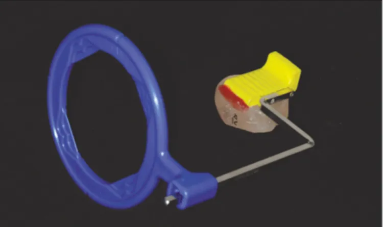
안정성 평가는 임플란트 식립 직후와 보철직전 및 부하를 가 한 후 1, 3, 6, 12개월에 Osstell�의 측정값(ISQ)으로 평가하였다 (Fig. 4).
이 방법은 1996년 Meredith 등23에 의해 도입되었으며, 여기에 이용되는 작은 transducer는 임플란트의 지대주에 수직으로 부착 되고, screw에 의해 연결된다. Transducer는 L-shaped cantilever beam을 가지며, frequency response analyzer에 의해 분석된다. L-shaped beam의 수직 부위의 piezoelectric crystal이 대략 2 - 20Hz 주파수로 임플란트와 transducer의 복합체를 흔들게 되면, 두 번째 piezo- electric crystal이 resonance frequency peak를 감지하여 analyzer가 implant stability quotient (ISQ)로 전환하게 된다. 이 값을 식립 초기 와 비교하여 지속적인 감소가 없는 경우 성공으로 평가한다.
4) 임플란트 식립 위치에 따른 분포 및 성공률
식립 위치에 따른 성공률을 상악 소구치부, 상악 대구치부, 하악 소구치부, 하악 대구치부의 4부위로 나누어 평가하였다.
A B
Fig. 3. A: Insertion, B: After 3 months. Periapical radiography with the use of customized XCP bite.
Fig. 4. Evaluation of implant mobilization with the use of Osstell�.
상악 소구치부위에 8개, 상악 대구치부위에 9개의 임플란트가 식립되었고, 하악 소구치부위와 대구치부위에는 각각 11개, 51 개의 임플란트가 식립되었다.
5) 보철 전후에 따른 성공률
임플란트의 실패는 임플란트 식립 이후 최종 보철물 장착 전 까지 발생하는 초기 실패와 보철물 장착과 교합력 부하 이후 발생하는 후기 실패로 구분한다. 그리하여 본 연구에서도 실 패시기에 따른 성공률을 관찰하였다. 임플란트의 초기 실패는 임플란트 수술시 과도하게 골이 상해를 받은 경우, 세균 감염, 초기 고정이 부족한 경우에 나타날 수 있고, 후기 실패는 임플 란트 보철의 실패, 임플란트 파절, 골유착의 소실, 점진적인 변 연골의 소실 등에 의해 나타날 수 있다.24
6) 통계처리
상악과 하악의 소구치부와 구치부에 따른 성공률을 비교하 는 데에는 교차분석방법을 사용하였고, 유의수준은 95% 신뢰 구간(α=.05)을 설정하여 Fisher's exact test 검정법으로 유의성을 검 증하였다. 통계처리 과정에는 SPSS (version 17.0 for windows, SPSS Inc., Chicago, IL, USA) 프로그램을 이용하였다.
결과
1. 시기별 상하악 평균 변연골 흡수량
주기적으로 촬영한 치근단 방사선 사진으로 변연골의 흡수 량을 측정하였다. 보철물 장착 후 1년간의 평가에서 상악에서 는 평균 1.537 mm, 하악에서는 평균 1.172 mm의 변연골 흡수가 관찰되었으며, 전체적인 상하악의 평균 변연골 흡수량은 1.255 mm로 관찰되었다(Fig. 5, Table 1).
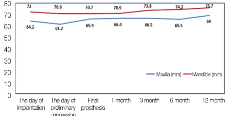
2. 시기별 상하악 평균 ISQ 값
수술 후 시간이 경과함에 따라 ISQ값은 미약하게 감소하였 다가 증가하는 경향을 보였으나, 상악과 하악 모두에서 식립 당일 이후 예비 인상일, 최종 보철물의 장착일과 주기적인 평 가 기간 동안 ISQ값의 큰 증가나 감소를 나타내지는 않았다 (Fig. 6, Table 2).
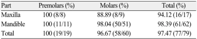
3. 임플란트 식립 부위에 따른 성공률
상악과 하악에서 대구치 부위에 식립된 임플란트 가운데 각
Table 1. Mean marginal bone loss in maxilla and mandible
Time The removal of suture silk The final impression taking After construction of final prosthesis
1 month 3 months 6 months 12 months
Maxilla (mm) 0 0.594 0.839 1.136 1.247 1.537
Mandible (mm) 0 0.404 0.651 0.803 1.000 1.172
Total (mm) 0 0.448 0.695 0.878 1.058 1.255
Table 2. Mean ISQ value in maxilla and mandible according to time
Time The day of The day of
Final prosthesis After construction of final prosthesis
implantation preliminary impression 1 month 3 months 6 months 12 months
Maxilla 64.2 61.2 65.9 66.4 66.5 65.5 69.0
Mandible 72.0 70.6 70.7 70.9 73.8 74.2 75.7
Total 70.4 68.5 69.6 70.1 72.3 72.5 74.2
Fig. 5. Mean marginal bone loss in maxilla and mandible. Fig. 6. Mean ISQ value in maxilla and mandible according to time.
1.8 1.6 1.4 1.2 1.0 0.8 0.6 0.4 0.2 0
The removal of suture silk
The final impression taking
1 month 3 month 6 month 12 month Maxilla (mm) Mandible (mm)
80 70 60 50 40 30 20 10 0
Maxilla (mm) Mandible (mm)
The day of implantation
The day of preliminary impression
Final prosthesis
1 month 3 month 6 month 12 month
각 하나의 임플란트가 실패하였다. 상악에서의 성공률은 94.12%였고, 하악에서의 성공률은 98.39%였다. 소구치 부위에 서는 100%의 성공률을 나타내었고, 대구치부위에서는 96.67%
의 성공률을 나타내었으나 두 부위 사이의 통계적인 유의성은 관찰되지 않았다(Table 3).
4. 보철 전후에 따른 성공률
이번 연구에서 상악과 하악 대구치부에 식립된 임플란트가 각각 한 개씩 실패하였으며 모두 초기 고정의 실패가 원인이 었다. 임플란트에 부하를 가한 후에는 1년 동안 실패가 관찰되 지 않았다.
고찰
최근의 연구에서 임플란트의 산화막에 Mg 성분이 포함된 경우 표면의 다기공성 형상을 통해 기공 속의 골형성 단백질 의 부착과 골조직 성장을 유도해서 기계적 결합력을 강화시킨 다는 결과를 보여주었다.25-27그리고 생활성 표면 화학 물질로 구성된 임플란트의 경우 임플란트와 골 계면간의 생화학적 골 결합을 유도함으로써 빠른 골유착 속도와 신생골 형성 속도를 보여 주었다.25-28특히, Sul 등28은 Mg 이온의 농도가 8.3 - 9.3%일 때 다른 농도에서 보다 통계적으로 유의성 있게 높은 회전 제거 력을 가진다고 보고하였으며, 위와 같은 결과의 주요 원인으 로서 임플란트 표면의 MgTiO을 매개로 하는 화학적인 작용을 들고 있다. 그러나 이러한 이점에도 불구하고 전향적인 임상 연구가 보고된 바가 적어 본 실험에서는 Mg 이온으로 표면 처 리한 Mg titanate implant를 사용하여 보철 후 1년 간 임상적인 성공 률에 관한 연구를 계획하여 수행하였다.
총 79개의 임플란트 가운데 2개가 실패하여 전체적인 임플란 트의 성공률은 97.47%였다. 또한 실험 기간에 따른 변연골의 흡수량을 평가하고 비교하기 위하여 일련의 방사선 사진을 촬 영한 결과 보철물 장착 후 1년의 경과 관찰 기간 중 단지 1.255 mm의 변연골의 흡수만이 관찰되었다. Albrektsson 등16에 의하면 임플란트 보철 후 동요도의 증가가 없고, 치근단 수직골 상실 이 저작기능 최초 1년까지 1.5 mm 미만, 저작기능 1년 후부터는 연간 0.2 mm 미만인 경우 임플란트의 임상적 성공으로 간주하 고 성공률이 85% 이상인 경우 유효성이 있다고 평가하므로, 이 번 연구에서 관찰된 변연골의 흡수량은 Mg titanate implant가 임
상적에서 성공적으로 이용될 수 있음을 보여준다.
임플란트의 안정성과 골유착을 평가하는 몇 가지 다양한 방 법들 가운데 공진주파수 분석(Resonance Frequency Analysis, RFA)이 생체 내 임플란트의 안정성을 정량적으로 측정하는데 유용한 방법으로 인정되고 있다.29,30RFA는 두 가지의 영향인 자, 즉 골 상방에 있는 임플란트의 길이(effective implant length), 임 플란트 주위 골의 강직도(stiffness)에 따라 RF값이 변한다.23RFA 는 임플란트와 주위 조직 계면간의 강직도와 안정성의 변화를 평가할 수 있으며, 임플란트의 성공과 실패를 구별할 수 있다.
그리고 긴 치유기간이 지난 임플란트는 조직학적으로 보다 견 고한 골유착을 나타내며, RF값 또한 높게 나오며, 이는 조직학 적 골유착 정도와 높은 상관관계를 나타낸다.31임플란트 주위 골의 흡수가 약 3 mm 정도 이상으로 진행되는 경우에 RF가 유 의차가 존재할 정도로 감소하며, 이는 골흡수의 정도와 임플 란트의 안정성을 평가하는데 민감도가 우수하다고 할 수 있 다.32현재 RFA를 정량화하여 임플란트의 안정성을 평가하고, 변환기와 임플란트 종류별로 RF값에 영향을 줄 수 있는 인자 들을 보상한 측정방법인 Osstell�(Integration Diagnostics AB, Gothenburg, Sweden)이 상용화 되었으며, 이 측정값인 ISQ값이 RF 값을 대신하여 사용되고 있다.15이 실험에서 상하악 모두에서 공진주파수 분석(RFA) 결과 ISQ값의 큰 변화가 없는 것으로 보 아 임플란트의 초기 안정성이 잘 유지되고 있는 것으로 사료 된다.
Sul 등33의 연구에 의하면 Mg titanate implant는 Tiunite implant와 Osseotite implant와의 비교에서도 우수한 결과를 보인다. 식립 후 3주 후, 6주 후 회전 제거력의 측정값을 비교하면, Tiunite implant보다는 조금 높으며 Osseotite implant에 비해서는 월등히 높 은 값이 측정된다. 3주 후 측정치는 27.1 (Mg), 15.4 (Os), 21.3 (Ti) Ncm이며, 6주 후 측정치는 37.5 (Mg), 21.4 (Os), 36.4 (Ti) Ncm이었 다. 새로운 골조직 형성에 있어서도 Mg titanate implant가 Tiunite implant와 Osseotite implant보다 높은 비율로 새로운 골형성이 이 루어졌다. 특히 Osseotite implant의 6주 후 측정값은 Mg titanate implant의 3주 후 측정값보다 낮은 수치를 보였다. 수치를 살펴 보면 3주 후는 29 (Mg), 15 (Os), 18 (Ti)%이며, 6주 후는 39 (Mg), 26 (Os), 31 (Ti)%를 보였다.
Mg titanate implant와 machine-turned implant를 비교한 다른 연구34 에서도 ISQ값은 각각 67.4와 64.0이었으며, 회전 제거력은 19.2와 10.1 Ncm로서 Mg titanate implant가 더 높은 값을 나타내어 본 연 구와 유사한 결과를 보여주었다.
이 연구는 임상시험을 통한 전향적 연구로서 환자마다 맞춤 형 resin bite를 제작하여 표준화된 치근단 방사선사진을 획득하 여 일정한 촬영각을 가지도록 노력하였으며, 각 시기마다 ISQ 를 측정하여 기능 중 임플란트의 안정성에 대한 좀 더 정확한 정보를 획득하고자 하였다. 그러나 보다 객관적인 분석을 위 해서는 다른 임플란트와의 비교연구가 필요하며, 성공여부의 판단을 위해 주변 치은 반응의 평가 및 환자만족도 등 보다 다 양한 평가 요소를 도입할 필요가 있을 것으로 사료된다.
Table 3. Survival rates according to implant insertion site
Part Premolars (%) Molars (%) Total (%)
Maxilla 100 (8/8) 88.89 (8/9) 94.12 (16/17) Mandible 100 (11/11) 98.04 (50/51) 98.39 (61/62) Total 100 (19/19) 96.67 (58/60) 97.47 (77/79)
% (No. of success/ No. of implants)
결론
본 연구는 2007년 9월부터 2010년 2월까지 경북대학교 병원에 서 실시된 Mg titanate implant에 대한 전향적 임상 연구로서 다음 과 같은 결론을 얻었다.
1. 총 38명의 환자에게 식립된 79개의 임플란트 중에서 2개 가 제거되어 성공률은 97.47%로 나타났다.
2. 식립 위치에 따른 성공률은 상악과 하악은 각각 94.12%, 98.39%이다. 그 중 소구치부는 100%, 대구치부는 96.67%의 값을 나타내었으나, 통계적으로 유의한 차이를 보이지는 않았다.
3. 임플란트가 실패한 시기를 보철 전과 후로 나누었을 때, 실 패한 임플란트는 모두 보철 전 초기 고정의 실패로 인한 것이었다.
4. 보철물 장착 후 1년간의 평가에서 변연골 흡수량은 전체 1.255mm로 양호한 결과를 보이며, 또한 상악과 하악 모두 ISQ값의 큰 증가나 감소는 나타나지 않았으므로 초기 안 정성이 유지된다고 평가할 수 있다.
본 실험의 결과로 미루어 Mg titanate implant는 임상적으로 양 호한 결과를 보여주었다.
참고문헌
1. Adell R, Lekholm U, Rockler B, Bra�nemark PI. A 15-year study of osseointegrated implants in the treatment of the eden- tulous jaw. Int J Oral Surg 1981;10:387-416.
2. Albrektsson T, Zarb G, Worthington P, Eriksson AR. The long- term efficacy of currently used dental implants: a review and pro- posed criteria of success. Int J Oral Maxillofac Implants 1986;
1:11-25.
3. Cox JF, Zarb GA. The longitudinal clinical efficacy of os- seointegrated dental implants: a 3-year report. Int J Oral Maxillofac Implants 1987;2:91-100.
4. Bra�nemark PI, Hansson BO, Adell R, Breine U, Lindstro¨m J, Halle′n O, Ohman A. Osseointegrated implants in the treat- ment of the edentulous jaw. Experience from a 10-year period.
Scand J Plast Reconstr Surg Suppl 1977;16:1-132.
5. Bra�nemark PI. Introduction to osseointegration. In Bra�nemark PI, Zarb GA, Albrektsson T. eds.: Tissue-Integrated Prosthese.
Osseointegration in Clinical Dentistry. Chicago; IL; Quintessence Publishing Co, Inc.; 1985.
6. Albrektsson TO, Johansson CB, Sennerby L. Biological as- pects of implant dentistry: osseointegration. Periodontol 2000 1994;4:58-73.
7. Zarb GA, Albrektsson T. Osseointegration: A requiem for the pe- riodontal ligament? Int J Periodont Res Dent 1991;11:88-91.
8. Albrektsson T, Dahl E, Enbom L, Engevall S, Engquist B, Eriksson AR, Feldmann G, Freiberg N, Glantz PO, Kjellman O, et al. Osseointegrated oral implants. A Swedish multicenter study of 8139 consecutively inserted Nobelpharma implants. J Periodontol 1988;59:287-96.
9. Adell R, Eriksson B, Lekholm U, Bra�nemark PI, Jemt T. Long- term follow-up study of osseointegrated implants in the treatment of totally edentulous jaws. Int J Oral Maxillofac Implants 1990;
5:347-59.
10. Johansson C, Albrektsson T. Integration of screw implants in the rabbit: a 1-year follow-up of removal torque of titanium implants.
Int J Oral Maxillofac Implants 1987;2:69-75.
11. Tjellstro¨m A, Jacobsson M, Albrektsson T. Removal torque of osseointegrated craniofacial implants: a clinical study. Int J Oral Maxillofac Implants 1988;3:287-9.
12. Adell R, Lekholm U, Rockler B, Bra�nemark PI, Lindhe J, Eriksson B, Sbordone L. Marginal tissue reactions at osseointegrated titanium fixtures (I). A 3-year longitudinal prospective study. Int J Oral Maxillofac Surg 1986;15:39-52.
13. Zarb GA, Schmitt A. The longitudinal clinical effectiveness of osseointegrated dental implants: the Toronto study. Part III:
Problems and complications encountered. J Prosthet Dent 1990;64:185-94.
14. Sunde′n S, Gro¨ndahl K, Gro¨ndahl HG. Accuracy and precision in the radiographic diagnosis of clinical instability in Bra�nemark dental implants. Clin Oral Implants Res 1995;6:220-6.
15. Barewal RM, Oates TW, Meredith N, Cochran DL. Resonance frequency measurement of implant stability in vivo on im- plants with a sandblasted and acid-etched surface. Int J Oral Maxillofac Implants 2003;18:641-51.
16. Albrektsson T, Bra�nemark PI, Hansson HA, Lindstro¨m J.
Osseointegrated titanium implants. Requirements for ensuring a long-lasting, direct bone-to-implant anchorage in man. Acta Orthop Scand 1981;52:155-70.
17. Carlsson L, Ro¨stlund T, Albrektsson B, Albrektsson T. Removal torques for polished and rough titanium implants. Int J Oral Maxillofac Implants 1988;3:21-4.
18. Gotfredsen K, Nimb L, Hjo¨rting-Hansen E, Jensen JS, Holme′n A. Histomorphometric and removal torque analysis for TiO2-blast- ed titanium implants. An experimental study on dogs. Clin Oral Implants Res 1992;3:77-84.
19. Cordioli G, Majzoub Z, Piattelli A, Scarano A. Removal torque and histomorphometric investigation of 4 different titanium surfaces: an experimental study in the rabbit tibia. Int J Oral Maxillofac Implants 2000;15:668-74.
20. Klokkevold PR, Nishimura RD, Adachi M, Caputo A.
Osseointegration enhanced by chemical etching of the titanium surface. A torque removal study in the rabbit. Clin Oral Implants Res 1997;8:442-7.
21. Wennerberg A, Albrektsson T, Andersson B, Krol JJ. A histo- morphometric and removal torque study of screw-shaped titanium implants with three different surface topographies. Clin Oral Implants Res 1995;6:24-30.
22. Ferguson SJ, Langhoff JD, Voelter K, von Rechenberg B, Scharnweber D, Bierbaum S, Schnabelrauch M, Kautz AR, Frauchiger VM, Mueller TL, van Lenthe GH, Schlottig F.
Biomechanical comparison of different surface modifications for dental implants. Int J Oral Maxillofac Implants 2008;23:1037-46.
23. Meredith N, Alleyne D, Cawley P. Quantitative determination of the stability of the implant-tissue interface using resonance fre- quency analysis. Clin Oral Implants Res 1996;7:261-7.
24. Tonetti MS, Schmid J. Pathogenesis of implant failures.
Periodontol 2000 1994;4:127-38.
25. Sul YT, Byon E, Wennerberg A. Surface characteristics of electrochemically oxidized implants and acid-etched implants:
surface chemistry, morphology, pore configurations, oxide thickness, crystal structure, and roughness. Int J Oral Maxillofac Implants 2008;23:631-40.
26. Sul YT, Jeong Y, Johansson C, Albrektsson T. Oxidized, bioac- tive implants are rapidly and strongly integrated in bone. Part 1- experimental implants. Clin Oral Implants Res 2006;17:521-6.
27. Sul YT, Johansson C, Byon E, Albrektsson T. The bone response of oxidized bioactive and non-bioactive titanium implants.
Biomaterials 2005;26:6720-30.
28. Sul YT, Johansson C, Wennerberg A, Cho LR, Chang BS, Albrektsson T. Optimum surface properties of oxidized im- plants for reinforcement of osseointegration: surface chem- istry, oxide thickness, porosity, roughness, and crystal structure.
Int J Oral Maxillofac Implants 2005;20:349-59.
29. Sennerby L, Meredith N. Resonance frequency analysis: mea- suring implant stability and osseointegration. Compend Contin Educ Dent 1998;19:493-8, 500, 502.
30. Meredith N, Book K, Friberg B, Jemt T, Sennerby L. Resonance frequency measurements of implant stability in vivo. A cross-sec- tional and longitudinal study of resonance frequency measure- ments on implants in the edentulous and partially dentate max- illa. Clin Oral Implants Res 1997;8:226-33.
31. Rasmusson L, Meredith N, Cho IH, Sennerby L. The influence of simultaneous versus delayed placement on the stability of ti- tanium implants in onlay bone grafts. A histologic and bio- mechanic study in the rabbit. Int J Oral Maxillofac Surg 1999;28:224-31.
32. Huang HM, Chiu CL, Yeh CY, Lee SY. Factors influencing the resonance frequency of dental implants. J Oral Maxillofac Surg 2003;61:1184-8.
33. Sul YT, Johansson C, Albrektsson T. Which surface properties enhance bone response to implants? Comparison of oxidized mag- nesium, TiUnite, and Osseotite implant surfaces. Int J Prosthodont 2006;19:319-28.
34. Sul YT, Johansson P, Chang BS, Byon ES, Jeong Y. Bone tissue responses to Mg-incorporated oxidized implants and machine- turned implantsin the rabbit femur. J Appl Biomater Biomech 2005;3:18-28.
The success rate of Mg-incorporated oxidized implants in partially edentulous patients: a prospective clinical study
Su-Jung Choi1, DDS, Jung-Ho Yoo1, DDS,
Ku-Bok Lee1, DDS, MSD, PhD, Jin-Wook Kim2*, DDS, MSD, PhD
1Department of Prosthodontics, 2Department of Oral and Maxillofacial Surgery, School of Dentistry, Kyungpook National University, Daegu, Korea
Purpose: This study examined the clinical success rate of Mg titanate implants (M Implant system, Shinhung, Korea), which employ a Mg coating method, by evaluating the marginal bone loss and implant stability using radiographs and Osstell�, over a 1 year. Materials and methods: The locations of the implants placement were divided into 4 areas; the maxillary and mandibular premolars and molars. In the maxilla, 8 and 9 implants were inserted in the premolar and molar areas, respectively. In the mandible, 11 and 51 implants were inserted in the premolar and molar areas. Marginal bone loss and ISQ of all implants (79) were measured after insertion, mounting the prosthetic appliance, and 1, 3, 6, and 12 months after loading. The marginal bone loss was measured from the radiograph using XCP bite, which was customized, and the implant stability measured using Osstell�. Fisher's exact test (α=.05) was used to compare the success rates of each region. Results: The mean marginal bone loss for the upper and lower jaws were 1.537 mm and 1.172 mm. The mobility showed a non-significant reduction or increase according with time. The success rates were accounted for 94.12% and 98.39% in the upper and lower jaws; the premolars and molars were accounted for 100% and 96.67%. The two cases of early failure resulted from failure of primary stability during implant inser- tion. The late failures were not observed for 1 year after adding a loading to the implants. Conclusion: The Mg titanate implant showed good primary stability and good clin- ical results in both healing and function. (J Korean Acad Prosthodont 2012;50:176-83)
Key words: Mg titanate implant; Marginal bone loss; Implant stability; Implant success rate; Prospective clinical evaluation
*Corresponding Author: Jin-Wook Kim
Department of Oral and Maxillofacial Surgery, School of Dentistry, Kyungpook National University, 2175, Dalgubeoldaero, Daegu, 700-705, Korea +82 53 600 7575: e-mail, [email protected]
Article history
Received July 12, 2012 / Last Revision July 18, 2012 / Accepted July 19, 2012 ORIGINAL ARTICLE