Levoatriocardinal Vein
Combined with Pulmonary Venous Varix Mimicking
Arteriovenous Malformations:
A Case Report
동정맥기형으로 오인되었던 폐정맥정맥류를 동반한 Levoatriocardinal 정맥: 증례 보고
Joo Hee Jeun, MD1 , Eun-Ju Kang, MD1* , Jeong-Hyun Jo, MD2 , Ki-Nam Lee, MD1
1Department of Radiology, Dong-A University Medical Center, Dong-A University College of Medicine, Busan, Korea
2Department of Radiology, Daedong Hospital, Busan, Korea
The levoatriocardinal vein is an uncommon pulmonary venous abnormality that connects the left atrium or pulmonary vein with the systemic vein. It is distinct from partial anomalous pul- monary venous return in that the former forms a connection with the left atrium through the normal pulmonary vein whereas the latter involves pulmonary venous drainage to the system- ic vein. Herein, we describe a case of the levoatriocardinal vein initially misdiagnosed as a pul- monary arteriovenous malformation using chest radiography and chest CT. The levoatriocardi- nal vein combined with pulmonary venous varix was confirmed using pulmonary angiography.
To the best of our knowledge, this unusual coexistence of the levoatriocardinal vein and pul- monary venous varix has not been reported in English literature.
Index terms Pulmonary Vein; Anomalous Pulmonary Venous Return; Varix
INTRODUCTION
Levoatriocardinal vein refers to a connection between the left atrium (LA) or the pul- monary vein and the systemic vein (1-4), and it is a rare pulmonary anomaly. The vein is usually accompanied by obstructive left heart lesions such as mitral or aortic atresia or stenosis (1). Levoatriocardinal vein can be confused with partial anomalous pulmo- nary venous return (PAPVR), which occurs when anomalous veins drain into a systemic
Received March 5, 2020 Revised May 19, 2020 Accepted July 3, 2020
*Corresponding author Eun-Ju Kang, MD Department of Radiology, Dong-A University Medical Center, Dong-A University
College of Medicine, 26 Daesingongwon-ro, Seo-gu, Busan 49201, Korea.
Tel 82-51-240-5367 Fax 82-51-253-4931 E-mail [email protected] This is an Open Access article distributed under the terms of the Creative Commons Attribu- tion Non-Commercial License (https://creativecommons.org/
licenses/by-nc/4.0) which permits unrestricted non-commercial use, distribution, and reproduc- tion in any medium, provided the original work is properly cited.
ORCID iDs Joo Hee Jeun https://
orcid.org/0000-0002-4012-8338 Eun-Ju Kang
https://
orcid.org/0000-0003-0937-3607 Jeong-Hyun Jo
https://
orcid.org/0000-0001-6964-5032 Ki-Nam Lee
https://
orcid.org/0000-0003-0848-3935
https://doi.org/10.3348/jksr.2020.0036 441 vein. The difference between the two conditions lies in the connection with the LA, into which the normal pulmonary vein of the levoatriocardinal vein drains directly (1).
Herein we present a case of pulmonary venous varix (PVV) connected to the left innomi- nate vein and also connected to the LA in the other direction,without a normal pulmonary vein. We describe this as a case of levoatriocardinal vein combined with PVV, even though there is no normal pulmonary vein between the systemic vein and LA. Chest radiography and CT are presented, as is angiography which depicted two sites of drainage into the left in- nominate vein and LA.
CASE REPORT
A 77-year-old woman visited the hospital for evaluation of abnormal chest radiography re- sults. She was a non-smoker and did not exhibit any respiratory symptoms. Her medical his- tory included an aortic valve replacement performed 7 years prior due to bicuspid aortic valve and aortic stenosis, and a transient ischemia attack (TIA) that had occurred 6 years ago.
Chest radiography depicted a well-defined nodular opacity approximately 1 cm in size on the left middle lung field (Fig. 1A). Contrast-enhanced chest CT depicted a corresponding fo- cally dilated vascular structure in the left lingular segment (Fig. 1B). The vascular structure exhibited drainage to the left innominate vein, and it was considered as PAPVR in the first place. The course from the dilated vasculature to the left innominate vein indicated that it entered the mediastinum at the left pulmonary artery and coursed upward, crossing the aor- tic arch inferiorly. Chest CT also revealed the absence of a normal left upper pulmonary ve- nous structure, which is typically situated between the left side of the pulmonary trunk and the left main bronchus (Fig. 1B). Volume-rendered CT revealed that the dilated portion con- tained another connected vascular structure other than the vasculature drained to left in- nominate vein (left image in Fig. 1C). It was not clear, however, into which vessel this struc- ture drained.
In a retrospective review of previous imaging, volume-rendered CT performed before the aortic valve replacement operation 7 years ago exhibited a smaller dilated vasculature (right image in Fig. 1C). It also depicted bidirectional drainage compatible with PAPVR and the oth- er drainage flow was still obscured. Considering the incidence rate of pulmonary congenital anomaly, we interpreted the focally dilated vasculature as a connection between the pulmo- nary artery and vein, defined as a pulmonary arteriovenous malformation. In addition, we consulted it to an interventional radiologist for pulmonary angiography and embolization.
In the arterial phase, pulmonary angiography via femoral artery puncture depicted normal pulmonary arteries with no delineation of arteriovenous malformation (left image in Fig. 1D).
In the venous phase, however, angiography revealed opacification of a round-shaped lesion corresponding with the dilated vessel visible on chest CT. And it also showed a solitary drain- age to the LA through left lower pulmonary vein (middle image in Fig. 1D). Selective segmen- tal pulmonary arteriography depicted two sites of drainage, into the left innominate vein and to the serpiginous vein, which was directed to the left lower pulmonary vein and subse- quently to the LA (right image in Fig. 1D). Because this connection between the LA and the systemic vein is a defining characteristic of levoatriocardinal vein rather than PAPVR, the pa-
A B
C
tient’s condition could be diagnosed as levoatriocardinal vein combined with PVV. An addi- tional skin puncture via the left basilic vein was conducted to approach the left innominate vein. Venography with the catheter tip near the left innominate vein did not indicate any flow toward the dilated portion, meaning that this portion was the source of the drainage flow. Approaching the focally dilated portion the flow receded, confirming bidirectional flow (left image in Fig. 1E). Embolization for the dilated vessel was recommended due to the pa-
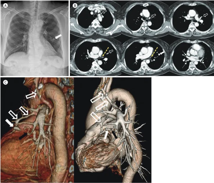
Fig. 1. Levoatriocardinal vein combined with pulmonary venous varix in a 77-year-old woman, presenting with chest abnormalities on screening.
A. Chest radiography depicts a well-defined, round increased opacity focus in the left middle lung field (arrow).
B. Contrast-enhanced CT images depict focally dilated vasculature in the left lingular segment corresponding to the round increased opacity focus on chest radiography (white arrow), with a drainage course to the left innominate vein (blank arrows). There is no normal left upper pul- monary vein between the pulmonary trunk and the left main bronchus (yellow dotted arrows), and the normal right pulmonary veins and left lower pulmonary vein (white thin arrows) are evident.
C. Three-dimensional volume-rendered CT image in the posterolateral view depicts a dilated sac (white arrow, left image) and smaller sac in the lateral view acquired 7 years before (whit arrow, right image). Further, a drainage pathway into the left innominate vein (blank arrows) is depicted. Another tortuous draining vasculature is visible (black arrows), but its drainage course is unclear in both images.
https://doi.org/10.3348/jksr.2020.0036 443 tient’s history of cerebral infarction, which meant that the structure had the potential to be the source of an embolism from systemic venous flow to the left heart, and also the size of the varix had increased substantially during the past 7 years. Coil embolization was per- formed to treat the PVV using microcoils (6 mm/14 cm × 1, 4 mm/14 cm × 3, Nester; Cook Medical, Bloomington, IN, USA and 8 mm/30 cm × 1, 6 mm/20 cm × 1, 5 mm/20 cm × 1, Concerto; Medtronic, Minneapolis, MN, USA). After embolization of the PVV a smaller con- Fig. 1. Levoatriocardinal vein combined with pulmonary venous varix in a 77-year-old woman, presenting with chest abnormalities on screening.
D. Left pulmonary angiography via a femoral vein puncture in the arterial phase does not depict any evi- dence of vascular anomalies (left image). Delayed left pulmonary angiography (middle image) demon- strates the convergence of the left pulmonary veins into the left lower pulmonary vein (white thin arrow) but no evidence of the left upper pulmonary vein. Note a round abnormal vascular opacification focus (white arrows) in the left lingular segment. Selective angiography of the segmental pulmonary artery (right image) depicts two sites of drainage into the left innominate vein (blank arrows), left lower pulmonary vein (black arrows), and finally to the left atrium.
E. Venography via a left basilic vein puncture also depicts bidirectional flow (blank arrow and black arrows) of the venous varix (left image). Embolization was performed at the venous varix (white arrow) with mi- crocoils (middle image), and another embolization with a microcoil was performed for an additional small connection (arrowhead). Left pulmonary angiography 3 days later via the left basilic vein puncture (right image) depicts systemic flow to the left innominate vein (blank arrow) and no flow to the left atrium, indi- cating adequate disconnection of flow.
D
E
nection between the left innominate vein and the LA was observed, and an additional embo- lization was performed using a microcoil (4 mm/10 cm × 1, Tornado; Cook Medical) (middle image in Fig. 1E). Pulmonary arteriography 3 days after the procedure depicted venous flow of the left innominate vein without any flow to the LA, confirming embolization of the PVV (right image in Fig. 1E). Angiography was used to diagnose the patient with coexisting levoat- riocardinal vein and PVV, because there was connection between the systemic vein and LA, leaving the absence of a normal pulmonary venous in between. The patient was treated with endovascular coil embolization. Follow-up chest radiography has indicated no further changes in embolization coils and no other notable symptoms for a period of 1 year follow- ing the procedure.
DISCUSSION
Levoatriocardinal vein is defined as a connection between the LA and a systemic vein (left image in Fig. 1F). The developing lungs are drained to the cardinal vein for the first 2 months, and the capillary plexus near the LA subsequently becomes the pulmonary vein (1, 2). The pulmonary vein connects to the LA while obliterating the normal connection to the cardinal vein. If the primitive connection with the cardinal vein is a consistent alternative pathway in the case of obstructive left heart lesions, the condition becomes levoatriocardinal vein with the LA and systemic vein connection derived from the cardinal vein (1-3). The levoatriocardi- nal vein most commonly drains to the left innominate vein and courses dorsally to the left pulmonary artery and laterally to the aortic arch (1, 4).
The other vascular anomaly that can be confused with levoatriocardinal vein is PAPVR.
PAPVR exhibits anomalous pulmonary vein drainage to a systemic vein (middle image in Fig. 1F) and is only detected in 0.4–0.7% of autopsies (5). If the vein is located in the left lung the most common drainage site is the left innominate vein or the coronary sinus (2). The pri- mary difference between levoatriocardinal vein and PAPVR is that the levoatriocardinal vein is connected with the normal pulmonary vein (1).
The current patient was initially diagnosed with PAPVR because there was clear drainage to the left innominate vein, whereas drainage to the LA was indeterminate in conventional chest CT. Subsequent angiography revealed another connection to the LA through serpigi- nous vasculature connected to the left lower pulmonary vein in the absence of a normal left upper pulmonary vein (right image in Fig. 1F). Because there was a connection to the LA through PVV, the term levoatriocardinal vein was more accurate to describe the condition.
We ultimately diagnosed the condition as PVV of levoatriocardinal vein.
It is known that levoatriocardinal vein usually drains toward a cephalad course, and less commonly drains bidirectionally (1). In the present case the vein drained bidirectionally to both the LA and the innominate vein. Further, the focally dilated portion of the venous struc- ture in the present case was between these two flows. That structure was also depicted as an increased oval-shaped opacity on chest radiography mimicking arteriovenous malformation, and it was determined to be venous dilatation via angiography. Focal dilatation of the pulmo- nary vein is a defining characteristic of PVV, and usually occurs near the entrance to the LA (3). To our knowledge there have been no reports of levoatriocardinal vein combined with
https://doi.org/10.3348/jksr.2020.0036 445 venous varix formation.
PVV is a rare condition involving anomalous pulmonary vessels, and there are two types, primary and secondary (6, 7). The secondary type is more common, and cardiovascular con- ditions such as mitral valve disease or another chronic heart disease are common causes (8).
The underlying chronic bicuspid aortic valve and aortic stenosis in the current case may have caused the varix formation. The characteristic features of the present case such as the bidirectional flow and and the unusual drainage passage to the LA, and consequent limita- tion of drainage flow may have been an another reason for the formation of PVV and tortu- ous vasculature. Varix formation is generally benign but in rare cases there are complications such as rupture and systemic emboli due to thrombosis within the varix (5). In addition, a le- voatriocardinal vein in the absence of hypoplastic left heart typically does not require man- agement (1). In the current case however the growing entity and the patient’s previous histo- ry of TIA suggested a high risk of complications, so treatment with embolization was performed.
In summary, we have described a rare case of levoatriocardianl vein combined with PVV.
The rare vasculature course involved was identified via angiography. A vascular anomaly was considered given the observation of a nodular opacity on a chest X-ray. Regular follow-ups to assess changes in shape or size should be conducted in the treatment of patients with the above-described condition and history.
Author Contributions
Conceptualization, all authos; investigation, all authos; supervision, K.E.; visualization, J.J.H., K.E.;
and writing—original draft, J.J.H.
RA
RV LV
LA LUPV
LLPV LIV
RV LV RA
LA
LLPV LIV
RV LV RA
LA
PVV Systemic drainage
Drainage to LA through LLPV F LIV
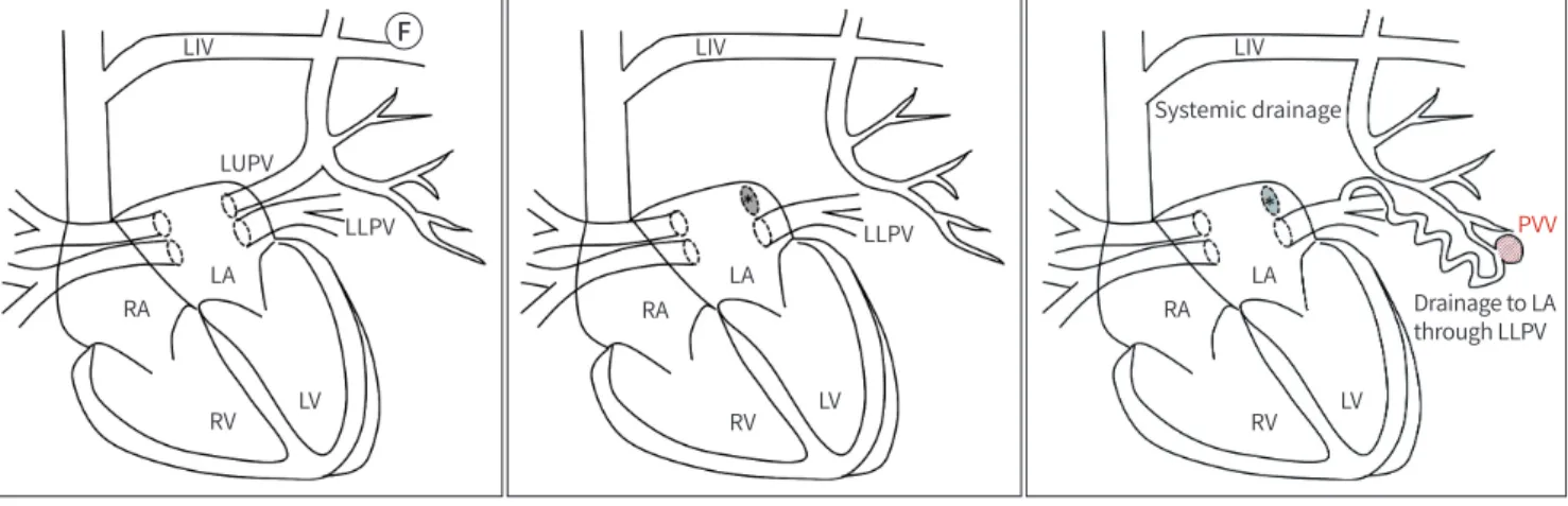
Fig. 1. A 77-year-old woman with abnormalities on screening chest radiography.
F. Schematic illustrations demonstrating pulmonary venous anomalies. In the levoatriocardinal vein, there is an abnormal connection be- tween the LA or a pulmonary vein and the systemic vein (left image). It has a normal pulmonary vein connected to the LA. In partial anoma- lous pulmonary venous return, a pulmonary vein drains into the systemic vein without normal pulmonary venous connection with the LA (*, middle image). This illustration shows drainage to the LIV. The right image shows a connection between the systemic vein and the LA of the present case. Unlike the typical levoatriocardinal vein, it is connected to the LA not through a normal pulmonary vein but through abnormal tortuous drainage vasculature directed to the LLPV. Further, it is also combined with PVV. Note the absence of the normal LUPV (*, right image).
LA = left atrium, LIV = left innominate vein, LLPV = left lower pulmonary vein, LUPV = left upper pulmonary vein, LV = left ventricle, PVV = pul- monary venous varix, RA = right atrium, RV = right ventricle
Conflicts of Interest
The authors have no potential conflicts of interest to disclose.
Funding None
REFERENCES
1. Agarwal PP, Mahani MG, Lu JC, Dorfman AL. Levoatriocardinal vein and mimics: spectrum of imaging find- ings. AJR Am J Roentgenol 2015;205:162-171
2. Dillman JR, Yarram SG, Hernandez RJ. Imaging of pulmonary venous developmental anomalies. AJR Am J Roentgenol 2009;192:1272-1285
3. Porres DV, Morenza OP, Pallisa E, Roque A, Andreu J, Martínez M. Learning from the pulmonary veins. Ra- diographics 2013;33:999-1022
4. Hyun D, Chae EJ, Seo JB, Kang JW, Do KH, Lee CW, et al. Partial anomalous pulmonary venous return via a levoatriocardinal vein in association with rheumatic mitral stenosis: MR demonstration and successful sur- gical repair. J Korean Soc Radiol 2010;63:339-343
5. Maillard JO, Cottin V, Etienne-Mastroïanni B, Frolet JM, Revel D, Cordier JF. Pulmonary varix mimicking pulmonary arteriovenous malformation in a patient with Turner syndrome. Respiration 2007;74:110-113 6. Hochhegger B, Zanetti G, Marchiori E. Pulmonary venous varix presenting as a pulmonary nodule. Ann
Thorac Surg 2017;103:e459
7. Hanson JM, Wood AM, Seymour R, Petheram IS. Anomalous unilateral single pulmonary vein: two cases mimicking arteriovenous malformations and a review of the literature. Australas Radiol 2005;49:246-251 8. Kumazoe H, Komori M, Ochiai R, Egashira R, Nakazono T, Kudo S. Pulmonary varix mimicking arteriove-
nous malformation. Clin Imaging 2008;32:61-64
동정맥기형으로 오인되었던 폐정맥정맥류를 동반한 Levoatriocardinal 정맥: 증례 보고
전주희1 · 강은주1* · 조정현2 · 이기남1
Levoatriocardinal 정맥은 폐정맥 기형의 드문 형태로, 좌심방 혹은 폐정맥과 체정맥을 연결 해 주는 구조이다. 이는 폐정맥이 체정맥으로 연결되는 기형을 뜻하는 부분 폐정맥 환류 이 상과 구분되는데, Levoatriocardinal 정맥은 정상 폐정맥을 통한 좌심방과의 연결성이 있다 는 차이점이 있다. 저자들은 폐정맥정맥류가 흉부 엑스선 및 전산화단층촬영에서 동정맥기 형으로 오인되었던 증례를 보고하고자 하며, 이는 혈관조영술을 통해 폐정맥정맥류와 동반 된 Levoatriocardinal 정맥으로 진단된 경우이다. 이러한 폐정맥정맥류를 동반한 Levoatrio- cardinal 정맥을 보이는 증례는 영문 문헌상 보고된 바 없기에 이를 보고하고자 한다.
1동아대학교 의과대학 동아대학교의료원 영상의학과,
2대동병원 영상의학과