weighted and T2-weighted images4). We present a report of local- ized PVNS arising from both anterior and posterior aspects of the proximal tibiofibular joint with bony erosion at the tip of the fibular in short periods.
case report
A 39-year-old female presented with a three month history of right knee pain and swelling. There was no history of trauma.
The pain was diffuse and dull in character. Physical examination revealed an ill-defined swelling at the proximal tibiofibular joint area. Tenderness was diffuse and mild in nature with a rubbery hard consistency and the mass was not mobile in any plane.
There was no neurological symptom or sign.
A private clinic made a provisional diagnosis of a ganglion or a lipoma and decided to do an excision biopsy under local anaes- thesia. However, it was discovered during surgery that the soft tissue mass was arising from the proximal tibiofibular joint and communicating with the anterior and posterior aspects of the joint. The partially resected specimen was sent for histopatho- logical examination and the results revealed PVNS. The patient was referred to this hospital for further evaluation and treatment.
Plain radiography showed a small sized bony erosion of the fibular tip (Fig. 1). MRI showed a well circumscribed lesion with a capsular margin in the posterior aspect of the proximal tibiofib-
Localized Pigmented Villonodular Synovitis of the Proximal Tibiofibular Joint
Jae Ho Kwon, MD
1, Jae Hwi Han, MD
1, Vivian RD’ Almeida, MS
1, Seong Hyun Kim, MD
2, Hai Jin Park, MD
2, and Kyung-Wook Nha, MD
1Departments of 1Orthopaedic Surgery and 2Dermatology, Inje University Ilsan Paik Hospital, Ilsan, Korea
Pigmented villonodular synovitis (PVNS) is a rare disease. It is a benign neoplastic process typically affecting young to middle-aged adults and most commonly involving the knee, hip, and shoulder joints. The symptoms include diffuse pain and swelling with discomfort. We report a rare case of localized PVNS originating at the proximal tibiofibular joint in a 39-year-old female patient with radiologic changes for short duration of time. The clinical history, plain radiographs, magnetic resonance imaging, and pathologic findings of the reported patient were reviewed. Complete surgical excision was performed and there was no evidence of recurrence after one-year follow-up.
Keywords: pigmented villonodular synovitis, tibiofibular joint
Case Report
Knee Surg Relat Res 2014;26(4):249-252 http://dx.doi.org/10.5792/ksrr.2014.26.4.249 pISSN 2234-0726 · eISSN 2234-2451
Knee Surgery & Related Research
Received December 9, 2013; Revised January 28, 2014;
Accepted February 21, 2014
Correspondence to: Kyung-Wook Nha, MD
Department of Orthopaedic Surgery, Inje University Ilsan Paik Hospital, 170 Juhwa-ro, Ilsanseo-gu, Goyang 411-706, Korea
Tel: +82-31-910-7312, Fax: +82-31-910-7319 E-mail: [email protected]
Pigmented villonodular synovitis (PVNS) is a rare, benign proliferating disorder of the synovium which can be locally ag- gressive affecting the joints, tendon sheaths and bursa. It is more commonly seen in young and middle-aged adults1). It is often monoarticular with the knee being the most common joint to be involved1,2). The other common sites of occurrence include the hip joints, the flexor tendon sheaths of the hand, the ankle and shoulder joints3). The sternoclavicular and tibiofibular joints are rare locations2). Patients frequently present with pain, joint effu- sions and swelling. The disease can present in the knee joint in two forms, diffuse and localized1,2). Plain radiography may show erosions in the adjacent bone. Magnetic resonance imaging (MRI) is useful to delineate the soft tissue extent, evaluate the bony in- volvement and the typical signal intensity changes seen on T1-
249
This is an Open Access article distributed under the terms of the Creative Commons Attribution Non-Commercial License (http://creativecommons.org/licenses/by-nc/3.0/) which permits unrestricted non-commercial use, distribution, and reproduction in any medium, provided the original work is properly cited.
Copyright © 2014 KOREAN KNEE SOCIETY www.jksrr.org
250
Kwon et al. Localized PVNSular joint with decreased heterogeneous signal intensity on both T1-weighted and T2-weighted images. The T2-weighted image showed a relatively hyper-intense signal compared to the skeletal muscle (Fig. 2A and 2B).
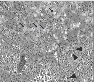
Complete excision and biopsy was done through a posterolat- eral incision (Fig. 3). Intraoperatively, the mass was yellowish brown in colour, arising from the proximal tibiofibular joint and circumscribing the joint. The mass was carefully isolated and excised totally. Pathological findings of the biopsy specimens showed proliferation of round to polygonal cells, with scant cytoplasm forming villous and fingerlike or rounded mass un- derlying the synovium. Multinucleated giant cells were scattered throughout the lesion. Hemosiderin-laden macrophages varied
in number, giving the synovium a dark-brown appearance. These findings findings confirmed the diagnosis of localized PVNS (Fig.
4). Postoperative recovery was uneventful and at the most recent follow-up at 12 months after surgery, the patient was fully asymp- tomatic with no evidence of recurrence on MRI.
discussion
Although PVNS is a benign disease, it can be locally destructive causing significant and permanent damage to the joint. The dis- tribution of lesion location depends on the lesion subtypes, either localized or diffuse, and on whether the lesion is intra-articular or extra-articular3,5). Granowitz et al.6) classified PVNS into diffuse
Fig. 1. Plain radiograph showing bony erosion at the tip of the fibular head (arrow).
Fig. 2. Enhanced coronal (A), sagittal (B) and axial (C) magnetic resonance images of the right knee showing a well circum- scribed lesion with a capsular margin in the posterior aspect of the proximal tibiofibular joint with increased heterogeneous signal intensity.
A B C
Fig. 3. Intraoperative appearance of the mass excision with a postero- lateral approach. a: pigmented villonodular synovitis lesion, b: common peroneal nerve.
a b
Calf Thigh
Knee Surg Relat Res, Vol. 26, No. 4, Dec. 2014
251
and localized types. Localized PVNS is an uncommon pathol- ogy characterized by the presence of nodular pedunculated mass protruding into the joint cavity7). Extra-articular lesions are rare and tend to occur in the same joint locations as intra-articular le- sions8).
This case report presents a rare site of occurrence of which only one citation is found in the English-language literature. Ryan et al.2) reported a case of PVNS at the proximal tibiofibular joint which was located just anterior to the tibiofibular joint. However, in our case, the mass was arising from the proximal tibiofibular joint extending to the extra-articular lesion and communicated with the anterior and posterior aspects of the joint.
The diagnosis of localized PVNS is difficult, and the time pe- riod between the onset of symptoms and the definitive diagnosis is usually two years4).
Plain radiographs are usually unremarkable. However, in about 9% to 25% of the total cases, extrinsic erosion, often with well-de- fined sclerotic margins of the underlying bone may be seen as in our case where the simple radiograph showed bony erosion at the tip of the fibular head. MRI is the method of choice for diagnosis and follow-up. It helps to delineate the location, size, and extent of the lesion4,9). The lesion usually has heterogeneous signal in- tensity depending on the amount of hemosiderin content and is characterized by decreased intensity on both the T1-weighted and T2-weighted images7,10). Albeit sensitive, MRI is not specific due to other conditions such as fibroxanthoma, hemangioma,
and desmoid tumors giving similar pictures7).
Histologically, the lesion is characterized by proliferation of po- lygonal mononuclear cells, histiocytes, scattered multinucleated giant cells, and foam cells with hemosiderin pigment deposits10). The recurrence rate of localized PVNS has been reported to range from 0% to 13%4,10), whereas the recurrence rate of diffuse PVNS has been reported to range from 8% to 35%7).
The location of PVNS described in this article, which originated at the proximal tibiofibular joint, is extremely rare. PVNS that in- volves small joints such as the tibiofibular joint can be easily mis- diagnosed as other mass lesions. Therefore, the clinicians should pay more attention and must consider different diagnosis for mass lesions around the knee joint. Complete surgical excision is the treatment of choice and the patient should be followed up for recurrence.
conflict of interest
No potential conflict of interest relevant to this article was re- ported.
acknowledgments
This article is supported by a 2015 Inje University Research Grant.
references
1. Tyler WK, Vidal AF, Williams RJ, Healey JH. Pigmented vil- lonodular synovitis. J Am Acad Orthop Surg. 2006;14:376- 85.
2. Ryan RS, Louis L, O’Connell JX, Munk PL. Pigmented villo- nodular synovitis of the proximal tibiofibular joint. Australas Radiol. 2004;48:520-2.
3. Yamashita H, Endo K, Enokida M, Teshima R. Multifocal localized pigmented villonodular synovitis arising separately from intra- and extra-articular knee joint: case report and literature review. Eur J Orthop Surg Traumatol. 2013;23 Suppl 2:S273-7.
4. Mandelbaum BR, Grant TT, Hartzman S, Reicher MA, Flan- nigan B, Bassett LW, Mirra J, Finerman GA. The use of MRI to assist in diagnosis of pigmented villonodular synovitis of the knee joint. Clin Orthop Relat Res. 1988;(231):135-9.
5. Jobe CM, Raza A, Zuckerman L. Pigmented villonodular synovitis: extrasynovial recurrence. Arthroscopy. 2011;27:
1449-51.
Fig. 4. Histopathological analysis of the biopsy specimen showing the typical appearance of pigmented villonodular synovitis with foam cells (arrows), hemosiderin-containing macrophages (arrowheads), and mul- tinucleated giant cells (asterisk) (H&E, ×200).
*
252
Kwon et al. Localized PVNS6. Granowitz SP, D’Antonio J, Mankin HL. The pathogenesis and long-term end results of pigmented villonodular synovi- tis. Clin Orthop Relat Res. 1976;(114):335-51.
7. Kim RS, Lee JY, Lee KY. Localized pigmented villonodular synovitis attached to the posterior cruciate ligament of the knee. Arthroscopy. 2003;19:E32-5.
8. Wagner ML, Spjut HJ, Dutton RV, Glassman AL, Askew JB.
Polyarticular pigmented villonodular synovitis. AJR Am J
Roentgenol. 1981;136:821-3.
9. De Ponti A, Sansone V, Malchere M. Result of arthroscopic treatment of pigmented villonodular synovitis of the knee.
Arthroscopy. 2003;19:602-7.
10. Perka C, Labs K, Zippel H, Buttgereit F. Localized pigmented villonodular synovitis of the knee joint: neoplasm or reactive granuloma? A review of 18 cases. Rheumatology (Oxford).
2000;39:172-8.