275
Aortic Valve Sclerosis on Echocardiography is a Good Predictor of Coronary Artery Disease in Patients With
an Inconclusive Treadmill Exercise Test
Dong-Bin Kim, MD, Hae-Ok Jung, MD, Doo-Soo Jeon, MD, Chan-Seok Park, MD, Sung-Won Jang, MD, Hoon-Joon Park, MD, Pum Joon Kim, MD, Sang Hong Baek, MD, Ki-Bae Seung, MD, Tai-Ho Rho, MD, Jae-Hyung Kim, MD and Kyu-Bo Choi, MD
Division of Cardiology, Department of Internal Medicine, The Catholic University of Korea College of Medicine, Seoul, Korea ABSTRACT
Background and Objectives : The treadmill exercise test (TMT) is used as a first-line test for diagnosing coronary artery disease (CAD). However, the findings of a TMT can be inconclusive, such as incomplete or equivocal re- sults. Aortic valve sclerosis (AVS) is known to be a good predictor of CAD. We determined the usefulness of as- sessing AVS on 2-dimensional (2D) echocardiography for making the diagnosis of CAD in patients with incon- clusive results on a TMT. Subjects and Methods : This prospective study involved 165 consecutive patients who underwent a TMT that resulted in inconclusive findings, 2D echocardiography to detect AVS, and coronary an- giography to detect CAD. Following echocardiography, AVS was classified as none, mild, or severe. CAD was de- fined as ≥70% narrowing of the luminal diameter on coronary angiography. Results : CAD was more common in patients with AVS than in patients without AVS (75% vs. 47%, respectively, p<0.01). Multiple logistic regres- sion analysis showed that AVS was the only independent predictor of CAD {odds ratio=8.576; 95% confidence interval (CI), 3.739-19.672}. The sensitivity, specificity, accuracy, positive predictive value, and negative predictive value of the presence of AVS for predicting CAD in a patient with an inconclusive TMT were 62%, 67%, 64%, 75%, and 53%, respectively. During a 1-year clinical follow-up, patients with and without AVS were similar in terms of event-free survival rates. Conclusion : If the results of TMT for patients with chest pain on exertion are incon- clusive, the presence of AVS on echocardiography is a good predictor of CAD.
(Korean Circ J 2009;39:275-279)KEY WORDS: Echocardiography; Treadmill test; Aortic valve; Sclerosis; Coronary artery disease.
Introduction
The treadmill exercise test (TMT) has been used to diagnose angina pectoris in many cardiovascular cen- ters as it is inexpensive, easily performed, and provides real-time results. Despite some inter-investigator varia- tions, a meta-analysis found that the sensitivity and specificity of a TMT for diagnosis of coronary artery disease (CAD) averaged 68% and 77%, respectively.
1)However, since a substantial number of patients with angina pectoris also have arthritic or lung diseases due to old age, they cannot reach the target heart beat and
the test is terminated without completion, and in young women the depression pattern of the ST segment occa- sionally shows an upslope, thus making the diagnosis difficult in such cases. Although the diagnosis could be confirmed by stress echocardiography or myocardial single photon emission computerized tomography (SP- ECT), such modalities are costly and difficult to per- form immediately when required. Another alternative is coronary multi-detector CT (MDCT) angiography;
however, it involves exposure to radiation in addition to the high cost.
While aortic valve sclerosis (AVS) detected using 2- dimensional (2D) echocardiography is known to be an independent risk factor for predicting the development of CAD,
2)3)it has not been investigated in association with a TMT.
The present study determined whether AVS is a pre- dictor of CAD in patients with suspected angina pec- toris in whom TMT findings were inconclusive.
Received: December 18, 2008 Revision Received: February 24, 2009 Accepted: March 25, 2009
Correspondence: Hae-Ok Jung, MD,Division of Cardiology, Department of Internal Medicine, The Catholic University of Korea College of Medi- cine, Banpo-dong, Seocho-gu, Seoul 137-701, Korea
Tel: 82-2-590-2075, Fax: 82-2-591-1075 E-mail: [email protected]
Subjects and Methods
The subjects
The study was conducted prospectively on 354 patients who visited our outpatient clinic with symptoms sus- picious of typical stable angina pectoris. All patients un- derwent TMT and 2D echocardiography. Coronary an- giography was performed on 317 patients, excluding pa- tients with negative TMT results. The exclusion criteria were patients with rheumatic valve diseases, patients with significant AV stenosis (a continuous wave Doppler speed of blood flow passing through the aortic valve
>2 m/s, or the area of the aortic valve orifice <2 cm
2), patients already diagnosed with angina pectoris by coro- nary angiography, atrial fibrillation, Wolff-Parkinson- White (WPW) syndrome, a history of cardiac surgery, myocardial infarction, heart failure, or left or right com- plete bundle branch block on electrocardiogram.
Treadmill exercise test
The TMT was conducted according to Bruce’s pro- tocol, and the results were interpreted based on the Ame- rican College of Cardiology/American Heart Associa- tion (ACC/AHA) guidelines and were scored as nega- tive, positive, incomplete, or equivocal.
4)Negative results showed no symptoms, and had no ST segment changes at target heart rate. Positive results showed typical symp- toms during the TMT regardless of reaching the target heart beat, with significant ST segment depression (based on baseline, ST segments showed >0.1 mV horizontal or downslope depression that was maintained for longer than 0.08 seconds), or ischemic ventricular arrhythmia.
Incomplete results were early TMT termination due to events other than the development of ischemic arrhy- thmia at <90% target heart beat. Patients who reached
> 90%, but <100% without typical symptoms and the atypical change of ST segments during the test, were evaluated to be equivocal results.
Patients with incomplete or equivocal TMT results were defined as inconclusive patients. There were 37 (10%) negative cases, 152 (43%) positive cases, and 165 (47%) inconclusive cases.
2-dimensional transthoracic echocardiography 2D transthoracic echocardiography was performed using a 3.5 MHz Sequoia C256 probe (Acuson Co., CA, USA) or a 3.5 MH Vivid 7 probe (GE Vingmed, Horten, Norway). 2D echocardiographic tests and Dop- pler tests were performed following the Standard Prac- tical Guideline for 2D echocardiography from the Ame- rican Society of Echocardiography.
5)The area of the aor- tic valve orifice on the sternal left margin was measured using a static image in which the 3 valves were most widely open, while the aortic valve blood flow speed was measured on the apical 5-chamber view using a con-
tinuous wave Doppler. These measurements were taken three times and the mean was used in analysis.
Aortic valve sclerosis measurement
Using a long axis view of the left sternum margin, valve thickness was defined as the value obtained by en- larging the area of the aortic valve, and measuring the thickest valve of the right coronary aortic valve as well as the non-coronary aortic valve during the systolic pe- riod. Using a short axis view of the left sternum mar- gin, when movements of the right coronary aortic valve and non-coronary aortic valve during the systolic pe- riod were examined by enlarging the aortic root, a valve opening inward concave was defined as normal, wher- eas a valve opening inward flat or inward convex was defined as showing restricted motion.
6)Definition of aortic valve sclerosis and severity A diagnosis of AVS was based on an aortic valve thick- ness >2 mm or restricted motion.
7)AVS severity was classified as mild or severe. Mild cases were defined as the cases that only one aortic valve thicker than 2 mm, and non-restricted motion. Severe cases were defined as patients that AVS in more than two aortic valves, or sclerosis detected in one valve that was thicker than 4 mm, or restricted motion.
6)Coronary artery disease diagnosis
Significant CAD was diagnosed when a coronary an- giogram performed through the right femoral artery showed stenosis >70% in diameter in more than one major epicardial artery.
Duke treadmill score
After the TMT, the Duke treadmill score was obtain- ed as a measure of CAD prognosis.
8)This score was based on the following expression: {exercise time (min)-5×
ST segment deviation (mm)-4×angina (0, 1, 2)}.
Statistical analysis
All results are presented as the mean and standard deviation. A commercial program was used for statis- tical analysis. Groups were compared based on the pre- sence or absence of AVS. For continuous variables, un- paired t-tests were applied, while Chi-square tests were used to compare categorical variables. When the patients were divided to three groups according to the presence or absence of AVS and analyzed, test for trend was used.
To assess factors associated with CAD, we used multi-
ple logistic regression analysis and calculated odds ratios
and 95% confidence intervals. The diagnostic value of
AVS was determined using a 2×2 table considering pa-
tients with and without CAD and patients with and
without AVS. Diagnostic sensitivity, specificity, accu-
racy, and positive and negative prediction rates were
determined. In addition, the survival rate for each group was obtained using Kaplan-Meier curves. Significance was defined as a p<0.05.
Results
This study analyzed 165 patients suspected of having angina pectoris with inconclusive TMT results. The 2D echocardiography and coronary angiography data from these patients were analyzed.
Patient characteristics according to aortic valve sclerosis
The AVS and non-AVS groups were similar in all ch- aracteristics except that the AVS group was older and had CAD more frequently (Table 1).
Coronary artery disease incidence according to aortic valve sclerosis
There were more non-AVS patients in the non-CAD than the CAD group. Conversely, there were more AVS patients in the CAD than the non-CAD group (Fig. 1).
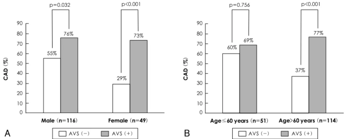
For both males and females, the incidence of CAD was higher in the AVS patients. When applying an age th- reshold of 60 years, we found that the incidence of CAD was higher only in AVS patients >60 years of age (Fig. 2).
Univariate analysis showed that AVS was the only risk factor for CAD in 165 patients who showed inconclusive results in the TMT (p<0.01). Multivariate logistic regres- sion analysis to assess independent risk factors includ- ed the traditional risk factors such as, age, gender, hy- pertension, diabetes, smoking, hyperlipidemia (low den- sity lipoprotein-cholesterol ≥140 mg/dL), and AVS.
The results showed that AVS was the only factor pre- dictive for CAD (odd ratio=8.576; 95% confidence interval, 3.739-19.672) (Table 2).
Table 1. Characteristics of patients with and without aortic valve sclerosis
No AVS (n=81) AVS (n=84) p Age (years)
061±10 67±8<0.01
Male (%) 70 71
<0.87Hypertension (%) 49 65
<0.06DM (%) 21 26
<0.47Smoking (%) 24 27
<0.60LDL-C (mg/dL) 104±25 110±24
<0.12CAD (%) 47 (n=38) 75 (n=63) <0.01 AVS: aortic valve sclerosis, DM: diabetes mellitus, LDL-C: low density lipoprotein-cholesterol, CAD: coronary artery disease
Table 2. Predictors of coronary artery disease according to mul- tivariate analysis
Variable Odds ratio (95% CI) p
Age>60 years 1.957 (0.756-5.065)
<0.16Male 1.959 (0.793-4.840)
<0.14Hypertension 1.337 (0.609-2.932)
<0.47Diabetes mellitus 1.381 (0.550-3.468)
<0.49Smoking 1.389 (0.549-3.513)
<0.49LDL-C≥140 mg/dL 1.169 (0.348-3.929)
<0.80Aortic valve sclerosis 8.576 (3.739-19.672) <0.01 CI: confidence interval, LDL-C: low density lipoprotein-chole- sterol
Fig. 2. Prevalence of coronary artery disease according to gender and age. A: prevalence of coronary artery disease in males and fe- males with and without aortic valve sclerosis. B: prevalence of coronary artery disease in patients aged ≤60 years and >60 years with and without aortic valve sclerosis. AVS: aortic valve sclerosis, CAD: coronary artery disease.
CAD (-)(n=64) CAD (+)(n=101) AVS (-)
AVS (+) 90
80 70 60 50 40 30 20 10 0
53%
25%
p<0.001
47%
75%
p<0.001
Fig. 1. Prevalence of coronary artery disease according to aor- tic valve sclerosis. CAD: coronary artery disease, AVS: aortic val- ve sclerosis.
Prevalence (%)
Male (n=116) Female (n=49) 76%
p=0.032
55%
29%
73%
p<0.001
90 80 70 60 50 40 30 20 10 0
CAD (%)
AVS (-) AVS (+)
Age≤60 years (n=51) Age>60 years (n=114) 69%
p=0.756
77%
p<0.001
90 80 70 60 50 40 30 20 10 0
CAD (%)
AVS (-) AVS (+) 60%
37%
A B
Coronary artery disease incidence according to aortic valve sclerosis severity
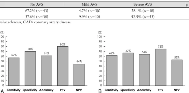
We found that the incidence of CAD increased as the severity of AVS increased (p for trend, p<0.001) (Table 3).
Diagnostic value of aortic valve sclerosis for coro- nary artery disease
Analysis of combined inconclusive and positive re- sult groups data showed that the sensitivity, specificity, accuracy, positive prediction rate, and negative predic- tion rates for the presence of AVS for CAD were 57%, 70%, 61%, 80%, and 44%, respectively (Fig. 3). In the inconclusive group, the sensitivity for CAD of severe AVS was 58% and the specificity was 70%.
Duke treadmill score
Patients were analyzed in three groups: non-AVS, mild AVS, and severe AVS. The Duke treadmill scores were 5.0±4.3, 4.9±3.7, and 3.9±4.2, respectively, but the apparent decreasing trend was not found to be significant.
Major cardiac events and mortality
Patients were followed-up clinically for an average of 11±6 months. No patient died during this time. The incidence of major clinical events represented by cor- onary revascularization or rehospitalization was 5% in the non-AVS group and 4% in the AVS group. The two groups did not differ in terms of event-free survival rates (Fig. 4).
Discussion
In the present study, 53% of patients had conclusive
TMT results, while 47% had inconclusive findings. For the latter group, additional tests, such as coronary an- giography, were performed based on risk factors for car- diovascular disease. However, a problem with perform- ing tests by considering risk factors is that unnecessary angiograms may be performed due to a desire to assess the presence or absence of lesions. According to the re- cent ENCOURAGE study,
9)mortality rates were simi- lar for stable angina pectoris patients undergoing either coronary angioplasty or drug treatment. Hence, drug treatment is a useful approach in patients unlikely to have CAD.
Previous studies reported an association between AVS and CAD,
10)and AVS patients had a higher inci- dence of CAD and consequently high mortality rate.
11)CAD is more frequently observed in AVS patients be-
Table 3. Results of coronary angiography in patients with differing aortic valve sclerosis severity
No AVS Mild AVS Severe AVS p for trend
CAD (-) 67.2% (n=43) 4.7% (n=3)0 28.1% (n=18)
CAD (+) 37.6% (n=38) 9.9% (n=10) 52.5% (n=53) <0.001
AVS: aortic valve sclerosis, CAD: coronary artery disease
Fig. 3. The diagnostic value of aortic valve sclerosis for coronary artery disease. A: in inconclusive and positive treadmill exercise test patients (n=317). B: in inconclusive treadmill exercise test patients (n=165). PPV: positive predictive value, NPV: negative predictive value, AVS: aortic valve sclerosis.
100 90 80 70 60 50 40 30 20 10 0
A
(%)
Sensitivity Specificity Accuracy PPV NPV 57%
70%
61%
80%
44%
100 90 80 70 60 50 40 30 20 10 0
B
(%)
Sensitivity Specificity Accuracy PPV NPV
62% 67% 64%
75%
53%
0 5 10 15 20 25 Months
1.0
0.9
0.8
0.7
0.6
Event-free survival
Fig. 4. Kaplan-Meier curves for event-free survival in patients with and without aortic valve sclerosis. Event-free survival was similar in both groups. AVS: aortic valve sclerosis.
p=0.80
AVS (-) AVS (+)
cause AVS is associated with aorta and carotid artery atherosclerosis. Severe AVS predicts advanced systemic atherosclerosis.
12-14)AVS and atherosclerosis show sim- ilar progression features, such as the presence of oxi- dized lipoprotein, inflammatory cells, microscopic cal- cification, and proteins produced by activated macro- phages in tissues. However, the increase in smooth mus- cle cells, calcium, and protein detected in atheroscle- rosis are not features of AVS.
15)According to a recent study, coronary angiography revealed the sensitivity was 64% and the specificity was 71% in the diagnostic value of AVS for CAD.
2)Simi- larly, a Korean study reported that AVS patients were examined by coronary angiogram and the results sh- owed that the sensitivity was 65% and the specificity was 66%.
3)Those two studies differed from the present study in that they involved patients with stable angina and acute coronary syndrome, whereas our study in- volved patients with stable angina. Despite this differ- ence, the present study found AVS had a sensitivity of 62% and a specificity of 67% for the diagnosis of CAD, similar to the previous reports.
The present study examined whether gender or age affected the association between AVS and CAD. Gen- der had no effect, while CAD was more common in AVS patients >60 years of age. We compared coronary angiography findings between inconclusive and all TMT patients and found there was no difference in terms of the diagnostic value of AVS for CAD (Fig. 3).
The Duke treadmill score did not differ between AVS and non-AVS patients, reflecting the change of ST seg- ments (the variable with the largest effect on the score) was not large in either group. AVS and non-AVS patients showed a similar incidence of major clinical events dur- ing the approximately 1-year clinical follow-up, which may reflect the short follow-up period. A follow-up study of AVS and stenosis patients by Otto et al.
11)found that differences in mortality rates became significant only after 5 years.
In conclusion, if the results of a TMT for patients with chest pain on exertion are inconclusive, the presence of AVS on echocardiography is a good predictor of CAD.
Limitation
Coronary angiograms were not performed on all pa- tients showing inconclusive TMT results. Coronary an- giography was not performed on patients with a low risk of CAD, or with financial problems. Such patients were excluded and it may possibly cause the errors in study results.
REFERENCES
1)
Gianrossi R, Detrano R, Mulvihill D, et al. Exercise-induced ST depression in the diagnosis of coronary artery disease: a meta- analysis. Circulation 1989;80:87-98.
2)
Sui SJ, Ren MY, Xu FY, Zhang Y. A high association of aortic valve sclerosis detected by transthoracic echocardiography with coronary arteriosclerosis. Cardiology 2007;108:322-30.
3)
Park YW, Kim DS, Jeong YS, et al. Association between aortic valve sclerosis and risk factors of coronary artery disease in pa- tients with suspected coronary artery disease. Korean Circ J 2006;
36:374-80.
4)
Gibbons RJ, Balady GJ, Bricker JT, et al. ACC/AHA 2002 guide- line update for exercise testing: summary article: a report of the American College of Cardiology/American Heart Association Task Force on Practice Guidelines ( Committee to Update the 1997 Ex- ercise Testing Guidelines ) . Circulation 2002;106:1883-92.
5)
Cheitlin MD, Armstrong WF, Aurigemma GP, et al. ACC/AHA/
ASE 2003 guideline update for the clinical application of echo- cardiography: summary article: a report of the American College of Cardiology/American Heart Association Task Force on Prac- tice Guideline ( ACC/AHA/ASE Committee to update the 1997 Guideline for the Clinical Application of Echocardiography ) . Circulation 2003;108:1146-62.
6)
Jeon DS, Lee MY, Kim CJ, et al. The severity of aortic valve sc- lerosis is associated with carotid intima media thickness/plaque in neurologically asymptomatic patients. Korean Circ J 2004;
34:1049-55.
7)
Tolstrup K, Crawford MH, Roldan CA. Morphologic characte- ristics of aortic valve sclerosis by transesophageal echocardio- graphy: importance for the prediction of coronary artery disease.
Cardiology 2002;98:154-8.
8)
Mark DB, Shaw L, Harrell FE Jr, et al. Prognostic value of a treadmill exercise score in outpatients with suspected coronary artery disease. N Engl J Med 1991;325:849-53.
9)
Boden WE, O’Rourke RA, Teo KK, et al. Optimal medical th- erapy with or without PCI for stable coronary disease. N Engl J Med 2007;356:1503-16.
10)
Pohle K, Maffert R, Ropers D, et al. Progression of aortic valve calcification: association with coronary atherosclerosis and car- diovascular risk factors. Circulation 2001;104:1927-32.
11)
Otto CM, Lind BK, Kitzman DW, Gersh BJ, Siscovick DS. As- sociation of aortic-valve sclerosis with cardiovascular mortality and morbidity in the elderly. N Engl J Med 1999;341:142-7.
12)
Agmon Y, Khandheria BK, Meissner I, et al. Aortic valve sc- lerosis and aortic atherosclerosis: different manifestations of the same disease?: insights from a population-based study. J Am Coll Cardiol 2001;38:827-34.
13)
Adler Y, Levinger U, Koren A, et al. Relation of nonobstructive aortic valve calcium to carotid arterial atherosclerosis. Am J Cardiol 2000;86:1102-5.
14)
Tolstrup K, Roldan CA, Qualls CR, Crawford MH. Aortic valve sclerosis, mitral annular calcium, and aortic root sclerosis as mar- kers of atherosclerosis in men. Am J Cardiol 2002;89:1030-4.
15)