Squamous Cell Carcinoma of the Pancreas with Invasion of Duodenum and Pylorus
5
0
0
전체 글
(2) 388. 대한외과학회지:제 71 권 제 5 호 2006. ꠏꠏꠏꠏꠏꠏꠏꠏꠏꠏꠏꠏꠏꠏꠏꠏꠏꠏꠏꠏꠏꠏꠏꠏꠏꠏꠏꠏꠏꠏꠏꠏꠏꠏꠏꠏꠏꠏꠏꠏꠏꠏꠏꠏꠏꠏꠏꠏꠏꠏꠏꠏꠏꠏꠏꠏꠏꠏꠏꠏꠏꠏꠏꠏꠏꠏꠏꠏꠏꠏꠏꠏꠏꠏꠏꠏꠏꠏꠏꠏꠏꠏꠏꠏꠏꠏꠏꠏꠏꠏꠏꠏꠏꠏꠏꠏꠏꠏꠏꠏꠏꠏꠏꠏꠏꠏꠏꠏꠏꠏꠏꠏꠏꠏꠏ. Fig. 1. Endoscopy shows a nearly collapsed pylorus due to intraluminally protruded mass arising from duodenum.. Fig. 2. Abdomen CT scan shows a 6 cm sized submucosal solid mass on pylorus of stomach or duodenum invaded the pancreas. 내시경 소견: 십이지장의 종괴가 내강으로 돌출되며 위 유문부를 누르는 양상으로 보였다(Fig. 1). 방사선 검사 소견: 복부 전산화 단층 촬영에서 유문부 및 십이지장 제1부에 걸친 6 cm의 점막 하 종양이 췌장표면을 침습한 것으로 판단되어 위장관에 발생한 간질종양이 췌장 침습을 한 것으로 생각하였다(Fig. 2). 수술 소견: 위전정부 후벽에 위치한 6×7 cm 크기의 구형 종괴가 췌장 체부 및 경부 침습으로 보여 위아전절제술과 함께 종괴가 붙어있는 췌장을 부분 절제하였다. 조직 병리학 소견: 종괴는 연노랑에서 회백색을 띠는 구 형의 과립형 단면을 보이며 단단하게 촉지되었고, 간질성 종양에서 특징적으로 나타나는 나선형과 섬유 다발성 구조. 는 보이지 않았다(Fig. 3). 술 후 병리 조직 검사상 십이지장 침습성 편평 세포암으로 진단되었고, 조직 슬라이드에서 십이지장의 점막층은 정상 소견을 보이고 점막하층에 종양 세포가 군데군데 흩어져 있는 소견이 보였다(Fig. 4A). 종양 세포가 근육층까지 침습된 소견과 함께 끈 또는 덩어리 모 양의 편평 세포 군집들과 케라틴이 관찰되었다(Fig. 4B). 편 평 세포를 확인하기 위해 cytokeratin 면역조직화학염색에서 갈색으로 염색되는 편평세포로 확인되어(Fig. 4C) 위장관의 침습성 편평 상피암종으로 진단되었다. 술 후 경과: 환자는 술 후 3일째부터 췌장액 누출이 있었 으며 입원기간 도중 점차적으로 식욕부진과 전신 쇠약 증 세를 보여 고영양 수액 요법을 유지하며 췌장액 누출에 대.
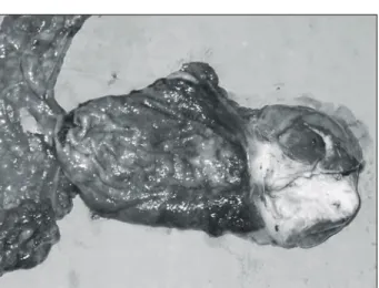
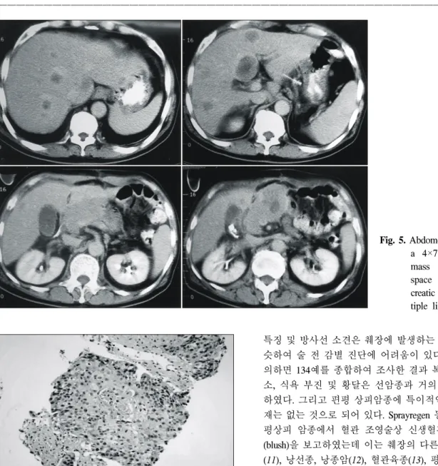
(3) 반주영 외:십이지장과 위유문부 침습을 보이는 췌장의 편평세포암. 389. ꠏꠏꠏꠏꠏꠏꠏꠏꠏꠏꠏꠏꠏꠏꠏꠏꠏꠏꠏꠏꠏꠏꠏꠏꠏꠏꠏꠏꠏꠏꠏꠏꠏꠏꠏꠏꠏꠏꠏꠏꠏꠏꠏꠏꠏꠏꠏꠏꠏꠏꠏꠏꠏꠏꠏꠏꠏꠏꠏꠏꠏꠏꠏꠏꠏꠏꠏꠏꠏꠏꠏꠏꠏꠏꠏꠏꠏꠏꠏꠏꠏꠏꠏꠏꠏꠏꠏꠏꠏꠏꠏꠏꠏꠏꠏꠏꠏꠏꠏꠏꠏꠏꠏꠏꠏꠏꠏꠏꠏꠏꠏꠏꠏꠏꠏ. 한 치료를 지속하였다. 배액관으로 췌장액 누출이 줄어들 어 식이 개시를 하였다. 환자는 간헐적인 식후 구토 증세 외에는 별다른 이상 소견은 없었다. 술 후 30일째 수술부위 의 종물이 촉진되어 시행한 복부 전산화 단층촬영에서 다 발성 간전이와 절제한 췌장 표면에서 4×7 cm 크기의 종괴 가 보여(Fig. 5) 세침 흡입세포 검사를 시행하였고 그 결과 췌장에서 발생한 편평상피 세포암으로 확진되었다(Fig. 6). 환자는 식욕부진과 전신 쇠약 증세로 지속적으로 입원 치 료를 받았고 술 후 50일째 악액질로 사망하였다.. 고. Fig. 3. Macroscopic appearance of the pale yellow to grayish white cut surface of the solid mass. It was not seen that whorling and fasciculated bundular stricture on stromal tumor.. A. C. 찰. 췌장의 편평 상피암종은 비내분비성 췌장암의 드문 변형 이다.(3) 췌장의 편평 상피 선암과 선암, 순수 편평 상피암 종은 연령분포, 췌장 내 암종의 위치, 악성도, 예후 등에 있어 서 유의한 차이가 없다고 받아들여지고 있다.(4) Kovi(5). B. Fig. 4. The compact tumor is composed of the area of metastatic squamous cell carcinoma, moderately differentiated of duodenum extending to pyloric ring and involving omentum (A: H&E, ×40), (B: H&E, ×100). In order to confirm squamous cell, cytokeratin stain was done. It shows brown colored squamous cell on bluish background (C: Cytokeratin, ×100)..
(4) 390. 대한외과학회지:제 71 권 제 5 호 2006. ꠏꠏꠏꠏꠏꠏꠏꠏꠏꠏꠏꠏꠏꠏꠏꠏꠏꠏꠏꠏꠏꠏꠏꠏꠏꠏꠏꠏꠏꠏꠏꠏꠏꠏꠏꠏꠏꠏꠏꠏꠏꠏꠏꠏꠏꠏꠏꠏꠏꠏꠏꠏꠏꠏꠏꠏꠏꠏꠏꠏꠏꠏꠏꠏꠏꠏꠏꠏꠏꠏꠏꠏꠏꠏꠏꠏꠏꠏꠏꠏꠏꠏꠏꠏꠏꠏꠏꠏꠏꠏꠏꠏꠏꠏꠏꠏꠏꠏꠏꠏꠏꠏꠏꠏꠏꠏꠏꠏꠏꠏꠏꠏꠏꠏꠏ. Fig. 5. Abdomen CT scan shows a 4×7 cm sized large mass in peripancreatic space after partial pancreatic resection and multiple liver metastasis.. Fig. 6. Microscopic appearance of squamous cell carcinoma with showing of the keratin pearl (H&E, ×100). 는 다른 원발 병소 없이 췌장에서 발생한 대부분의 순수한 편평 상피암의 경우에 주의 깊은 조직병리 검사로 선암부 위를 발견할 수 있으리라 제시했으나 본 증례에서는 순수 편평상피암종만 확인되었다. 췌장에는 정상적으로 편평 세포 성분이 없으므로 평편 상피암종의 발생에 대해서는 여러 가지 가설이 있으나 증 명된 것은 없다. 최근에는 선암종에 의해 만성 췌장염과 췌 관의 폐색이 발생하고 이로 인한 지속적인 염증으로 선암 종에서 편평 상피암종으로의 이형성화를 가장 가능성 있는 가설로 제시하고 있으며, 또한 이소성의 편평 상피가 존재 하고 여기에서 종양이 발생한다는 보고도 있으며, 선세포 와 편평 상피세포로 모두 분화할 수 있는 미분화 세포로부 터 암종이 발생한다는 보고도 있다.(6-8) 환자들의 임상적. 특징 및 방사선 소견은 췌장에 발생하는 선암종과 거의 비 슷하여 술 전 감별 진단에 어려움이 있다. Madura 등(9)에 의하면 134예를 종합하여 조사한 결과 복부 동통, 체중 감 소, 식욕 부진 및 황달은 선암종과 거의 유사하다고 보고 하였다. 그리고 편평 상피암종에 특이적인 진단 검사도 현 재는 없는 것으로 되어 있다. Sprayregen 등(10)은 췌장의 편 평상피 암종에서 혈관 조영술상 신생혈관 형성과 압입형 (blush)을 보고하였는데 이는 췌장의 다른 암종인 도세포암 (11), 낭선종, 낭종암(12), 혈관육종(13), 평활근 육종(14), 혈 관종(11), 선편평 암종(10)에서도 보이는 소견이다. 췌장의 편평상피 암종은 선암종과 유사하게 일차적으로 국소 림프 절로 전이를 보이고 간전이와 폐전이를 보인다.(12,15) 본 증례의 경우 처음에는 위장관에 발생한 간질종양이 췌장 침습을 한 것으로 생각되었는데 이것은 컴퓨터 촬영상의 오인과 췌장효소치의 상승이 없었기 때문이었다. 췌장의 편평 상피 암종의 조직학적 특징은 상피 세포 내 불규칙한 모양의 cord와 nest 양상과 풍부한 세포질 내 케라 틴펄 소견이다.(16) 환자는 술 후 병리 조직학적 소견상 췌 장의 편평 세포암으로 진단된 후 술 후 30일째 다발성 간전 이와 췌장의 절제 표면에서 종괴가 촉진되는 등의 종양의 빠른 성장이 있었다. Charbit 등(17)은 편평 상피 암종의 이 배화 시간은 선암종의 절반 정도로 짧다고 보고하였고 여 러 문헌의 보고에 의하면 췌장의 편평 세포암종이 인접 장 기를 침범한 경우에는 80% 이상에서 진단 당시에 절제가 불가능하고 5년 생존율은 1% 정도로 보고하고 있다. 또한 췌장의 편평 상피 암종에서의 항암화학요법이나 방사선 요.
(5) 반주영 외:십이지장과 위유문부 침습을 보이는 췌장의 편평세포암. 391. ꠏꠏꠏꠏꠏꠏꠏꠏꠏꠏꠏꠏꠏꠏꠏꠏꠏꠏꠏꠏꠏꠏꠏꠏꠏꠏꠏꠏꠏꠏꠏꠏꠏꠏꠏꠏꠏꠏꠏꠏꠏꠏꠏꠏꠏꠏꠏꠏꠏꠏꠏꠏꠏꠏꠏꠏꠏꠏꠏꠏꠏꠏꠏꠏꠏꠏꠏꠏꠏꠏꠏꠏꠏꠏꠏꠏꠏꠏꠏꠏꠏꠏꠏꠏꠏꠏꠏꠏꠏꠏꠏꠏꠏꠏꠏꠏꠏꠏꠏꠏꠏꠏꠏꠏꠏꠏꠏꠏꠏꠏꠏꠏꠏꠏꠏ. 법은 그 효과가 입증되지 않았다.(18) 본 증례에서도 환자 는 술 후 50일째 다발성 간전이와 악액질로 사망하였다. 본 증례는 술 전 복부 전산화 단층촬영 결과 유문부 및 십이지장 제1부에 걸쳐 발생한 점막하 종괴가 췌장 표면을 침습한 것으로 생각되어 수술한 예로 수술 후 조직검사 및 조기 재발 탐지를 통해 췌장의 편평상피암종으로 밝혀진 예로서 향후 이런 증례를 통해서 췌장에서 발생한 암종이 위장관에 전이했을 가능성에 대해서도 생각해 보아야 하겠 다.. REFERENCES. 8) 9). 10). 11) 12). 1) Baylor SM, Berg JW. Cross-classification and survival characteristic of 5,000 cases of cancer of the pancreas. J Surg Oncol 1973;5;335-58. 2) Beyer KL, Marshall JB, Metzler MH, Poulter JS, Seger RM, Diazarias AA. Squamous cell carcinoma of the pancreas: report of an unusual case and review of the literature. Dig Sci 1992;37:312-8. 3) Cubilla AL, Fitzgerald PJ. Morphological patterns of primary nonendocrine human pancreas carcinoma. Cancer Res 1975; 35:2234. 4) Makiyama K, Takuma K, Zea-Iriarte WL, Ikuno N, Kawatomi M, Mori N, et al. Adenosquamous carcinoma of the pancreas. J Gastroenterol 1995;30:798. 5) Kovi J. Adenosquamous carcinoma of the pancreas: a light and electron microscopic study. Ultrastruct Pathol 1982;3:17-23. 6) Cihak RW, Kawashima T, Steer A. Adenoacanthoma (adenosquamous carcinoma) of the pancreas. Cancer 1972;29:1133-40. 7) Jamieson JD, Ingber DE, Mureson V, Hull BE, Sarras MP Jr,. 13) 14) 15). 16) 17). 18). Maylie-Pfenninger MF, et al. Cell surface properties of normal, differentiating, and neoplastic pancreatic acinar cells. Cancer 1981;47;1516-27. Lawrence DH. Squamous cell carcinoma of the pancreas. ColoMed 1934;31:172-5. Madura JA, Jarman BJ, Doherty MG, Yum MN, Howard TJ. Adenosquamous carcinoma of the pancreas. Arch Surg 1999; 134:599-603. Sprayregen S, Schoenbaum S, Messinger NH. Angiographic features of squamous cell carcinoma of the pancreas. J Cancer Assoc Radiol 1975;2:122-4. Gray RK, Rosch J, Grollman JH Jr. Arteriography in the diagnosis of islet-cell tumor. Radiology 1970;97:39-44. Abrams RM, Beranbaum ER, Beranbaum SL, Ngo NL. Angiographic stuides of benign and malignant cystadenoma of the pancreas. Radiology 1967;89:1028-32. Rosch J, Bret J. Arteriography of the pancreas. Am J Roentgenol 1970;94:182-93. Meaney TF, Buonocore E. Arteriographic manifestations of pancreatic neoplasm. Am J Roentgenol 1965;95:720-6. Halpert B, Makk L, Jordan GL. A retrospective study of 120 patients with carcinoma of the pancreas. Surg Gynecol Obstet 1965;121:91-6. Sears HF, Kim Y, Strawitz J. Squamous cell carcinoma of the pancreas. J Surg Oncol 1980;14:261-5. Charbit A, Malaise EP, Tubiana M. Relation between the pathological nature and the growth rate of human tumors. Eur J Cancer 1971;7:307-15. Ravry M, Moertel CG, Schutt AJ, Hahn RG, Reitemeier RJ. Treatment of advanced squamous cell carcinoma of the gastrointestinal tract with bleomycin. Cancer Chemother Rep 1973;54:493-5..
(6)
수치

관련 문서
The expression levels of apoptotic related proteins and caspase dependent proteins by the ostreolysin purified from P.ostreatus on cell viability in FaDu human
Concentration-Dependent inhibition of cell viability by adenosine in human oral fibroblast and FaDu human head and neck squamous cell carcinoma · · ·
It also suggest that cell proliferation inhibits by a novel signal transduction for adenosine effect in human FaDu hypopharynx squamous cell carcinoma cells....
Differential Expression of Desmoglein1, Desmoglein3, Epithelial Membrane Antigen, Ber-EP4 and CD10 in Basal Cell Carcinoma and Squamous Cell Carcinoma
1 John Owen, Justification by Faith Alone, in The Works of John Owen, ed. John Bolt, trans. Scott Clark, "Do This and Live: Christ's Active Obedience as the
Patients with breast cancer; ovarian cancer; renal cell carcinoma; pancreatic neuroendocrine cancer; colorectal cancer; head and neck cancer; non-small cell lung
International consensus guidelines for management of intraductal papillary muci- nous neoplasms and mucinous cystic neoplasms of the pancreas.. Pancreatology : official journal
Method We obtained blood samples from 49 patients who underwent surgical treatment for esophageal squamous cell cancer (ESCC) We performed ctDNA analysis with
