57 http://dx.doi.org/10.4196/kjpp.2013.17.1.57
Received October 31, 2012, Revised November 28, 2012, Accepted December 4, 2012
Corresponding to: Seong-Geun Hong, Department of Physiology, Institute of Health Sciences and Medical Research Center for Neural Dysfunction, Gyeongsang National University School of Medicine, 92, Chilam-dong, Jinju 660-751, Korea. (Tel) 82-55-772-8041, (Fax) 82-55-772-8049, (E-mail) [email protected]
This is an Open Access article distributed under the terms of the Creative Commons Attribution Non-Commercial License (http://
creativecommons.org/licenses/by-nc/3.0) which permits unrestricted non-commercial use, distribution, and reproduction in any medium, provided the original work is properly cited.
ABBREVIATIONS: Akt, Protein Kinase B (PKB); AMPK, 5' adenosine monophosphate (AMP)-activated protein kinase; ASIC, acid-sensing ion channel; ENaC (ENaC-α), (α-subunit of) epithelial Na+ channel;
ERK, extracellular-signal-regulated kinases or classical MAP kin- ases; HS, hypoxic solution; KO2 channels, oxygen-sensitive K+ cha- nnels; NMDG, N-methyl-D-glucamine; N*Po, channel activity; PI3K, phosphatidylinositol 3-kinase; PI(s), phosphoinositide(s); PI(3,4)P2, phosphoinositol 3,4-biphosphate; PI(3,4,5)P3, phosphoinositol 3,4,5- triphosphate; Po, open probability; pS, picoSiemen (10-12 Siemen);
TTX, tetrodotoxin.
Acute Hypoxia Activates an ENaC-like Channel in Rat Pheo- chromocytoma (PC12) Cells
Yeon Ju Bae, Jae-Cheal Yoo, Nammi Park, Dawon Kang, Jaehee Han, Eunmi Hwang, Jae-Yong Park, and Seong-Geun Hong
Department of Physiology, Institute of Health Sciences and Medical Research Center for Neural Dysfunction, Gyeongsang National University School of Medicine, Jinju 660-751, Korea
Cells can resist and even recover from stress induced by acute hypoxia, whereas chronic hypoxia often leads to irreversible damage and eventually death. Although little is known about the response(s) to acute hypoxia in neuronal cells, alterations in ion channel activity could be preferential. This study aimed to elucidate which channel type is involved in the response to acute hypoxia in rat pheo- chromocytomal (PC12) cells as a neuronal cell model. Using perfusing solution saturated with 95%
N2 and 5% CO2, induction of cell hypoxia was confirmed based on increased intracellular Ca2+ with diminished oxygen content in the perfusate. During acute hypoxia, one channel type with a conductance of about 30 pS (2.5 pA at╶ 80 mV) was activated within the first 2~3 min following onset of hypoxia and was long-lived for more than 300 ms with high open probability (Po, up to 0.8). This channel was permeable to Na+ ions, but not to K+, Ca+, and Cl╶ ions, and was sensitively blocked by amiloride (200 nM). These characteristics and behaviors were quite similar to those of epithelial sodium channel (ENaC). RT-PCR and Western blot analyses confirmed that ENaC channel was endogenously expressed in PC12 cells. Taken together, a 30-pS ENaC-like channel was activated in response to acute hypoxia in PC12 cells. This is the first evidence of an acute hypoxia-activated Na+ channel that can contribute to depolarization of the cell.
Key Words: Acute hypoxia, Amiloride, Epithelial Na+ channel, PC12 Cells, Rats
INTRODUCTION
Hypoxia induces critical stress to cells under ischemic conditions, resulting in membrane depolarization, in- creased cellular Ca2+, alterations in pH, and finally cell death without recovery to normoxia [1-3].
Upon exposure to hypoxia, chemo-sensitive cells, such as those in the carotid body and adrenal medulla, rapidly elicit appropriate changes in both signal and cell function [4].
Oxygen-sensitive K+ (KO2) channels have been identified in rat pheochromocytoma (PC12) cells from the adrenal me- dulla [5,6]. Inhibition of this current by hypoxia results in cellular depolarization as well as increased intracellular Ca2+ through voltage-operated Ca2+channels [4-7]. The
Kv1.2 gene was suggested to encode the α subunit of slow- inactivating KO2 channels in PC12 cells [7]. Thus, PC12 cells have been used as a model system to study O2-sensitive channels and their function in response to low O2 levels.
Although KO2 channels are considered to be the main modulator of depolarization in response to hypoxia, other types of ion channels may be involved. For example, hypo- xia could potentially increase the background inward Na+ current. Therefore, the depolarization induced by hypoxia may not be solely due to the inhibition of K+ efflux but also the net influx of cations largely carried by Na+ [8]. Thus, although the initial cause of depolarization is reduction of K+ current, TTX-sensitive Na+ influx is critical for depolari- zation as it is unaffected by hypoxia [9,10].
Recently, amiloride-sensitive and voltage-independent Na+ channels such as ENaC, which currents are enhanced by the extracellular pH drop, have been suggested as the pro-
58 YJ Bae, et al
ton sensor involved in the transduction of ischemic signals in CNS neurons [11]. In addition to their pathophysiological role, ENaC-like Na+ channels are tightly regulated in non-ep- ithelial cells in order to avoid cell death due to Na+ loading and a reduced electrochemical membrane gradient [12].
Depolarization through ENaC activated by hypoxia/is- chemia remains unclear. A number of signaling pathways that modulate ENaC activity in response to hypoxia have been described, including the Ca2+-calmodulin pathway, AMPK pathway, ERK1/2 pathway, stress-activated protein kinase (also known as the p38 kinase) pathway, and phos- phatidylinositol 3-kinase (PI3K)-Akt pathway [13].
Among them, the PI3K/Akt pathway has diverse effects in response to hypoxia. PI3K mediates neuroprotection aga- inst cerebral infarction [14], and it produces phosphoinosi- tides, especially PI(3,4)P2 and PI(3,4,5)P3, both of which ac- tivate ENaC [15]. As an endogenous marker of tumor hypo- xia [16], IκB kinase-β directly interacts and thereby aug- ments ENaC activity [17]. These prior studies suggest that ENaC mediates Na+ influx for the maintenance or induction of depolarization in response to hypoxia. Unfortunately, there has been no report on neuronal ENaC activated by onset of hypoxia.
In this study, we observed that acute hypoxia activated ENaC, which depolarized cells by direct influx of Na+, in rat PC12 cells, as well as reduced K+ outflow through an oxygen-sensing K+ channel.
METHODS Cell culture
PC12 cells were cultured in RPMI-1640 (GIBCO) medium containing 10% fetal bovine serum, 5% horse serum, 100 U/ml of penicillin, and 100 μg/ml of streptomycin under 5% CO2 at 37oC, and they were transferred every 2~3 days.
For electrophysiological recording, cells were subcultured in a 35-mm Petri dish containing round coverslips with a 10 mm diameter.
Solutions
For electrophysiological measurements, standard bath and pipette solutions contained 120 mM NaCl, 5 mM KCl, 1 mM MgCl2, 1.8 mM CaCl2, 25 mM NaHCO3, 1.2 mM KH2PO4, and 10 mM HEPES, adjusted to pH 7.4 using NaOH or HCl.
To decrease Na+ content in the pipette solution, NaCl was substituted using 120 mM NMDG-Cl. To test ion selectivity, bath solution contained 100 mM potassium glutamate, 8 mM KCl, 5 mM NaCl, and 10 mM HEPES.
Hypoxic conditions
Hypoxic conditions were generated by bubbling bathing sol- ution with mixed gas containing 95% N2 and 5% CO2. The oxygen concentration was measured with an O2-sensing mi- croelectrode and analyzed as mmHg unit and percentage.
Calcium imaging
For calcium imaging, 2 day-cultured cells were loaded with 5 μM Fluo-3 AM (Invitrogen) and incubated for 30 min at 37oC in bath solution containing 120 mM NaCl, 5 mM KCl, 1 mM MgCl2, 1.8 mM CaCl2, 25 mM NaHCO3,
1.2 mM KH2PO4, and 10 mM HEPES (pH 7.4). Intracellular Ca2+ was analyzed by the Fluo-3 AM fluorescence dye de- tection method using commercial software (MetaMorph V7.7, Molecular Devices, USA).
Electrophysiology
Single-channel currents were recorded using a patch- clamp amplifier (Axopatch 200B, Molecular Devices, USA).
Patch pipettes were made from borosilicate glass capillaries (Warner, USA) coated with Sylgard elastomer (Dow Corning, USA). Single-channel currents were sampled at 5 kHz and filtered at 2 kHz using an analog-digital inter- face (Digidata 1440A, Molecular Devices, USA). Voltage pulse was modified from the ramp protocol used by Choi et al. [18]. Experiments were performed at room temper- ature. Currents were analyzed using Clampfit software (V10.3, Molecular Devices, USA).
RT-PCR
Total RNA was isolated from PC12 cells using TRIZOL reagent (Invitrogen) according to the manufacturer’s pro- tocol. One microgram of total RNA was reverse-transcribed using SuperScript II RT (Invitrogen), 1μl of oligo dT pri- mer, and 2.5 mM dNTPs at 50oC for 1 h. cDNA was ampli- fied using the following primer sequences: rENaC-α, 5’-AATCCTTACAGATACACTGAA-3’ and 5’-TTCCTTT- CTCATACTGATGGA-3’; rENaC-β, 5’-CAAGAAGAAGGC- CATGTGGT-3’ and 5’ GTACTGGAAGGGGCTGGAAT-3’;
rENaC-γ, 5’-CAAAGATCCATCATGGAGAGC-3’ and 5’-AT TGGTCAACTGAGTGTCTGT-3’.
Protein extraction and Western blotting
Lung tissue from rat was lysed with RIPA buffer (50 mM Tris-HCl, pH 7.4, 150 mM NaCl, 5 mM EDTA, 1 mM PMSF, and 1% NP-40) containing protease-inhibitor cocktail and homogenized three times for 5 min in a homogenizer (FastPrep-24). PC12 cells were lysed with the same buffer.
Whole cell lysates were incubated on ice for 30 min and cleared at 20,000×g for 20 min at 4oC. Proteins were sepa- rated by 8% SDS-PAGE and blotted onto PVDF mem- branes. Blots were incubated overnight at 4oC using ENaC- α antibody (1:250; Santa Cruz Biotechnology).
RESULTS
To confirm the hypoxic conditions induced by the hypoxic solution (HS) saturated with 95% N2 and 5% CO2, changes in intracellular Ca2+([Ca2+]i) and oxygen levels in PC12 cells were compared with those induced by CoCl2, which is wide- ly used as a chemical hypoxic agent.
When 200 μM CoCl2 was applied for 3 min, [Ca2+]i was significantly elevated (266.4±39.2% compared to control, n=5). Similarly, N2 mixed gas increased [Ca2+]i about 2.8 fold within 3 min (280.2±10.0%, n=6) following bubbling (Supplementary Fig. 1). The oxygen level was remarkably reduced to about 40% within 3 min following onset of HS perfusion (normal: 152.76 mmHg and hypoxia: 91 mmHg, n=3). However, N2 gas did not alter the pH level in the experimental solution. These results show that application of N2 gas induced acute hypoxia.
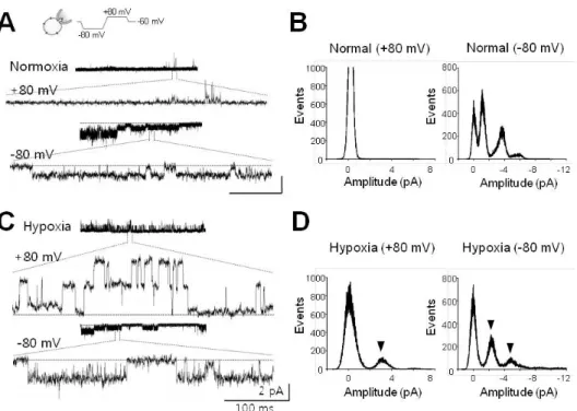
Fig. 1. Single channel currents activated by acute hypoxia. (A) Single channel currents recorded at +80 mV (upper) and╶80 mV (lower) under normoxic conditions. Channel currents in expanded scale were quoted from each representative recording. (B) All-point histograms analyzed from activities shown in (A). Histograms at +80 mV and╶80 mV were separately presented. (C) Channel currents recorded under hypoxic conditions. These activities were recorded in the same cell after switching to hypoxic solution (HS). (D) Histograms analyzed with recording data shown in (C). Arrowheads indicate the peaks activated by infusion of HS. Current recordings were made in cell-attached (CA) configuration with voltage waveforms as presented at the top of (A). Dashed lines (in A, C) denote closed level. Calibration bars are equal to 100 ms and 2 pA.
Acute hypoxia-induced channel with 30-pS conduc- tance
In this study, we tried to identify a channel responsible for direct depolarization in response to acute hypoxia. For activation of such channels, single channel currents were recorded before and after hypoxic stress using a cell-at- tached (CA) configuration, which can protect the environ- ment of the channel.
After forming CA configuration under symmetrical Na+- rich (120 mM NaCl) conditions, diverse channels with am- plitudes ranging from less than 0.5 pA to ~3.5 pA were observed. Channels could be detected at╶80 mV, but not at the depolarization potential (+80 mV, refer to Fig. 1A).
Hypoxic stress did not affect channel activities, except for one channel with a conductance of 30-pS and an amplitude of 2.5 pA at +80 or╶80 mV (Fig. 1C). This channel was activated ~2 min after application of HS to the bath sol- ution (arrowheads in Fig. 1D). Further, the channel could be detected at a rate of ~13% (one of seven to eight cells tested) under hypoxic conditions, and it showed long-lived opening behavior (mean open time, to 310.7±52.4 ms, n=4) and open probability (Po) was 0.88±0.02 (n=4).
The 30-pS channel has Na+ selectivity
Interestingly, the 30-pS channel was activated in re- sponse to acute hypoxia. Patch mode was switched to in- side-out (IO) configuration from CA in order to identify
which ion passes through. Under symmetrical Na+-rich con- ditions, channel activation induced by hypoxia was main- tained after excision of the membrane patch, implying that the channel was permeable to Na+ or Cl╶ (Fig. 2A and 2B).
This activation disappeared after return to normoxia (data not shown), suggesting that the channel could work in the absence of cytosolic components. In the present study, we did not observe activation of the channel in IO patch mode from CA mode upon exposure to HS.
In the next step, to determine whether or not the channel is permeable to either Na+ or Cl╶, the effects of chloride on channel currents were examined using glutamate. Hypoxia-in- duced 30-pS channels were not affected by removal of Cl╶ (Fig.
2D). For the experiments shown in Fig. 2C and D, we clamped the cell interior at +80 mV in order to record currents elicited either by internal (bath) K+ or external (pipette) Cl╶. To examine cation selectivity, IO membrane patches were excised from cells containing 30-pS channels activated by hypoxia. Channel activity was switched off by recovery to normoxia (upper trace in Fig. 3A) but vigorously re- activated by subsequent application of HS (middle in Fig.
3A and right panel in 3B). When Na+ was removed by re- placement with eqimolar K+ or N-methyl-D-glucamine (NMDG) in the bath solution (i.e. cytoplasmic side), channel activity was completely abolished (Fig. 2C and 3A). As shown in the lower trace in Fig. 3A, channel activity (N*Po) in the presence of NMDG, but not Na+, was remarkably reduced to 18.07±6.13% (n=3) compared to that under hypoxic Na+-rich conditions. Channel inhibition was also observed upon the removal of Na+ in the pipette solution (extracel-
60 YJ Bae, et al
Fig. 2. Hypoxia-activated channel currents impermeable to anions. (A) Preservation of channel activity induced by hypoxia in excised cell-free membrane patch. This channel was silent upon return to normoxia (upper tracings) after switching to inside-out (IO) configuration.
This channel was reactivated following perfusion of HS (lower tracings). (B) All-point histograms analyzed from activities recorded in IO patch before (left) and after perfusion of HS (right). These experiments were performed on membrane patches, which were found to contain hypoxia-activated channels in CA mode. Arrowhead shows the peak activated by hypoxia. (C) Effects of potassium and choride ions on hypoxia-activated channel. Right panel shows inhibition of 30-pS channels by application of potassium glutamate (K-glu). (D) Ineffectiveness of Cl╶ on hypoxia-elicited activation of 30-pS channel. Right panel shows no effect without Cl╶ in the pipette. For experiments in (C) and (D), membrane patches were clamped at +80 mV in order to record currents elicited either by internal (bath) K+ or external (pipette) Cl╶. Dashed lines (in A, C) denote closed level. Calibration bars are equal to 100 ms and 2 pA
Fig. 3. Hypoxia-activated channel selective to Na+. (A) Representative tracings of 30-pS channels blocked by removal of Na+. Upper two traces were recorded in the presence of Na+ in normal (upper) and hypoxic bathing solution (middle), respectively, whereas the lower one was recorded in the presence of impermeable NMDG instead of Na+ as indicated by the drawings. (B) Na+-dependence of peaks corres- ponding to activation of channels under hypoxic conditions. Arrowhead designates typical peak for channels with an amplitude of 2.5 pA (at 80 mV) under hypoxic conditions. Note that removal of Na+ abolished typical peaks in the histogram labeled as Hypoxia+NMDG-Cl.
(C) Summarized bar chart representing the channel activity reduced by removal of Na+ under hypoxic conditions. All data were obtained from single channel recordings in IO patches clamped at +80 mV. Asterisk (*) stands for differences from control (p<0.001).
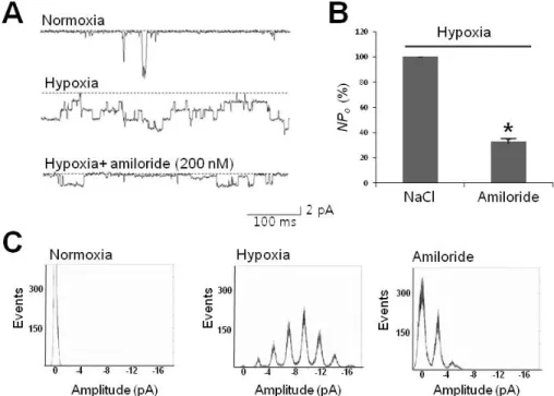
Fig. 4. Hypoxia-activated channels sensitive to amiloride. (A) Representative tracings of 30-pS channels blocked by amiloride, a neuronal Na+ channel blocker. Tracings represent that channel activity due to hypoxia (second tracing) was sensitively inhibited by 200 nM amiloride (third tracing). (B) Summarized bar chart representing the channel activity reduced by amiloride under hypoxic conditions. All data were obtained from single channel recordings at╶80 mV. (C) All-point histogram showing the abolishment of peaks typically activated under hypoxic conditions. Note that the middle histogram shows hypoxia-elicited peaks in regular 2.5-pA intervals. Asterisk (*) stands for differences from control (p<0.01)
lular side). Since the solutions used in the present study contained Ca2+ (1.8 mM), it is unlikely that Ca2+ passed through the 30-pS channel. As shown in Fig 3A, channel activity was significantly (p<0.001) reduced under Na+-free conditions containing Ca2+. These results suggest that the channel was predominantly Na+-selective.
Finally, to determine the channel preference for Na+, we examined whether or not another monovalent such as K+ passes through. After adjustment to K+-rich (100 mM K-glutamate) conditions, channel activity completely dis- appeared under low Na+ content (20 mM), as shown in Fig.
2C, suggesting that K+ did not pass through. Therefore, the hypoxia-activated 30-pS channel was predominantly Na+- selective.
The Na+-selective 30-pS channel is blocked by lido- caine and amiloride
Since the channel activated by acute hypoxia in PC12 cells was a Na+ channel, we explored its pharmacology us- ing neuronal Na+ channel blockers such as lidocaine and amiloride. Channel activity was dramatically reduced to 80% by 100 μM lidocaine (Supplementary Fig. 2).
Regarding Na+ channels in neurons, most voltage-in- dependent Na+ channels are rapidly inactivated, with mean open times at best from 16 ms to 0.7 ms [19,20]. On the contrary, voltage-independent Na+ channels such as epi- thelial Na+ channel (ENaC)/degenerin family members are open longer than voltage-dependent Na+ channels, with rel- atively high Po up to 0.9 [21]. According to such simple ki- netic criteria, we found that the 30-pS channel shared prop-
erties with ENaC, including conductance of 30 pS, 310 ms-long mean open time, and Po of 0.88 at╶80 mV, as stat- ed with Fig. 1, which are close to those of epithelial α-subunit Na+ (ENaC-α) channel [22].
To determine whether or not the 30-pS channel shares common pharmacology with ENaC, we applied amiloride, which is widely used to specifically block ENaC and acid-sensing ion channel (ASIC). As shown in Fig. 4, less than 1 min following treatment with a nanomolar amount of amiloride (200 nM), channel activity was significantly reduced to about 30.58±10.62% (n=3) compared to that of control (p<0.05). Since ASICs require just a few tens of μM amiloride to achieve 50% inhibition (IC50), and also Ca2+-permeability [23], the 30-pS channel was determined to be an ENaC rather than the ASIC family member.
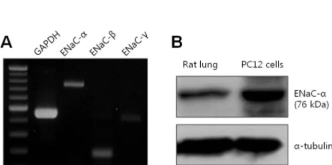
ENaC is endogenously expressed in PC12 cells Previous results have strongly suggested that the 30-pS channel activated by hypoxia demonstrates properties resem- bling ENaC. Therefore, ENaC RNA and protein were con- firmed by using RT-PCR and Western blotting in order to de- termine that ENaC is endogenously expressed in PC12 cells.
Using constructed subunit primers of ENaC consisting of α, β, and γ subunits, we confirmed the presence of RNAs corresponding to three subunits in PC12 cells (Fig.
5A). Finally, we demonstrated the endogenous protein ex- pression of ENaC using antibody specific to rat ENaC-α as well as RNA expression using RT-PCR. As shown in Fig.
5B, ENaC protein was detected by Western blotting using ENaC-α-specific antibody and lung tissue as a positive con-
62 YJ Bae, et al
Fig. 5. Expression of ENaC in PC12 cells. (A) mRNA expression levels of ENaC-α subunit (747 bp), β subunit (178 bp), and γ subunit (405 bp) in PC12 cells. (B) Detection of ENaC-α subunit protein by Western blotting in rat lung (left lane) and PC12 cells (right lane). ENaC-α subunit detected by human polyclonal antibody.
trol (left lane in Fig. 5B). These results confirm that the 30-pS channel could be a member of the ENaC family acti- vated by acute hypoxia in rat PC12 cells.
Taken together, the results in the present study suggest the presence of an ENaC-like channel activated by acute hypoxia in neuronal rat PC12 cells.
DISCUSSION
The present study provides evidence for the presence of a Na+ channel activated by acute hypoxia in PC12 cells.
This result was supported by the following: the 30-pS chan nel was activated under hypoxic conditions and its activity disappeared upon return to normoxia (Figs. 1 and 2).
Second, the channel was selective for Na+, whereas it was unaffected by Ca2+, Cl╶, or K+. Finally, the channel was blocked by lidocaine, a neuronal Na+ channel blocker.
In contrast to voltage-gated Na+ channels, which are rap- idly inactivated, opening of the hypoxia-activated channels lasted longer than 300 ms, and the inactivation process was not observed when the recording lasted for longer than 10 sec (Po=0.88, refer to Fig. 1C) [21]. The hypoxia-activated channel shared properties similar to those of ENaC, a mem- ber of the vertebrate ENaC/ASIC family [24]. On the other hand, this channel revealed differences in kinetics and pharmacology from those of ASIC. Further, it was sensi- tively blocked with a low dose (200 nM) of amiloride, where- as ASICs are less sensitive (up to hundreds μM) and rap- idly inactivated [25,26]. Since the pH level did not respond to bubbling of anoxic N2 gas, it was identified as an ENaC rather than ASIC. This suggestion was supported by the expression of ENaC protein in PC12 cells (Fig. 5), although direct evidence is required that heterologously expressed ENaC is also sensitive to acute hypoxia.
The Na+ channel conductance activated by acute hypoxia was larger than those reported in studies on endogenous ENaCs. The conductance was about 30 pS (2.5 pA at╶80 mV) in the present study, whereas a unit conductance of ENaC has been reported at best to be less than 10 pS [27-29]. If the hypoxia-activated Na+ channel belongs to the ENaC family, then a larger conductance would conflict with those of ENaCs which are believed to form a heteromeric channel composed of four subunits α, β, γ, and δ [21].
Only a few reports have reported conductance levels of 21~40 pS from recordings performed in the planar lipid bi- layer or on homomeric ENaC-α [30-32], implying that the conductance could be modulated by β- and γ-subunits as
well as cytoplasmic factor(s). For instance, reduction of di- sulfide bonds with dithiothreitol from ENaC incorporated into planar lipid bilayers reduced single channel con- ductance from 40 pS to 13 pS [30]. Considering this redox modulation of ENaC conductance, it is likely that oxidation can elevate unit ENaC conductance.
The mechanism underlying ENaC activation during acute hypoxia is difficult to explain based solely on this study. At present, there is a general agreement that inter- mediate or long-term hypoxia inhibits ENaC activity in the alveolar epithelia, whereas the effect of acute hypoxia on neuronal ENaC unclear. This channel is upregulated by proteolytic enzymes and phosphoinositides (PIs), including PI(4,5)P2 and PI(3,4,5)P3. Both PIs can interact with the N terminus of ENaC-β as well as the proximal C terminus of ENaC-γ, thereby enhancing ENaC activity. Depletion of membrane PI(4,5)P2 inhibits ENaC activity [33]. On the other hand, hypoxia provokes intense Ca2+ mobilization as well as the level of PI(4,5)P2, stimulating calpain proteolytic activity [34]. Thus, it can be stated that acute hypoxia acti- vates proteolytic enzymes such as calpain and prostasin, which, in turn, trigger the activation of ENaC [35]. In order to mimic hypoxia, we did not add ATP to the bath.
Therefore, the effect of PIs on the channel activity was greatly limited since ATP is essential to the modulation of membrane-delimited PI kinases such as PI3K or PLC.
Throughout the experiments, activation of the 30-pS chan- nel was not spontaneous and usually occurred 2~3 min af- ter exposure to hypoxia. These channels in the excised patch were re-activated without any alteration in unit con- ductance in response to hypoxia, whereas they became si- lent under normoxia (refer to Figs. 2~4). These findings strongly suggest that channel conductance can be sepa- rately regulated along with channel activity in response to hypoxia.
There have been many studies on K+ channel activity in response to acute hypoxia [4,5,7]. In a study on Kv1.2 and Kv2.1 in PC12 cells, hypoxia inhibited oxygen-sensitive K+ (KO2) current, resulting in membrane depolarization and peak inhibition within 2 min, which correlated well with the time course of membrane depolarization. The amplitude of depolarization depended on the grade of hypoxia, i.e. lev- el of low PO2 [6]. However, it was also revealed that ex- posure to hypoxia for more than 2 min caused further depo- larization (see details in Fig. 2A and 2B in reference 6).
For persistent hypoxia, membrane depolarization could continue due to the influx of cations through another chan- nel in addition to the closure of KO2 channels. In this con- text, 30-pS Na+ channels are proper since they are activated at least 2~3 min following exposure to hypoxia, facilitating further depolarization.
The main focus of this study was to identify a channel other than a KO2 channel activated by acute hypoxia. Our study was performed in PC12 cells using solution half satu- rated with oxygen (91 mmHg O2) in normal solution (152 mmHg O2). This is a disadvantage of our study compared to those performed under almost anoxic conditions (<20%
of normal). However, our conditions are more physiological, considering that the O2 level cannot reach an anoxic state within the few minutes of acute ischemic injury. Under these conditions, the present study first identified that the Na+ channel was activated by acute hypoxia and has a sin- gle channel conductance of 30 pS with ENaC-like pro- perties. However, it is unclear whether or not this channel protects from hypoxia/ischemia. With respect to hypoxia,
further study on the ENaC-like channel can provide ther- apeutic implications for neurons in response to hypoxia.
SUPPLEMENTARY MATERIALS
Supplementary data including one figure can be found with this article online at http://pdf.medrang.co.kr/paper/
pdf/Kjpp/Kjpp017-01-09-s001.pdf.
ACKNOWLEDGEMENTS
This study was supported by the Basic Science Research Program through the National Research Foundation of Korea (NRF) funded by the Ministry of Education, Science and Technology (No. 2012-0000300).
REFERENCES
1. Cross JL, Meloni BP, Bakker AJ, Lee S, Knuckey NW. Modes of neuronal calcium entry and homeostasis following cerebral ischemia. Stroke Res Treat. 2010;2010:316862.
2. Fung ML. Role of voltage-gated Na+ channels in hypoxia- induced neuronal injuries. Clin Exp Pharmacol Physiol. 2000;
27:569-574.
3. Ratan RR, Siddiq A, Smirnova N, Karpisheva K, Haskew- Layton R, McConoughey S, Langley B, Estevez A, Huerta PT, Volpe B, Roy S, Sen CK, Gazaryan I, Cho S, Fink M, LaManna J. Harnessing hypoxic adaptation to prevent, treat, and repair stroke. J Mol Med (Berl). 2007;85:1331-1338.
4. Conrad PW, Conforti L, Kobayashi S, Beitner-Johnson D, Rust RT, Yuan Y, Kim HW, Kim RH, Seta K, Millhorn DE. The molecular basis of O2-sensing and hypoxia tolerance in pheo- chromocytoma cells. Comp Biochem Physiol B Biochem Mol Biol. 2001;128:187-204.
5. Conforti L, Millhorn DE. Selective inhibition of a slow- inactivating voltage-dependent K+ channel in rat PC12 cells by hypoxia. J Physiol. 1997;502:293-305.
6. Zhu WH, Conforti L, Czyzyk-Krzeska MF, Millhorn DE.
Membrane depolarization in PC-12 cells during hypoxia is regulated by an O2-sensitive K+ current. Am J Physiol. 1996;
271:C658-665.
7. Conforti L, Bodi I, Nisbet JW, Millhorn DE. O2-sensitive K+ channels: role of the Kv1.2 -subunit in mediating the hypoxic response. J Physiol. 2000;524:783-793.
8. Kim D. K+ channels in O2 sensing and postnatal development of carotid body glomus cell response to hypoxia. Respir Physiol Neurobiol. 2013;185:44-56.
9. López-López JR, González C, Ureña J, López-Barneo J. Low pO2 selectively inhibits K channel activity in chemoreceptor cells of the mammalian carotid body. J Gen Physiol. 1989;93:
1001-1015.
10. López-López JR, González C, Perez-Garcia MT. Properties of ionic currents from isolated adult rat carotid body chemo- receptor cells: effect of hypoxia. J Physiol. 1997;499:429-441.
11. Ji HL, Benos DJ. Degenerin sites mediate proton activation of deltabetagamma-epithelial sodium channel. J Biol Chem. 2004;
279:26939-26947.
12. Wesch D, Miranda P, Afonso-Oramas D, Althaus M, Castro- Hernández J, Dominguez J, Morty RE, Clauss W, González- Hernández T, Alvarez de la Rosa D, Giraldez T. The neuronal- neuronal-specific SGK1.1 kinase regulates δ-epithelial Na+ channel independently of PY motifs and couples it to phos- pholipase C signaling. Am J Physiol Cell Physiol. 2010;299:
C779-790.
13. Seta KA, Spicer Z, Yuan Y, Lu G, Millhorn DE. Responding to hypoxia: lessons from a model cell line. Sci STKE. 2002;
2002:re11.
14. Abe E, Fujiki M, Nagai Y, Shiqi K, Kubo T, Ishii K, Abe T, Kobayashi H. The phosphatidylinositol-3 kinase/Akt pathway mediates geranylgeranylacetone-induced neuroprotection against cerebral infarction in rats. Brain Res. 2010;1330:151-157.
15. Tong Q, Gamper N, Medina JL, Shapiro MS, Stockand JD.
Direct activation of the epithelial Na+ channel by phospha- tidylinositol 3,4,5-trisphosphate and phosphatidylinositol 3,4- bisphosphate produced by phosphoinositide 3-OH kinase. J Biol Chem. 2004;279:22654-22663.
16. Chen Y, Shi G, Xia W, Kong C, Zhao S, Gaw AF, Chen EY, Yang GP, Giaccia AJ, Le QT, Koong AC. Identification of hypoxia-regulated proteins in head and neck cancer by proteomic and tissue array profiling. Cancer Res. 2004;64:
7302-7310.
17. Lebowitz J, Edinger RS, An B, Perry CJ, Onate S, Kleyman TR, Johnson JP. Ikappab kinase-beta (ikkbeta) modulation of epithelial sodium channel activity. J Biol Chem. 2004;279:
41985-41990.
18. Choi SW, Ahn JS, Kim HK, Kim N, Choi TH, Park SW, Ko EA, Park WS, Song DK, Han J. Increased expression of atp-sensitive K+ channels improves the right ventricular tole- rance to hypoxia in rabbit hearts. Korean J Physiol Pharmacol.
2011;15:189-194.
19. Wood JN, Boorman JP, Okuse K, Baker MD. Voltage-gated sodium channels and pain pathways. J Neurobiol. 2004;61:55-71.
20. Catterall WA, Goldin AL, Waxman SG. International Union of Pharmacology. XLVII. Nomenclature and structure-function relationships of voltage-gated sodium channels. Pharmacol Rev. 2005;57:397-409.
21. Kellenberger S, Schild L. Epithelial sodium channel/degenerin family of ion channels: a variety of functions for a shared structure. Physiol Rev. 2002;82:735-767.
22. Hammarström AK, Gage PW. Hypoxia and persistent sodium current. Eur Biophys J. 2002;31:323-330.
23. Waldmann R, Champigny G, Bassilana F, Heurteaux C, Lazdunski M. A proton-gated cation channel involved in acid- sensing. Nature. 1997;386:173-177.
24. Alexander SPH, Mathie A, Peters JA. Guide to receptors and Channels (GRAC), 5th edition. Br J Pharmacol. 2011;164 Suppl 1:S1-324.
25. Ohbuchi T, Sato K, Suzuki H, Okada Y, Dayanithi G, Murphy D, Ueta Y. Acid-sensing ion channels in rat hypothalamic vasopressin neurons of the supraoptic nucleus. J Physiol. 2010;
588:2147-2162.
26. Chu XP, Miesch J, Johnson M, Root L, Zhu XM, Chen D, Simon RP, Xiong ZG. Proton-gated channels in PC12 cells. J Neurophysiol. 2002;87:2555-2561.
27. Garty H, Palmer LG. Epithelial sodium channels: function, structure, and regulation. Physiol Rev. 1997;77:359-396.
28. Kellenberger S, Hoffmann-Pochon N, Gautschi I, Schneeberger E, Schild L. On the molecular basis of ion permeation in the epithelial Na+ channel. J Gen Physiol. 1999;114:13-30.
29. Anantharam A, Palmer LG. Determination of epithelial Na+ channel subunit stoichiometry from single-channel conduc- tances. J Gen Physiol. 2007;130:55-70.
30. Ismailov II, Berdiev BK, Bradford AL, Awayda MS, Fuller CM, Benos DJ. Associated proteins and renal epithelial Na+ channel function. J Membr Biol. 1996;149:123-132.
31. Kelly O, Lin C, Ramkumar M, Saxena NC, Kleyman TR, Eaton DC. Characterization of an amiloride binding region in the alpha-subunit of ENaC. Am J Physiol Renal Physiol. 2003;285:
F1279-1290.
32. Ma HP, Al-Khalili O, Ramosevac S, Saxena S, Liang YY, Warnock DG, Eaton DC. Steroids and exogenous γ-ENaC subunit modulate cation channels formed by α-ENaC in hu- man B lymphocytes. J Biol Chem. 2004;279:33206-33212.
33. Ma HP, Chou CF, Wei SP, Eaton DC. Regulation of the epithelial sodium channel by phosphatidylinositides: experi- ments, implications, and speculations. Pflugers Arch. 2007;455:
169-180.
34. Yamashima T, Saido TC, Takita M, Miyazawa A, Yamano J,
64 YJ Bae, et al
Miyakawa A, Nishijyo H, Yamashita J, Kawashima S, Ono T, Yoshioka T. Transient brain ischaemia provokes Ca2+, PIP2 and calpain responses prior to delayed neuronal death in monkeys.
Eur J Neurosci. 1996;8:1932-1944.
35. Donaldson SH, Hirsh A, Li DC, Holloway G, Chao J, Boucher RC, Gabriel SE. Regulation of the epithelial sodium channel by serine proteases in human airways. J Biol Chem. 2002;277:
8338-8345.