Synchronous Triple Primary Lung Cancers: A Case ReportHyun Jung Yoon, MD
5
0
0
전체 글
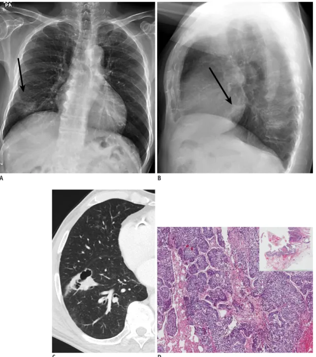
(2) Synchronous Primary Lung Cancers. A. B. C. D. Fig. 1. 72-year-old man with synchronous triple primary lung cancers.. A, B. Posteroanterior (A) and lateral (B) chest radiographs reveal 4.0-cm ill-defined mass in right lower lobe (arrows). Lung window images (window level, -500 Hounsfield unit [HU]; window width, 1500 HU) of CT scan (2.5-mm slice thickness) of thorax and corresponding microscopic features at low magnification (hematoxylin and eosin stain, x 5 [x 1 in inset]). C. Anterior basal segment of right lower lobe is showing 4.2 cm irregular cavitary lesion. D. This lesion was confirmed to be squamous cell carcinoma with moderate differentiation exhibiting nests of polygonal cells with pink cytoplasm and distinct cell borders.. adenocarcinoma (tumor 2), and an invasive nonmucinous adenocarcinoma (tumor 3). There was no significant enlargement of lymph nodes in the mediastinum or hilar areas and no other abnormalities in both lungs. The positron emission tomography (PET)/CT revealed a mild to high 18F-fluorodeoxyglucose uptake in the masses (maximum standardized uptake value [SUVmax] of 4.4, 1.7, and 0.7 for kjronline.org. Korean J Radiol 15(5), Sep/Oct 2014. tumor 1, 2, and 3, respectively) in the right lung. Combined results obtained from abdominal ultrasonography, brain magnetic resonance imaging, radionuclide bone scanning and PET/CT scanning revealed no metastatic lesions. A video-assisted thoracoscopic right lower lobectomy, mediastinal lymph node dissection and a wedge resection of the right upper lobe were performed without a preoperative 647.
(3) Yoon et al.. E. F. G. H. Fig. 1. 72-year-old man with synchronous triple primary lung cancers.. E. 3.8-cm parenchymal low-attenuating consolidative lesion is at posterior-basal segment of right lower lobe. F. This lesion was confirmed to be invasive mucinous adenocarcinoma composed of glands lined by tall columnar mucin-containing cells. G. 2.5-cm well-defined part-solid nodular lesion is at posterior segment of right upper lobe. H. It revealed adenocarcinoma with moderate differentiation and acinar pattern showing acini of polyhedral cells.. percutaneous needle biopsy for diagnosis. On the pathologic examination, two lesions were found in the right lower lobe; tumor 1 at the anterior basal segment measured 2.4 x 2.0 cm in size and was confirmed to be a squamous cell carcinoma with moderate differentiation (Fig. 1D) and tumor 2 at the posterior basal segment was 5.1 x 4.9 cm in size and was confirmed to be an invasive mucinous adenocarcinoma (Fig. 1F). Tumor 3 at the posterior segment 648. of the right upper lobe measured 1.5 x 1.0 cm in size and was confirmed to be an invasive adenocarcinoma with a moderate differentiation and an acinar pattern (Fig. 1H). Regional lymph nodes were examined and two metastatic lymph nodes were found in the right upper paratracheal and subcarinal area involving a squamous cell carcinoma. Thus, the final TNM stage was set as pT2bN2M0. A genetic mutation analysis was performed for the largest invasive Korean J Radiol 15(5), Sep/Oct 2014. kjronline.org.
(4) Synchronous Primary Lung Cancers. mucinous adenocarcinoma and it was found to be a wild type of epidermal growth factor receptor/K-ras and anaplastic lymphoma kinase. After the surgery, the adjuvant chemotherapy was planned and the patient was discharged.. DISCUSSION Multiple primary lung cancers are divided into simultaneous (synchronous) and sequential (metachronous) tumors. Synchronous tumors are significantly rarer than metachronous tumors (1). The incidence of synchronous multiple primary lung cancer has been variably reported to range from 0.26% to 1.33% (6-8). Ferguson (9) summarized the overall incidence of histologically categorized, synchronous double primary lung cancers; over 51.3% of individual patients had identical histologic subtypes and the remaining 48.7% had different histologic subtypes. A squamous cell carcinoma is the most common cancer, comprising over 70% of synchronous cancers with identical histologic subtypes and nearly 85% of patients with tumors of different histologic subtypes. An adenocarcinoma was present in a relatively smaller percentage of patients with synchronous primary lung cancer. Nevertheless, it is not easy to evaluate the exact incidence due to the difficulties in distinguishing a synchronous multiple primary lung cancer from a single pulmonary neoplasm with intrapulmonary metastases or pulmonary metastases originating from primary cancers in different organs. However, in order to plan the appropriate treatment based on the stage it is clinically relevant during the initial workup and imaging study to assess if the second tumor is a synchronous tumor or a metastasis (10). Generally accepted diagnostic criteria include the demonstration of synchronous masses with different histology and the proof that tumors arise from separate and distinct endobronchial foci, if histologically similar (9). The Martini and Melamed (11) criteria are based on tumor characteristics (inclusive of, but not limited to, morphology, location, presence or absence of carcinoma in situ, vascular invasion and metastasis), but lack the power to differentiate between metastasis and a second primary lung cancer (12). Malignant characteristics of CT imaging of primary lung cancer in various lesions may provide valuable clues for the preoperative distinction of synchronous primary tumors from primary lung tumors with intrapulmonary metastases or other metastases at the initial workup. Not all patients can be stratified in accordance kjronline.org. Korean J Radiol 15(5), Sep/Oct 2014. with the above diagnostic criteria of synchronous multiple primary lung cancers (11). Thus, separate biopsies need to be performed for different pulmonary masses. We reported a patient with three synchronous, independent and histologically variant lung cancers. All three tumors had different and distinct CT imaging features and all lesions displayed the malignant characteristics of a primary lung cancer. On CT, the solid nodule/mass with irregular cavitation was diagnosed as a squamous cell carcinoma, the most common histological type of lung cancer to cavitate (82% of cavitary primary lung cancer) (3, 5). An unchanged parenchymal consolidative lesion with low attenuation was diagnosed as an invasive mucinous adenocarcinoma (4) whereas a well-defined, part-solid nodule appearing as mixed areas of ground-glass opacity and solid attenuation was likely to be a nonmucinous adenocarcinoma (2). A subsequent percutaneous needle biopsy to exclude the possibility of metastasis was not necessary due to the confirmed differences in radiologic features. Even though there are many case reports regarding synchronous multiple primary lung cancers in the literature, very few cases of triple tumors with different histologies have been investigated yet (6, 13-15). Moreover, no previously reported synchronous lung cancers have been investigated in terms of radiologic findings. To our knowledge, this is the first report on three synchronous primary lung cancers with radiologic-histopathologic correlation.. REFERENCES 1. Sulkes A, Naparstek Y, Shalit M, Kopolovic J. Second primary lung carcinoma. J Surg Oncol 1980;15:375-380 2. Aoki T, Tomoda Y, Watanabe H, Nakata H, Kasai T, Hashimoto H, et al. Peripheral lung adenocarcinoma: correlation of thinsection CT findings with histologic prognostic factors and survival. Radiology 2001;220:803-809 3. Chaudhuri MR. Primary pulmonary cavitating carcinomas. Thorax 1973;28:354-366 4. Lee SM, Goo JM, Park CM, Lee HJ, Im JG. A new classification of adenocarcinoma: what the radiologists need to know. Diagn Interv Radiol 2012;18:519-526 5. Vourtsi A, Gouliamos A, Moulopoulos L, Papacharalampous X, Chatjiioannou A, Kehagias D, et al. CT appearance of solitary and multiple cystic and cavitary lung lesions. Eur Radiol 2001;11:612-622 6. Ferguson MK, DeMeester TR, DesLauriers J, Little AG, Piraux M, Golomb H. Diagnosis and management of synchronous lung. 649.
(5) Yoon et al.. cancers. J Thorac Cardiovasc Surg 1985;89:378-385 7. Deschamps C, Pairolero PC, Trastek VF, Payne WS. Multiple primary lung cancers. Results of surgical treatment. J Thorac Cardiovasc Surg 1990;99:769-777; discussion 777-778 8. Wu SC, Lin ZQ, Xu CW, Koo KS, Huang OL, Xie DQ. Multiple primary lung cancers. Chest 1987;92:892-896 9. Ferguson MK. Synchronous primary lung cancers. Chest 1993;103(4 Suppl):398S-400S 10. Huang J, Behrens C, Wistuba I, Gazdar AF, Jagirdar J. Molecular analysis of synchronous and metachronous tumors of the lung: impact on management and prognosis. Ann Diagn Pathol 2001;5:321-329 11. Martini N, Melamed MR. Multiple primary lung cancers. J. 650. Thorac Cardiovasc Surg 1975;70:606-612 12. Ostrovnaya I, Olshen AB, Seshan VE, Orlow I, Albertson DG, Begg CB. A metastasis or a second independent cancer? Evaluating the clonal origin of tumors using array copy number data. Stat Med 2010;29:1608-1621 13. Carey FA, Donnelly SC, Walker WS, Cameron EW, Lamb D. Synchronous primary lung cancers: prevalence in surgical material and clinical implications. Thorax 1993;48:344-346 14. Sarper A, Ozbilim G, Demircan A. Metachronous triple cancer: esophageal carcinoma 4 years later the synchronous bilateral bronchogenic carcinoma. Eur J Cardiothorac Surg 2003;24:303 15. Tokat AO, Ozkan M, Güngör A. [Synchronous lung carcinoma: a case report]. Tuberk Toraks 2003;51:70-73. Korean J Radiol 15(5), Sep/Oct 2014. kjronline.org.
(6)
수치
관련 문서
(in case of partially miscible and form a low-boiling azeotrope).
This book contains hundreds of complete, working examples illustrating many common Java programming tasks using the core, enterprise, and foun- dation classes APIs.. Java Examples
a prospective analysis of 50 cases. Preoperative staging of non-small-cell lung cancer with positron-emission tomography. Staging of non- small-cell lung cancer
Effects of Group Piano Playing on International Marriage Immigrant Women's Self-Efficacy; A Case Study.. Woo,
Results: 44 of 1048 patients with gastric cancer(4.1%) had synchronous and metachronous cancers. The average time interval between gastric cancer and secondary primary cancer
Generally, in case that we predict the vibration by blasting, we compute it on the basis of the charge weight per delay but in case of shocking
à For each subentity, create a table that includes the attributes of that entity set plus the primary key of the higher level
At the end of the study, a reevaluation of each study case was performed with the same questionnaire. The result shows there has been a meaningful result in the group
