Effect of Developmental Lead Exposure on the Expression of Hippocampal NMDA Receptor Subunit mRNA
Tae-Wan Kim*, In-Sung Chung*, Jae-Hoon Bae
1), Dong-Hoon Shin, Mi-Young Lee, Joon-Sik Kim
2)Department of Occupational & Environmental Medicine and Institute for Medical Science Keimyung University Dongsan Medical Center
Department of Physiology1), Pediatrics2)School of Medicine and Institute for Medical Science Keimyung University
─Abstract ─
해마신경세포 발생단계별 N M DA 수용체 아단위 m R NA 발현에 대한 연의 영향
계명대학교 동산의료원 산업의학과 및 의과학연구소, 계명대학교 의과대학 생리학교실1 ), 소아과학교실2 )및 의과학연구소
김태완*・정인성*・배재훈1 )・신동훈・이미영・김준식2)
목적: in vivo 및 in vitro에서 해마 신경세포의 발생단계별 NMDA 수용체 아단위 mRNA 발현에 대한 연 폭 로 영향을 알아보고자 하였다.
방법: 흰쥐 해마 신경세포의 발생단계별 NMDA 수용체 NR2A, NR2B 아단위 mRNA 발현에 대한 연의 영향 은 정상군과 연 폭로군의 출생 후 7일, 14일, 22일 흰쥐의 해마에서 in situ hybridization으로 mRNA 발현 정도를 d e n s i t o m e t e r로 측정하여 비교하였고, 연과 N M D A의 세포독성은 해마 신경세포 일차배양 후 도립현미 경을 이용한 형태학적인 관찰과 LDH 활성도를 이용하여 측정하였다.
결과: 연 과 N M D A에 의한 세포독성에 대한 in vitro 실험에서 형태학적 소견과 LDH 활성도에서 해마 미성숙 신경세포와 성숙 신경세포사이의 차이가 있었으므로, 연과 NMDA 독성효과는 해마 신경세포의 발달 단계에 따 라 차이가 있다. 정상군의 해마에서의 NR2A mRNA 발현은 출생 후 연령이 증가함에 따라 점진적으로 증가하 였으나, NR2B mRNA 발현은 연령의 증가에 따른 변화가 없었다. 연 폭로에 희한 NR2A mRNA 발현은 유 의하게 감소하였으나(p<0.05), NR2B mRNA 발현은 변화가 나타나지 않았다. 만성적 연 폭로는 N R 2 A를 포 함하는 NMDA 수용체를 감소시킬 수 있음을 알 수 있다.
결론: 연은 해마신경세포의 발생단계에서 NMDA 수용체 아단위 특히 NR2A mRNA 발현의 변화를 야기하여 시냅스 신호 전달에 영향을 나타냄을 알 수 있었다. Hippocampus
Key Words: Lead, NMDA receptor, Subunit, mRNA, Hippocampus
<접수일: 2005년 9월 22일, 채택일: 2005년 11월 4일>
교신저자: 김 준 식 (Tel: 053-250-7525) E-mail: [email protected]
✽ T.W. Kim and I.S. Chung contributed equally to this work
Corresponding author: Joon-Sik Kim (Tel:053-250-7525) E-mail: [email protected]
✽ This work was supported by the research promoting grant from the Keimyung University Dongsan Medical center in 1999.
I N T R O D U C T I O N
Lead is the most ubiquitous toxic metal and is detectable in practically all phases of the inert environment and in all biological systems. Lead is used for the production of batteries, pigments, solder, plastics, cable sheathing, ammunition, and a variety of other extruded products (Fischbein, 1998). Environmental sources include lead-based indoor paint in old dwelling, lead in contaminated drinking water, lead in air from combustion of lead-containing industrial emis- sion, hand-to-mouth activities of young children living in polluted environments, lead-glazed pot- tery and, less commonly, lead dust brought home by industrial workers on their clothes and shoes (Goyer, 1996).
Lead is the major environmental and occupa- tional health hazard affecting several organ sys- tems. Among these, the nervous system is most vulnerable (Nehru & Iyer, 1994). Children are considered a particularly sensitive risk group for lead because, on a body weight basis, their dietary intake, absorption and total retention is markedly higher than adults. In addition, the blood brain barrier in young children is not yet fully developed and their specific behavioral characteristics, such as hand-to-mouth and play- ground activities, put them into higher risk group (Winneke & Kramer, 1997). The most critical or sensitive effects in infants and chil- dren are involved in the nervous system (Goyer, 1 9 9 6 ) .
The toxic effects of lead on the CNS have been known for many years and range from acute encephalopathy when humans or experimental animals are exposed to high levels of the lead (Byers & Lord, 1943; Pentschew, 1965), to sub- tle changes in behavior and cognition associated with low-level exposures (Needleman et al, 1990). Experimental evidences indicate that chronic exposure to low-level lead in children during development, results in the impairment of learning and memory function (Bellinger et al, 1987; McMichael et al, 1988).
The hippocampus plays a pivotal role in learn-
ing and memory, and is markedly developed in postnatal period, thus it is main target in lead- induced neurotoxicity in children. However, the mechanism of lead-induced neurotoxicity remains u n e x p l a i n e d .
The great importance of neurodevelopmental status for lead-induced neurotoxicity has been demonstrated by the experiments concerning the effect of lead on N-methyl-D-aspartate (NMDA) receptor-mediated function (Alkondon et al, 1990; Ujihara & Albuquerque, 1992). NMDA receptor has also been shown to participate in the induction of long-term potentiation in the hippocampus, a cellular phenomenon thought to be involved in some forms of learning and mem- ory (Madison et al, 1991).
Functional NMDA receptors are heteromeric complexes containing NR1 and NR2 (NR2A- NR2D) subunits (Dingledine et al, 1999), with the NR2 subunit determining many functional properties of NMDA receptors in heterologous systems (Cull-Candy et al, 2001). In the fore- brain structures including cortex and hippocam- pus, expression of NR2B mRNA was much high- er than in cerebellum and remained relatively constant over time (Zhong et al, 1994). In addi- tion, neonatal rats, levels of NR2B mRNA were much higher than levels of NR2A mRNA, but these subunits were expressed at similar levels in adults. This suggests that in neonatal rats most NMDA receptors maybe composed of NR1 and NR2B subunits, whereas in adult animals NMDA receptors may be composed of NR1 and NR2A or NR1 and NR2B subunits. More com- plex pattern of expression including multiple NR2 subunits may also exist (Sheng et al, 1994).
Therefore, lead-induced changes in hippocam- pal NMDA receptor subunit mRNA expression may lead to abnormality of natural NMDA stoi- chiometry and interfere with the physiological function of the NMDA receptor in the develop- ment of defined neuronal connections (Zhang et al, 2002).
The present study investigated the expression, at the mRNA level with in situ hybridization, of
NR2A and NR2B subunits in vivo postnatal 14 and 22 days following chronic lead exposure in gestation 0 day. In addition, this study exam- ined the effects of lead in vitro hippocampal cells to understand the differential cytotoxicity depending on the neuronal development.
M ATERIALS AND METHODS 1. Animal experimental procedure:
Adult female Sprauge-Dawley rats were indi- vidually housed in plastic cages with bedding at ( 2 2±2℃) under a light/dark cycle of (12L/12D).
The method of lead exposure in the present study was that animal exposed 750 ppm lead acetate in drinking water. Pregnant rats were randomly assigned to exposure groups containing either 0 or 750 ppm lead acetate. Food intake was allowed ad libitum. Treatment started from the beginning of gestation and continued after parturition, which was carefully verified. The litter size was culled to 8 to 10 pups. The date of birth was considered to be Day 0 (zero). Pups were weaned at 21 days of age and were weighed. Pups were euthanized by decapitation of three to five different litters at each age group at postnatal day 7, 14 or 22, only one ani- mal per litter is used . The brains were careful- ly removed from the skull, placed on an ice- chilled plate, rinsed with ice cold sterile saline and fixed in the 4% paraformaldehyde for 12 hr. The brains were stored at -70℃ until used for in situ hybridization studies. Prior to wean- ing, all pups from a litter were euthanized at a particular age. The brains were paraffin-embed- ded and serial coronal sections 7 mm thick were obtained.
2. Plasmids and in vitro transcription:
The cDNA fragments containing c-terminals of NR2A (aa; 893-1101) and NR2B (aa; 1167-1482 including 84 base pair 3-end untranslating region) which were subcloned into a PCR 2.1 were kindly provided by Dr. Moon (Dongguk
University, Korea). The plasmids containing fragments of NR subunits cDNAs were lin- earized at BamH Ⅰ and Hind Ⅲ. The ribo probe were generated with T7 (antisense) or Sp6 (sense) polymerase using an in vitro transcrip- tion kit (Bohringer Mannheim, Germany) in the presence of digoxigenin-labeled UTP according to m a n u f a c t u r e r’s instructions.
3. In situ hybridization:
Paraffin wax was removed with xylene and the sections were rehydrated with ethanol (99%, 95%, 90%, 80% and 70% in order). After wash- ng with 0.1% diethylpyrocarbonate (DEPC) water, the sections were dipped consecutively in 0.05 mol/L Tris-HCl (pH 7.4) for 2 min, 0.2 N HCl for 10 min and 0.1% DEPC water for 5 min at room temperature. Then, they were pretreat- ed with 10 ㎍/mL proteinase K at 37 ℃ for 30 min. After washing with 0.1% DEPC water, the sections were fixed with 0.4% paraformaldehyde in 0.1 mol/L phosphate buffered solution (PBS) for 10 min at 4 ℃. Prehybridization was per- formed at 50 ℃ for 3 hr in a buffer containing 4
× SSC, 10% dextran sulfate, 50% formamide, 2 mmol/L EDTA, 1 × Denhart solution, and 500
㎍/mL herring sperm DNA.
Hybridization was done overnight at 50 ℃ in a prehybridization buffer supplemented with 1—5
㎍/mL digoxigenin-labelled anti-sense probe.
After removal of the hybridization buffer, the sections were consecutively washed twice at 42 ℃ for 15 min in 2 × SSC, at 42 ℃ for 10 min in 0.2 × SSC, and room temperature for 5 min in 0.1 × SSC. After rinsing in solution Ⅰ〔1 0 0 mmol/L Tris (pH 7.5), 150 mmol/L NaCl〕for 5 min at room temperature, they were treated with solution Ⅱ (1% BSA in solution Ⅰ) for 1 h at room temperature, and then incubated with alkaline phosphatase conjugated digoxigenin antibody (1:500 in solution Ⅱ). After rinsing again with solution Ⅰ at room temperature for 15 min, the sections were treated with solution
Ⅲ〔100 mmol/L Tris (pH 9.5), 100 mmol/L NaCl, 50 mmol/L MgCl2〕. Detection of digoxi-
genin-labelled RNA-mRNA hybrid was accom- plished with nitro blue tetrazolium and 5-bromo- 4-chloro-3-indolyl phosphate. After color reac- tion, the sections were rinsed with 10 mmol/L Tris-HCl (pH 8.0), 1 mmol/L EDTA and with crystal mount.
4. Semi-quantitative Measurments :
To measure the intensity of mRNA hybridiza- tion signal, a scanning densitometer (Scion image software version 1.59, USA) was used after background subtraction. We analyzed hybridized sections at the level of -2.56 mm from the bregma, according to the atlas of Paxinos and Watson (1986). In the hippocampus, the identification of the CA1 region was con- firmed in HE stained sections. Hybridization results were categorized by 2 successive similar features. Positive hybridization signal of slides were calculated at five portions from each hip- pocampal regions.
5. Primary hippocampal neuronal cell cul- t u r e :
Primary neuronal cultures were prepared from fetal hippocampi. Dissected hippocampal tissue was digested with trypsin (1 mg/mL) for 10 min at 37 ℃ and mechanically dissociated by tritura- tion through a 25-gauge sterile needle in Neurobasal medium (Life Technologies, Paisley, UK) containing 10% fetal bovine serum (HyClone Laboratories, UT, USA). Cells were collected by centrifugation and resuspended in Neurobasal medium containing 10% foetal bovine serum supplemented with L-glutamine (0.5 mmol/L) and gentamicin (10 g/mL). Cell viabili- ty was assessed by trypan blue exclusion (gener- ally 98%). Cells were plated at a density of 5 × 10⁴ cells/well in poly-D-lysine-coated 24-well plates (Biocoat, Becton Dickinson Ltd, NJ, USA) and maintained under a humidified atmosphere, at 37 ℃/5% CO2. After 24 hr, the medium was replaced with Neurobasal medium containing B- 27 growth supplement (Life Technologies,
Paisley, UK), L-glutamine (0.5 mmol/L), gen- tamicin (10 g/mL) and cytosine arabinoside (5 mol/L) to allow for optimal neuronal growth and to prevent proliferation of non-neuronal cells.
Cells were maintained for a further 21 days prior to treatment. Immunohistochemical and morphological analyses with an anti-glial fibril- lary acidic protein (GFAP, Sigma Chemicals, UK) antibody or anti-neurone-specific enolase (NSE) antibody indicate that this method of hippocampal cell culture generates an NSE-posi- tive and GFAP-negative neuronal culture com- prised largely of pyramidal cell-like, bipolar and stellate neurones. Less than 2% of cells are GFAP-positive. The cells were stable for at least 3 weeks after plating and their viability was not affected by treatment.
6. Measurement of cell death:
Neuronal cell death was assessed by morpho- logical examination of cells using phase-contrast microscopy and quantified by measuring lactate dehydrogenase (LDH) released by damaged or destroyed cells into the bathing medium 16—24 h after Lead and/or NMDA treatments, otherwise indicated somewhere else. Basal LDH levels were determined and substracted from the levels in experimental conditions to yield the LDH signal specific to experimemntal injuries. Absorbance was measured at 570 nm.
7. Statistical analysis:
Data are expressed as the mean ± SEM and analyzed for statistical significance using Wilcoxon rank sum test or Kruskal-Wallis test.
P value of <0.05 was considered significant.
R E S U LTS
1. NR2A and NR2B mRNA expression in the hippocampus after in vivo developmental lead exposure
During postnatal days from day 7 to 22, the
expression of NR2A, NR2B subunit mRNAs could be observed in three subfields of the hip- pocampus, i.e., CA1, CA3 and dentate gyrus (DG).
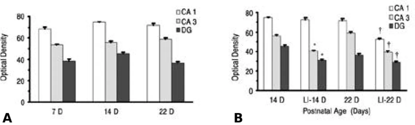
In the CA1, CA3 and DG subfields of the hip- pocampus, NR2A mRNA levels were lowest at 7 days of age and increased to peak at 22 days of age in control animals. Developmental Pb expo- sure resulted in significant decreases in NR2A mRNA levels in the CA1 subfield of the hip- pocampus at 14 days of age. At 22 days of age, significant decreases in NR2A mRNA levels were observed in the CA1, CA3 and DG subfields in Pb-exposed rats(Fig. 1).
In control animals, the expression of NR2B
mRNA in the CA1, CA3 and DG subfields of the hippocampus was highest after birth and were stable as the animals aged. No significant effect of developmental Pb exposure on NR2B mRNA expression was observed the CA1 subfield of the hippocampus at 14 days of age, but developmen- tal Pb exposure on NR2B mRNA expression was significantly decreased in the CA1, CA3 and DG subfield of the hippocampus at 22 days of age(Fig. 2).
2. The morphological changes of hippocam- pal cells after in vitro lead or NMDA e x p o s u r e
Fig. 1. Developmental regional expression of NR2A subunit mRNA levels in the hippocampus of normal postnatal days 7 (7D), 14 (14D), 22 (22D) ( A) and lead intoxicated rats (LI) (B) in this study. Each value is the mean ± SEM of three to five different litters at each age group, only one animal per litter is used. DG: dentate gyrus; *: P<0.05 relative to normal 7D; ‡, §: P<0.05 relatively to normal 14D, 22D respectively
A B
Fig. 2. Developmental regional expression of NR2B subunit mRNA levels in the hippocampus of normal postnatal days 7(7D), 14(14D), 22(22D)(A) and lead intoxicated rats(LI)(B) in this study. Each value is the mean ± SEM of three to five different litters at each age group, only one animal per litter is used. DG: dentate gyrus; *, †: P<0.05 relative to normal 14D, 22D respectively.
A B
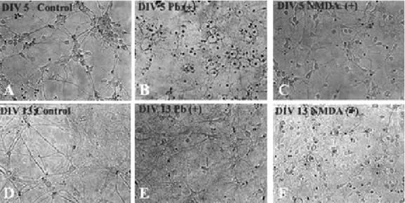
In the control, individual cells made up pyra- midal-like neurons days in vitro 5 (DIV 5) and synapses of neurons are stronger DIV 13 than DIV 5. In the Pb 50 μmol treated cultures, indi- vidual cells degenerated with loss of their process and making halo inside of the cells in the DIV 5, however, there was not clearly dif- ferent from that seen in control at DIV 13. In the NMDA 300 μmol/L for 30 min treated cul- tures, there was not clearly different from that seen in control at DIV 5, but there was cellular degeneration in the neurons DIV 13 (Fig. 3).
3. Cytotoxicity of hippocampal cells after in vitro Lead or NMDA exposure:
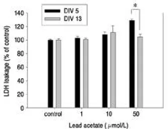
LDH leakage was employed as indices of in vitro development of hippocampal neuronal cell death. Assays were performed at 24 hr after Pb treatments. At the concentration of 50 μm o l / L Pb, significant LDH release of hippocampal neu- rons were induced in the DIV 5, but not in the DIV 13(p<0.05)(Fig. 4).
In Figure 5, the effects of NMDA and Pb dur- ing developmental stage were experimented on leakage of LDH from cultured hippocampal cells.
Assay was performed 24 hr after Pb 10 μm o l / L and/or NMDA 300 μmol/L treatment for 30 min.
In contrast to Pb-induced LDH releases, NMDA-
induced LDH releases in DIV 13 was higher than in DIV 5 (P<0.05). At concentration of 10 μm o l / L Pb, LDH release of hippocampal neurons were not significantly induced in the DIV 5 and 13.
Combined treatment of Pb and NMDA also showed similar result as NMDA only treatment.
This result implies that the LDH was released solely from the NMDA in the DIV 13.
D I S C U S S I O N
The genes encoding the NMDA receptor have been cloned and two subunit families, NR1 and NR2, have been characterized in the rat brain (Moriyoshi et al, 1991; Monyer et al, 1992). All NMDA receptors appear to function as het- eromeric assemblies composed of multiple NR1 subunits in combination with at least one type of NR2 (Stuart, 2001). The changes in the pro- tein expression of the major subunits of the NMDA receptor occur during the aging process and these subunit alterations may explain some of the changes that are seen in NMDA receptor functions during aging (Magnusson et al, 2002).
A gradual replacement NR2B by NR2A during postnatal development has been implicated in the spreading of NMDAR-EPSC decay - a phe- nomenon often linked with the ability of neu- ronal circuits to exhibit experience-dependent
Fig. 3. Photomicrograph of hippocampal neuronal cells DIV 5 (A, B, C) and 13 (D, E, F) of normal controls (A, D), 50 μmol/L Pb exposed (B, E), and 300 μmol/L NMDA for 30 min treated (C, F) rats (× 20). Micrographs are taken 20 h after Pb or NMDA treatments and are representative of 4 separate experiments. DIV: days in vitro.
synaptic plasticity (Constantine-Paton & Cline, 1998). NMDA receptor dependent long term potentiation in the CA1 region and DG of the hippocampus have been shown to be impaired by developmental lead exposure (Altmann et al, 1994; Gilbert et al, 1996).
In this study, we examined the developmental expression of NMDA receptor NR2A and NR2B subunit mRNAs in the hippocampal subfields (CA1/CA3/DG) of rats chronically exposed to low levels of Pb. NR2A mRNA subunit expression increased as a function of age in the CA1 and CA3 subfields, but NR2B mRNA not decreased as the animal aged. These findings are consis- tent with those obtained previously study (Guilarte & McGlothan, 1998; Zhang et al, 2002). Exposure of Pb during development resulted in significant decreases in NR2A sub- unit mRNA expression in the rat hippocampus.
Lead-induced changes in NR2A mRNA level during postnatal period may lead to changes in the levels or types of NMDA receptors which may alter normal hippocampal physiology and the formation of synapses. Disruptions of those process by Pb during early brain development may lead to long-lasting changes in hippocampal
synaptic plasticity (Nihei et al, 2000).
Additionally, decrease of NR2A-containing NMDA receptors which are mediate shorter duration EPSP than the NR2B-containing NMDA receptors following lead exposure might be decrease the detection of temporal precise sig- naling which are closely associated with the exactness of the signal transmission.
Omelchenko et al (1996) have also shown that the sensitivity of NMDA receptor inhibition by lead is dependent on the subunit combination and receptor assemblies composed of NR1/2A subunits appear to be the most sensitive to inhi- bition by lead. However, This study can not experiment about effect of lead of NMDA recep- tor subunits combination. The further evalua- tion of effects of lead on NMDA receptor sub- units combination will be needed. Because many of the physiological and pharmacological proper- ties of NMDA receptors depend on specific sub- units (Williams, 1993; Williams et al, 1994), it is possible that lead exposure results in alter- ations of the expression of NMDA receptor sub- units significantly influence on the synaptic sig- nal transmission.
In the CNS, lead toxicity is more common in
Fig. 4. Effects of various concentration of lead acetate on leak- age of LDH from cultured hippocampal cells at DIV 5 and 13. LDH levels were expressed as percentage of control release of LDH; n = 4 and determined from the bathing media. Assay were performed at 20 h after lead acetate treatment. DIV: days in vitro; *: P<0.05 signifi- cant different between DIV 5 and 13.
Fig. 5. Effects of NMDA (300 μmol/L) for 30 min and lead acetate (Pb, 10 μmol/L) on leakage of LDH from cul- tured hippocampal cells at DIV 5 and 13. LDH levels are expressed as percentage of control release of LDH;
n = 4. Assay are performed at 24 h after lead acetate and/or NMDA treatment. DIV: days in vitro; *: P<0.05 significant different between DIV 5 and 13.
children than adults and may produce either overt symptoms of acute encephalopathy such as ataxia, headache, convulsions, and coma or less- er deficits including learning disorders and hyperactive behavior (Blackman, 1937;
Needleman et al, 1990). In children, lead blood level as low as 1.3 to 13 μmol/L, may affect CNS development, leading to mental retarda- tion, impaired visual motor coordination and permanent cognitive deficits (Goldstein, 1992).
Because plasma lead gains ready access to the extracellular environment of nerve and glial cells, it is suggested that the concentration of lead in the extracellular space of the CNS is comparable to that of plasma (Cory-Slechta, 1988). Some experimental studies, using rodents and primates, have confirmed significant inter- action between the exposure timing of lead and the nature and persistence of neurotoxic effects (Cory-Slechta et al, 1983; Cory- Slechta, 1988;
Shigeta et al, 1989). In this study, LDH leakage in immature cells was more significantly higher than mature cells at a concentration of 50 μ mol/L lead. In addition, our findings showed that there was lead-induced cellular degenera- tion in immature hippocampal cells after expo- sure to lead is more toxic than that of the mature hippocampal cells. These results suggest that lead-induced cytotoxicity is different between the immature and mature hippocampal cell and partly depends on the developmental stage of neuronal cells.
In this study, this level of lead exposure was comparable to those prevalent in pediatric popu- lation. And the developmental blood level of lead in animals exposed to 750 ppm in the diet were 2 0—30 ㎍/dL range in earlier time points (7 and 14 days), but at 21 days of age doubling in blood levels of lead was measured following by return to 20—30 ㎍/dL at 28 and 35 days of age.
They suggested that finding may be associated with a decrease in the intestinal absorption of the ingested lead in the older animals. Brain lead level were highest at 7 and 14 days of age followed by a steady decrease at the aged ani- mals because of the maturation of the blood-
brain barrier limiting the amount of lead enter- ing the brain. Therefore the lead exposure pro- tocol used in the present study could not com- promise the amount of loss and the nutritional status of the rats.
NMDA induced age-dependent hippocampal cell toxicity was shown in Figure 4 and 5. There was different cellular degeneration and LDH leakage between the lead or NMDA-induced mature and immature hippocampal cells. The toxic effects on immature hippocampal cells to lead were more potent than that of the mature cells whereas mature hippocampal cells were more susceptible to NMDA exposure compared with that of the immature hippocampal cells. These results sug- gest that the toxic effects in hippocampal cells after lead exposure depend on the neuronal development, which are closely associated with the composition and properties of the NMDA.
In summmary, this study demonstrates that chronic Pb exposure during brain development alters the levels of specific NMDA receptor sub- unit mRNA in the rat hippocampus. It is sug- gested that possibility that the altered composi- tion and function of NMDA receptor after chronic lead exposure during development can affect the normal synaptic signal transmission which are partly attributed by the neuronal maturation.
S U M M A RY
Objectives: The purpose of the present study was to examine the differential effects of lead (Pb) exposure on the expression of specific NMDA receptor subunit mRNAs on the hip- pocampal cells depending on the neuronal devel- opmental stage.
Methods: Expression of the NR2A and NR2B subunits of the NMDA receptors mRNA on the hippocampal neurons was measured by in situ hybridization in the control and Pb treated groups. Pb-treated and NMDA cytotoxicity was assessed by morphological examination and LDH m e a s u r e m e n t s .
Results: Hippocampal NR2A subunit mRNA
expression was gradually increased with increas- ing age, and was significantly decreased after Pb exposure. The expression of NR2B subunit mRNA was not changed during development in the rat hippocampus and the developmental effect of Pb exposure on NR2B expression was minimal. These results indicate that chronic Pb exposure may decrease the levels of NR2A-con- taining NMDA receptors and may thereby alter normal synaptic signal transmission. Pb or NMDA-induced cytotoxicity in vitro differed sig- nificantly between the immature and the mature hippocampal cells.
Conclusions: This study demonstrates that chronic Pb exposure during brain development alters the levels of specific NMDA receptor sub- unit mRNA in the rat hippocampus. These results suggest that chronic Pb exposure may attenuate the precise neuronal synaptic trans- mission through the differential alteration of the composition of the NMDA receptor subunit on the hippocampus depending on neuronal develop- mental stage.
R E F E R E N C E S
Alkondon M, Costa ACS, Radhakrhishman V, Aronstam RS, Albuquerque EX. Selective blockade of NMDA-activated channel currents may be implicated in learning deficits caused by lead. FEBS Lett 1990;261:124-30.
Altmann L, Gutowski M, Wiegand H. Effects of maternal lead exposure on functional plasticity in the visual cortes and hip- pocampus of immature rats. Brain Res Dev Brain Res 1994;81:50-6.
Bellinger D, Leviton A, Waternaux C. Longitudinal analyses of prenatal and postnatal lead exposure and early cognitive development. N Engl J Med 1987;316:1037-43.
Blackman SA. The lesions of lead encephalitis in children. Bull Johns Hopkins Hosp 1937;61:1-61.
Byers RK, Lord EE. Late effect of lead poisoning on mental development. Am J Dis Child 1943;66:471-94.
Constantine-Paton M, Cline HT. LTP and activity-dependent synaptogenesis: the more alike they are, the more different they become. Curr Opin Neurobiol 1998;8:139-48.
Cory-Slechta DA. Chronic low-level lead exposure: behavioral consequences, biological exposure indices, and reversibility.
Sci Total Environ 1988;71:433-40.
Cory-Slechta DA, Weiss B, Cox C. Delayed behavioral toxici-
ty of lead with increasing exposure concentration. Toxicol Appl Pharmacol 1983;71:342-52.
Cull-Candy SG, Brickley SG, Farrant M. NMDA receptor sub- units: diversity, development, and disease. Curr Opin Neurobiol 2001;11:327-35.
Dingledine R, Borges K, Bowie D, Traynelis SF. The gluta- mate receptor ion channels. Pharmacol Rev 1999;51:7-61.
Fischbein A. Occupational and environmental lead exposure.
In Rom WN (Ed): Environmental and Occupational Medicine. 3rd ed. New York, Lippincott-Raven, 1998. pp 973-96.
Gilbert ME, Mack CM, Lasley SM. Chronic developmental lead exposure increases the threshold for long-term potentia- tion in rat dentate gyrus in vivo. Brain Res 1996;736:118-24.
Goldstein GW. Neurologic concepts of lead poisoning in chil- dren. Pediatr Ann 1992;21:384-8.
Goyer RA. Toxic effects of metals. In Klaassen CD (Ed):
Toxicology. The basic science of poisons. 5th ed. New York, McGraw-Hill, 1996. pp 703-9.
Guilarte TR, McGlothan JL. Hipocmpal NMDA receptor mRNA undergoes subunit specific changes during develop- mental lead exposure. Brain Res 1998;790:98-107.
Madison DV, Malenka RC, Nicoll RA. Mechanismes underly- ing long term potentiation of synaptic transmission. Ann Rev Neurosci 1991;14:379-97.
Magnusson KR, Nelson SE, Young AB. Age-related changes in the protein expression of subunits of the NMDA receptor.
Brain Res Mol Brain Res 2002;99:40-5.
McMichael AJ, Baghurst PA, Wigg NR. Port Pirie Cohort study: environmental exposure to lead and children’s abili- ties at the age of four years. N Engl J Med 1988;319:468-75.
Monyer H, Sprengel R, Shoepfer R, Herb A, Higuchi M, Lomeli H, Burnashev N, Sakmann B, Seeberg PH.
Heteromeric NMDA receptors: molecular and functional dis- tinction of subtypes. Science 1992;256:1217-21.
Moriyoshi K, Masu M, Ishii T, Shigemoto R, Mizuno N, Nakanishi S. Molecular cloning and characterization of the rat NMDA receptor. Nature 1991;354:31-7.
Needleman HL, Schell A, Bellinger D, Levinton A, Allred EN.
The long-term effects of exposure to low doses of lead in childhood. New Engl J Med 1990;322:83-8.
Nehru B, Iyer A. Effect of selenium on lead-induced neurotoxi- city in different brain regions of adult rats. J Environ Pathol Toxicol Oncol 1994;13:265-8.
Nihei MK, Desmond NL, McGlothan L, Kuhlmann AC, Guilarte TR. N-Methyl-D-Aspartate receptor subunit changes are associated with lead-induced deficits of long- term potentiation and spatial learning. Neuroscience 2000;99(2):233-242.
Omelchenko IA, Nelson CS, Marino JL, Allen CN. The sensi- tivity of N-methyl- D-aspartate receptors to lead is dependent
on the receptor subunit composition. J Pharmacol Exp Ther 1996;278:15-20.
Paxinos G, Watson C. The Rat Brain in Stereotaxic Coordinates. London, Academic press, 1986.
Pentschew A. Morphology and morphogenesis of lead encephalopathy. Acta Neuropathol 1965;5:133-60.
Sheng M, Cummings J, Roldan LA, Jan YN, Jan LY. Changing subunit composition of heteromeric NMDA receptor during development of rat cortex. Nature 1994;368:144-7.
Shigeta S, Miyake K, Misawa T. Critical period of brain devel- opment in learning caused by lead exposure in rats. Tokai J Exp Clin Med 1989;14:147-52.
Stuart GJ. Determinants of spike timing-dependent synaptic plasticity. Neuron 2001;32:966-8.
Ujihara H, Albuquerque EX. Developmental change of the inhibition by lead of NMDA-activated currents in cultured hippocampal neurons. J Pharmacol Exp Ther 1992;263:868- 75.
Williams K. Ifenprodil iscriminates subtypes of the N-methyl- D-aspartate receptor: selectivity and mechanisms at recombi- nant heteromeric receptors. Mol Pharmacol 1993;44:851-9.
Williams K, Zappia AM, Pritchett DB, Shen YM, Molinoff PB.
Sensitivity of the N-methyl-D-aspartate receptor to polyamines is controlled by NR2 subunits. Mol Pharmacol 1994;45:803-9.
Winneke G, Kramer U. Neurobehavioral aspects of lead neuro- toxicity in children. Cent Eur J Public Health 1997;5:65-9.
Zhang XY, Liu AP, Ruan DY, Liu J. Effect of developmental lead exposure on the expression of specific NMDA receptor subunit mRNAs in the hippocampus of neonatal rats by digoxigenin-labeled in situ hybridization histochemistry.
Neurotoxicol Teratol 2002;24:149-60.
Zhong J, Russell SL, Pritchett DB, Molinoff PB, Williams K.
Expression of mRNAs encoding subunits of the N-methyl-D- aspartate receptor in cultured cortical neurons. Mol Pharmacol 1994;45:846-53.