Glucagon-Like Peptide-1 Enhances Glucokinase
Activity in Pancreatic -Cells through the Association of Epac2 with Rim2 and Rab3A
Jae-Hyung Park, Sun-Joo Kim, Sung-Hee Park, Dae-Gu Son, Jae-Hoon Bae, Hyoung Kyu Kim, Jin Han, and Dae-Kyu Song
Department of Physiology (J.-H.P., S.-J.K., S.-H.P., J.-H.B., D.-K.S.) and Chronic Disease Research Center (J.-H.P., S.-J.K., S.-H.P., D.-G.S., J.-H.B., D.-K.S.), Keimyung University School of Medicine, Daegu 704-701, Korea; and Department of Physiology, Cardiovascular and Metabolic Disease Center (H.K.K, J.H.), Inje University School of Medicine, Jin-Gu, 614-735, Busan, Korea
Glucokinase (GK), which phosphorylatesD-glucose, is a major glucose sensor in-cells for glucose- stimulated insulin secretion (GSIS) and is a promising new drug target for type 2 diabetes (T2D). In T2D, pancreatic-cells exhibit defective glucose sensitivity, which leads to impaired GSIS. Although glucagon-like peptide-1-(7–36)-amide (GLP-1) is known to enhance-cell glucose sensitivity, the effect of GLP-1 on GK activity is still unknown. The present study demonstrated that GLP-1 pre- treatment for 30 min significantly enhanced GK activity in a glucose-dependent manner, with a lower Michaelis-Menten constant (Km) but unchanged maximal velocity (Vmax). Thus, GLP-1 acutely enhanced cellular glucose uptake, mitochondrial membrane potential, and cellular ATP levels in response to glucose in rat INS-1 and native-cells. This effect of GLP-1 occurred via its G protein- coupled receptor pathway in a cAMP-dependent but protein kinase A-independent manner with evidence of exchange protein activated by cAMP (Epac) involvement. Silencing Epac2, interacting molecule of the small G protein Rab3 (Rim2), or Ras-associated protein Rab3A (Rab3A) significantly blocked the effect of GLP-1. These results suggested that GLP-1 can further potentiate GSIS by en- hancing GK activity through the signaling of Epac2 to Rim2 and Rab3A, which is the similar pathway for GLP-1 to potentiate Ca2⫹-dependent insulin granule exocytosis. The present finding may also be an important mechanism of GLP-1 for recovery of GSIS in T2D. (Endocrinology 153: 574 –582, 2012)
I
n pancreatic -cells, defective insulin secretion in re- sponse to glucose is a characteristic of overt type 2 di- abetes (T2D) (1, 2). Impaired glucose sensitivity may fur- ther threaten-cell survival due to a disturbed utilization of glucose as a cellular energy source (3). This impairment of diabetic -cells may be due to disturbed glucose me- tabolism in mitochondria (4). In addition, some diabetic animals, such as db⫹/db⫹ mice and obese Zucker rats, exhibit an impairment of substantial-cell glucose uptake(5), which may result from gradual reductions of glucoki- nase (GK, hexokinase type IV) and glucose transporter 2 protein levels (6). Loss-of-function and gain-of-function mutations of GK lead to several types of hereditary T2D, such as maturity-onset diabetes of the young and per- manent neonatal diabetes mellitus (7) and hypoglyce- mia (8), respectively, suggesting that GK activity and GK protein levels are critical for normal glucose sensi- tivity of-cells (9).
ISSN Print 0013-7227 ISSN Online 1945-7170 Printed in U.S.A.
Copyright © 2012 by The Endocrine Society
doi: 10.1210/en.2011-0259 Received March 7, 2011. Accepted November 9, 2011.
First Published Online December 6, 2011
Abbreviations: Epac, Exchange protein activated by cAMP; FBS, fetal bovine serum; GK, glucokinase; GLP-1, glucagon-like peptide-1-(7–36)-amide; GLP-1R, G protein-coupled GLP-1 receptor; GLUT2, glucose transporter type 2; GSIS, glucose-stimulated insulin secretion; G-6-P, glucose 6-phosphate; KRPH, Krebs-Ringer phosphate-HEPES;⌬m, mitochondrial membrane potential; NAD, nicotinamide adenine dinucleotide; 8-pCPT-2⬘- O-Me-cAMP-AM, 8-(4-chlorophenylthio)-2⬘-O-methyladenosine-3⬘,5⬘-cyclic monophos- phate-acetoxymethyl ester; PKA, protein kinase A; Rab3A, Ras-associated protein Rab3A;
Rim2, interacting molecule of the small G protein Rab3; siRNA, small interfering RNA;
Sp-6-Bnz-cAMP, N6-benzoyl-adenosine-3⬘,5⬘-cyclic monophosphorothioate, Sp-isomer;
T2D, type 2 diabetes; TMRE, tetramethylrhodamine ethyl ester perchlorate; Vmax, maximal velocity.
574 endo.endojournals.org Endocrinology, February 2012, 153(2):574 –582
Glucagon-like peptide-1-(7–36)-amide (GLP-1), an in- cretin hormone secreted by endocrine L cells of the intes- tinal tract, increases glucose-stimulated insulin secretion (GSIS) and cell survival in normal and diabetic-cells (10).
GLP-1 and its analogs have been implicated in the resto- ration of glucose-resistant-cells to a state of glucose com- petence (11–13). In this process, activation of the G pro- tein-coupled receptor of GLP-1 (GLP-1R) inhibits ATP- sensitive K⫹(KATP) (14) and voltage-dependent K⫹(15) channels and activates voltage-dependent Ca2⫹channels (16) in a glucose-dependent manner. Several reports have suggested that GLP-1R signaling further mobilizes cytosolic Ca2⫹from the endoplasmic reticulum, thereby stimulating mitochondrial glucose metabolism (17). Moreover, to po- tentiate Ca2⫹-mediated insulin granule exocytosis, GLP-1
signaling directly targets insulin granule-associated proteins, such as Rim, an interacting molecule of the small G protein Rab3, and Piccolo, a Ca2⫹-binding cytoskeletal matrix pro- tein that associates with the active zone (18 –20). Although prolonged treatment with the GLP-1R agonist exendin-4 has been shown to increase GK protein levels in-cells (21), it is unknown whether GLP-1 affects GK activity to sensitize
-cells to glucose.
We have previously reported (22) that GLP-1 acutely restores 2-deoxyglucose uptake impaired by glucosamine, a glucose uptake inhibitor (23, 24), in rat INS-1 and native
-cells in a cAMP-dependent but protein kinase A (PKA)- independent manner. In the present study, we further showed that the effect of GLP-1 was due to its potentiation of GK activity through GLP-1R signaling employing
FIG. 1. Effect of GLP-1 on 2-deoxy-[3H]glucose uptake, cellular ATP levels and⌬m in INS-1 cells. A, After incubation in KRPH buffer for 30 min, INS-1 cells were pretreated with 100 nMGLP-1, 100 nMexendin-4, or 100 nMexendin-9 for 20 min and then exposed to a mixture of 2-deoxy- [3H]glucose (2DG) and 2, 7, or 15 mMunlabeled 2-deoxyglucose for 10 min. B, After incubation in KRPH buffer for 30 min, INS-1 cells were pretreated with or without 100 nMGLP-1 for 20 min and then exposed to 2, 7, or 15 mMglucose for 10 min. Cellular ATP concentration was assayed by luciferase measurement. C, After establishment of a stable⌬m baseline, INS-1 cells were stimulated with 7 or 15 mMglucose for 5 min, and then 100 nMGLP-1 was applied. After 10 min, the protonophore carbonyl cyanide p-(trifluoromethoxy)phenylhydrazone (FCCP, 2M) was added. Images were collected at 3-sec intervals, and results are plotted as the change in fluorescence intensity in arbitrary units.
Representative data were chosen from six independent experiments. D, Area under the curve (AUC) of each area was depicted as the percentage of each control value. Values represent mean⫾SE; n⫽ 6–8 per group. **, P ⬍ 0.01; ***, P ⬍ 0.001, comparison with each control value.
cAMP-regulated guanine nucleotide exchange factors, which are also known as exchange proteins activated by cAMP (Epac) (19, 25). The present results also suggest that cAMP-dependent Epac2 activation involves interacting molecule of the small G protein Rab3 (Rim2) and Ras- associated protein Rab3A (Rab3A) when potentiating GK activity, which is a similar pathway to that of GLP-1 when facilitating Ca2⫹-dependent insulin granule exocytosis.
Materials and Methods
Reagents
Fetal bovine serum (FBS) was purchased from Life Technol- ogies, Inc. (Carlsbad, CA). RPMI 1640 medium was purchased
from Welgene (Daegu, Korea). The nonspecific dipeptidyl pep- tidase IV inhibitor (10M) purchased from Linco Research (St.
Charles, MO) was coadministered with GLP-1. LY294002 was purchased from Cell Signaling Technology (Beverly, MA). H-89 and AG1478 were purchased from Cayman Chemical (Ann Ar- bor, MI). MDL-12330A and PP1 were purchased from Biomol (Plymouth Meeting, PA). 8-(4-Chlorophenylthio)-2⬘-O-methy- ladenosine-3⬘,5⬘-cyclic monophosphate-acetoxymethyl ester (8- pCPT-2⬘-O-Me-cAMP-AM) and N6-benzoyl-adenosine-3⬘,5⬘- cyclic monophosphorothioate, Sp-isomer (Sp-6-Bnz-cAMP) were purchased from Biolog (Bremen, Germany). 2-Deoxy- [3H]glucose (10 Ci/mmol) was purchased from PerkinElmer Life and Analytical Science (Waltham, MA). All other chemicals were obtained from Sigma-Aldrich (St. Louis, MO).
INS-1 cell culture and isolation of islets
Rat insulinoma INS-1 cells were cultured in RPMI 1640 me- dium supplemented with 10% FBS, 2 mM L-glutamine, 1 mM
sodium pyruvate, 50M-mercaptoethanol, 50 U/ml penicillin, and 0.1 mg/ml streptomycin in a humidified atmosphere with 5% CO2at 37 C. Islets of Langerhans were isolated from the pancreas of male Sprague Dawley rats using a collagenase di- gestion technique. To collect dispersed islet cells, the islets were further triturated and incubated in RPMI 1640 medium with 11.1 mMglucose, 10% FBS in a humidified incubator at 37 C with 5% CO2for 24 h. The dispersed islet cells were stained with insulin and glucagon antibodies to confirm purity of-cells. All procedures were submitted and approved by the Institutional Guideline Committee for Animal Experiments.
Measurement of 2-deoxy-[3H]glucose uptake INS-1 cells cultured in 35-mm tissue culture dishes at a density of 1⫻ 106cells per well were washed with and incubated in Krebs-Ringer phosphate-HEPES (KRPH) buffer [10 mMphos- phate buffer (pH 7.4), 1 mMMgSO4, 1 mMCaCl2, 136 mMNaCl, 4.7 mMKCl, and 10 mMHEPES (pH 7.6)] containing 0.2% BSA and 2 mMglucose for 30 min. After pretreatment with or without 100 nMGLP-1 for 20 min, glucose uptake was determined by adding a mixture of 2-deoxy-[3H]glucose (1Ci; final concen- tration, 0.1M) and 2, 7, or 15 mMunlabeled 2-deoxyglucose.
After 10 min incubation, the reaction was stopped by three quick washes with ice-cold PBS. The total treatment time of GLP-1 was 30 min. The cells were then lysed in PBS containing 0.2MNaOH, and glucose uptake was assessed by scintillation counting.
ATP measurement
Cellular ATP concentration was assayed by luciferase mea- surement (Invitrogen, Carlsbad, CA) according to the manufac- turer’s protocol. INS-1 or dispersed islet-cells in 35-mm tissue culture dishes at a density of 1⫻ 106cells per well were incubated for 30 min in KRPH buffer containing 0.2% BSA and 2 mM
glucose. After pretreatment with or without 100 nMGLP-1 for 20 min, cells were exposed to 2, 7, or 15 mMglucose for 10 min at 37 C. The total treatment time of GLP-1 was 30 min. The cells were then harvested, and all procedures to measure ATP were performed at 28 C.
GK activity determination
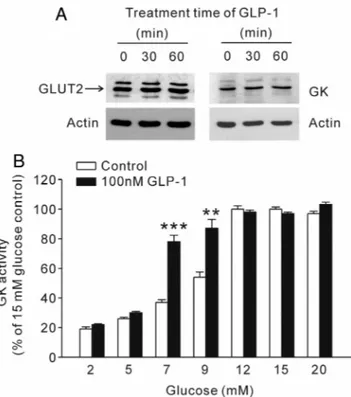
GK activity was determined by following the accumulation of reduced nicotinamide adenine dinucleotide (NAD) at 340 nm FIG. 2. Effect of GLP-1 on GLUT2 and GK in INS-1 cells. A, Cells were
exposed to 100 nMGLP-1 for 30 or 60 min. GLUT2 and GK expression levels were examined with Western blot analysis (representative data chosen from four independent experiments). B, After treatment with or without 100 nMGLP-1 for 30 min, GK activity was determined at concentrations of 2, 5, 7, 9, 12, 15, and 20 mMglucose in INS-1 cell lysates. Values represent mean⫾SE; n⫽ 6 per group. **, P ⬍ 0.01;
***, P⬍ 0.001, comparison with each control value.
Table 1. Kinetic parameters for GK in INS-1 cells Vmax
(mol/kg DNA䡠90 min) Km (mMglucose)
With GLP-1 7.86⫾ 0.21 12.7⫾ 0.21
Without GLP-1 7.98⫾ 0.16 10.6⫾ 0.49a After treatment with or without 100 nMGLP-1 for 30 min, GK Vmax
and Kmwere determined at concentrations of 2–100 mMglucose in INS-1 cell lysates. Vmaxand Kmvalues of GK were calculated as described in the text. Values represent mean⫾SE; n⫽ 6 per group.
aP⬍ 0.05 represents a comparison with control value without GLP-1.
using a spectrophotometer. In this assay, GK phosphorylates glucose to yield glucose 6-phosphate (G-6-P), which is trans- formed into 6-phosphogluconate, and NAD is then reduced by G-6-P dehydrogenase from Leuconostoc mesenteriode (Sigma).
This last reaction depends directly on GK activity. Briefly, INS-1 or dispersed islet-cells were incubated for 30 min in KRPH buffer containing 0.2% BSA and 2 mMglucose. After pretreat- ment with or without 100 nM GLP-1 for 30 min, cells were harvested and lysed in 500l lysis buffer [1 Mphenylmethyl-
sulfonyl fluoride, 50 mMTris (pH 7.6), 4 mMEDTA, 150 mMKCl, 4 mMMg2SO4, and 2.5 mMdithiothreitol] and centrifuged at 4 C for 10 min at 12,000⫻ g to remove mitochondrial-bound hexokinase. DNA content was measured using 10-l aliquots of the extract. The supernatant (10l) was added to 200l reaction buffer containing 50 mMHEPES/HCl (pH 7.6), 100 mMKCl, 7.4 mMMgCl2, 15 mM-mercaptoethanol, 0.5 mM NAD⫹, 0.05% BSA (wt/vol), 3
g/ml G-6-P dehydrogenase, and 5 mMATP with glucose (2–100 mM). The assay was conducted for 90 min at 30 C, and the re- action was stopped by adding 1 ml of a 500 mMNaHCO3buffer (pH 9.4). Absorbance was measured in triplicate samples (excita- tion 350 nm/emission 460 nm), and the mean value was used as a single observa- tion. For the standard curve, 0.3–3.0 nmol G-6-P was used in the reaction buffer containing 100 mMglucose. Maximal velocity (Vmax) and Michaelis-Menten constant (Km) of GK were ob- tained by Lineweaver-Burk plot.
Confocal microscopy to measure mitochondrial membrane potential (⌬m)
Mitochondria were labeled using the mitochondria-specific dye tetramethylrhodamine ethyl ester per- chlorate (TMRE; Invitrogen). INS-1 or dispersed islet-cells were incubated for 30 min in KRPH buffer containing 0.2%
BSA, 2 mM glucose, and 10 nMTMRE.
Confocal microscopy was performed on living cells using a Carl Zeiss confocal mi- croscope (LSM 5 EXCITER; Carl Zeiss, Jena, Germany) connected to an axio-ob- server Z1 inverted microscope using a C- Apochromat⫻40 objective (1.2 numeri- cal aperture). Fluorescent images were generated at 37 C, and all solutions con- tained 10 nM TMRE during recordings.
TMRE fluorescence was excited at 514 nm and recorded at 590 nm. For measure- ments of the time-dependent TMRE fluo- rescence changes, data were acquired ev- ery 3 sec. The relationship between TMRE fluorescence and⌬m is governed by the Nernst equation. The ⌬m-dependent component of TMRE will accumulate in a Nernstian fashion that can be described by the intensity of its fluorescence (26).
The non-⌬m-dependent component of TMRE, also known as the binding com- ponent, was ignored because it is fixed and voltage independent (26). Image anal- ysis was completed with LSM 5 EXCITER software (Carl Zeiss).
Oxygen consumption
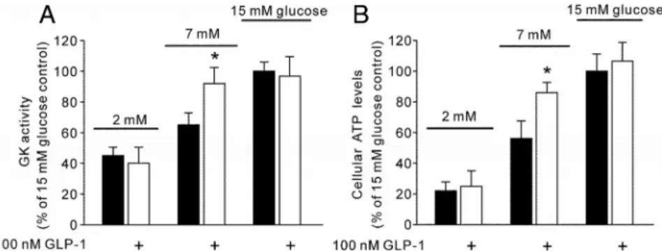
Oxygen uptake by dispersed islet cells was measured with a Clark-type oxygen FIG. 3. Effect of GLP-1 on GK activity and cellular ATP levels in dispersed islet-cells. A, After
treatment with or without 100 nMGLP-1 for 30 min, GK activity was determined at
concentrations of 2, 7, and 15 mMglucose in dispersed islet-cell lysates. B, After incubation in KRPH buffer for 30 min, dispersed islet-cells were pretreated with or without 100 nM GLP-1 for 20 min and then exposed to 2, 7, or 15 mMglucose for 10 min. Cellular ATP concentration was assayed by luciferase measurement. Values represent mean⫾SE; n⫽ 6–8 per group. *, P⬍ 0.05, comparison with each control value.
FIG. 4. Effect of GLP-1 on⌬⌿m and respiration in dispersed islet-cells. A, After establishment of a stable⌬m baseline, dispersed islet -cells were stimulated with 7 or 15 mMglucose for 5 min, and then 100 nMGLP-1 was applied. After 10 min, p-
(trifluoromethoxy)phenylhydrazone (FCCP; 2M) was added. Images were collected at 3-sec intervals, and results were plotted as the change in fluorescence intensity in arbitrary units.
Representative data were chosen from six independent experiments. B, Area under the curve (AUC) of each area was depicted as the percentage of each control value. C, Oxygen consumption was measured with a Clark-type oxygen electrode. After treatment with 7 or 15 mMglucose for 7 min, dispersed islet cells were exposed to 100 nMGLP-1 for 10 min. Oxygen uptake is expressed in nanomoles of O2per minute per milligram of protein. Values represent mean⫾SE; n⫽ 6–8 per group. **, P ⬍ 0.01, comparison with each control value.
electrode (Instech, Plymouth Meeting, PA) in a 600-l air- saturated chamber at 37 C. Respiration medium consisted of 10 mMphosphate buffer, 1 mMMgSO4, 1 mMCaCl2, 136 mM
NaCl, 4.7 mMKCl, and 10 mMHEPES, 0.2% BSA (pH 7.4).
Endogenous (basal) respiration was measured for 3 min. After treatment with 7 or 15 mMglucose for 7 min, dispersed islet cells were exposed to 100 nMGLP-1 for 10 min. Oxygen uptake is expressed in nanomoles of O2per minute per mil- ligram of protein.
Western blot analysis
INS-1 cells were incubated for 30 min in KRPH buffer con- taining 0.2% BSA and 2 mMglucose. After pretreatment with or without 100 nMGLP-1 for 30 or 60 min, the cells were washed twice in ice-cold PBS, and total cellular proteins were extracted in lysis buffer [10 mMTris-Cl (pH 7.4), 130 mMNaCl, 5%
(vol/vol) Triton X-100, 5 mMEDTA, 200 nMaprotinin, 20 mM
leupeptin, 50 mMphenanthroline, and 280 mMbenzamidine- HCl] for 20 min at 4 C. The protein concentrations were mea- sured using the Bio-Rad (Hercules, CA) protein assay. Cellular lysates were separated by SDS-PAGE and electrotransferred to
an Immobilon-P membrane (Millipore, Billerica, MA). The membranes were then probed with specific antibodies as follows:
anti-GK (Santa Cruz), anti-glucose transporter type 2 (GLUT2) (Santa Cruz) targeting amino acids in the extracellular loop, anti-Epac2 (Santa Cruz), anti-Rim2 (Abcam, Cambridge, UK), anti-Rab3A (Santa Cruz), and anti--actin (Sigma). The immunoreactive bands were visualized with a horseradish peroxidase-conjugated secondary antibody (Santa Cruz) us- ing enhanced chemiluminescence (Amersham Biosciences, Little Chalfont, UK). The experiments were repeated at least three times.
Small interfering RNA (siRNA) transfection
Knockdown of Epac2, Rim2, and Rab3A expression in INS-1 cells was performed using BLOCK-iT RNAi duplexes (Invitro- gen). The siRNA sequences for the target genes were as follows:
GCTACTACAGGAGCCAGCCATTCAA (Epac2), AGACAA TGATCTGTAACACCTTCCC (Rim2), and GCGCCAAGGA CAACATTAATGTCAA (Rab3A). Cells were transfected with 50 nMsiRNA using the Lipofectamine RNAiMAX reagent (Invitrogen) according to the manufactur- er’s instructions. Stealth RNAi negative control (Invitrogen) was used as a control for off-target effects. The knockdown ef- ficiency was assessed by Western blot analysis. The cells were treated as indi- cated in the figure legends and processed for 2-deoxyglucose uptake, cellular ATP level, or GK activity analyses.
Statistical analysis
The results are expressed as mean⫾SE. SPSS version 14.0 (SPSS Inc., Chicago, IL) was used for the statistical analyses. The area under the curve was calculated us- ing Microcal (Northampton, MA) Origin software version 7.0. Comparisons be- tween two groups were performed with the Student’s two-tailed t test for paired or unpaired data. For comparisons of more than two groups, significance was tested using ANOVA with Bonferroni correc- tion to deal with relatively small numbers of samples. P values⬍0.05 were consid- ered significant.
Results
GLP-1-mediated increase in 2-deoxy-[3H]glucose uptake
As shown in Fig. 1A, a 20-min pre- treatment with 100 nMGLP-1 increased 2-deoxyglucose uptake in INS-1 cells when using 7 mM2-deoxyglucose, and the effect was not detected when using 15 mM2-deoxyglucose. The effect of GLP-1 at 7 mM2-deoxyglucose was replicated
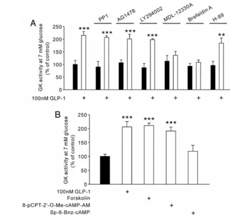
FIG. 5. Involvement of cAMP and Epac in the GLP-1 effects on GK activity in INS-1 cells. A, INS-1 cells were incubated in KRPH buffer for 30 min with the following inhibitors as indicated: c-Src inhibitor PP1 (10M), epidermal growth factor receptor kinase inhibitor AG1478 (250 nM), phosphoinositide 3-kinase inhibitor LY294002 (20M), adenylate cyclase inhibitor MDL-12330A (10M), nonselective Epac inhibitor brefeldin A (10g/ml), or nonselective PKA inhibitor H-89 (10M). The cells were further incubated for 30 min with or without 100 nMGLP-1. GK activity was determined at 7 mMglucose in INS-1 cell lysates. B, INS-1 cells were incubated in KRPH buffer for 30 min. The cells were treated with the following agents for 20 min: GLP-1, cAMP-elevating agent forskolin (100M), Epac-selective activator 8-pCPT-2⬘-O-Me-cAMP-AM (100M), or PKA-selective activator Sp-6-Bnz-cAMP (100M). GK activity was determined at 7 mMglucose in INS-1 cell lysates. Values represent mean⫾SE; n⫽ 6–8 per group. **, P ⬍ 0.01; ***, P ⬍ 0.001, comparison with each control value.
by the GLP-1R agonist exendin-4. However, pretreatment with the GLP-1R antagonist exendin-9 had no effect.
GLP-1-mediated increase in cellular ATP level and
⌬m
Cellular ATP levels were measured in INS-1 cells at various concentrations of glucose with or without pre- treatment with 100 nMGLP-1 (Fig. 1B). The ATP levels at 7 mMglucose were greater in the GLP-1-pretreated group.
The effect of GLP-1 was not detected when using 15 mM
glucose, which was consistent with the 2-deoxyglucose uptake results. Consistently, acute treatment with 100 nM
GLP-1 further increased⌬m in response to 7 mMglucose but not 15 mMglucose (Fig. 1, C and D).
GLP-1-mediated increase in GK activity
The effect of GLP-1 on the expression levels of GLUT2 and GK is shown in Fig. 2A. Pretreatment with GLP-1 for 60 min had no effect on GLUT2 and GK expression levels in INS-1 cells. To evaluate whether GLP-1 directly en- hances GK activity, we measured GK activity in response to glucose (2–20 mM) with or without a 30-min GLP-1 pretreatment (Fig. 2B). As expected, GK activity was en- hanced by GLP-1 when compared with the treatment without GLP-1. Although the Vmax value for 100 nM
GLP-1 was not significantly changed, the Km value was decreased by a 30-min pretreatment with 100 nMGLP-1 (Table 1).
The GLP-1 effects in rat native-cells
We further tested whether the effect of GLP-1 observed in INS-1 cells could also be observed in rat native-cells (Figs. 3 and 4). Expectedly, GK activity (Fig. 3A), cellular ATP levels (Fig. 3B), and⌬m (Fig. 4, A and B) measured in dispersed islet-cells showed the same response as those in INS-1 cells. We also examined the effect of GLP-1 on oxygen consumption at different glucose concentrations in dispersed islet -cells. Acute treatment with 100 nM GLP-1 further increased oxygen consumption in response to 7 mMglucose but not 15 mMglucose (Fig. 4C). After finishing all experiments, the dispersed islet cells were stained with insulin and glucagon antibodies to confirm purity of-cells (data not shown).
GLP-1 enhances GK activity through cAMP and Epac To investigate the signaling mechanism for the en- hancement effect of GLP-1 on GK activity in INS-1 cells, experiments were performed with 7 mMglucose in the presence of various inhibitors of the GLP-1 signaling path- way. As shown in Fig. 5A, the effect of GLP-1 on GK activity was not observed in the presence of the adenylate cyclase inhibitor MDL-12330A (10M) or the nonselec- tive Epac signaling inhibitor, brefeldin A (10g/ml) (27, 28). The following reagents were used to pharmacologi- cally confirm the major involvement of cAMP/Epac in reg- ulating GK activity in INS-1 -cells without GLP-1:
cAMP-elevating agent forskolin (100M), Epac-selective activator 8-pCPT-2⬘-O-Me-cAMP-AM (100 M), and PKA-selective activator Sp-6-Bnz-cAMP (100 M). The enhancing effects on GK activity most closely mimicking those of GLP-1 were obtained with forskolin and 8-pCPT- 2⬘-O-Me-cAMP-AM (Fig. 5B).
GLP-1 promotes GK activity through Epac2, Rim2, and Rab3A
In -cells, Epac2 is more abundant than Epac1 (29).
Recently, Epac2, Rim2, and the Ca2⫹sensor Piccolo have
FIG. 6. Effect of GLP-1 on GK activity, 2-deoxy-[3H]glucose uptake, and cellular ATP levels in Epac2-, Rim2-, or Rab3A-knockdown INS-1 cells. INS-1 cells were transfected with BLOCK-iT RNAi (Epac2, Rim2, or Rab3A siRNA) for 72 h. A, Western blots of transfected cells were quantified. B, Transfected cells were incubated in KRPH buffer for 30 min. After treatment with or without 100 nMGLP-1 for 30 min, GK activity was determined at 7 mMglucose in INS-1 cell lysates. C, Transfected cells were incubated in KRPH buffer for 30 min. After treatment with or without 100 nMGLP-1 for 20 min, the cells were exposed to a mixture of 2-deoxy-[3H]glucose (2DG) and 7 mM
unlabeled 2-deoxyglucose for 10 min. D, Transfected cells were incubated in KRPH buffer for 30 min. After treatment with or without 100 nMGLP-1 for 20 min, the cells were exposed to 7 mMglucose for 10 min. Cellular ATP concentration was assayed by luciferase measurement. Values represent mean⫾SE; n⫽ 6–8 per group.
**, P⬍ 0.01; ***, P ⬍ 0.001, comparison with each control value.
been reported to form a macromolecular complex in the presence of Ca2⫹, which interacts with the GTP-bound form of Rab3A located on the cytoplasmic surface of in- sulin secretory granules to regulate priming and exocytosis (30). In addition, -cell GK has been reported to be an integral component of insulin secretory granules (31). To confirm the association of the effect of GLP-1 on GK ac- tivity with the up-regulation of GLP-1 signaling, we sup- pressed the expression of Epac2, Rim2, or Rab3A in INS-1 cells using siRNA. As shown in Fig. 6A, the expression of each protein was reduced compared with the expression levels in control cells. As expected, the enhancement ef- fects of GLP-1 on GK activity (Fig. 6B), 2-deoxyglucose uptake (Fig. 6C) and cellular ATP levels (Fig. 6D) were dramatically reduced in each protein-depleted cell. These data indicate that Epac2, Rim2, and Rab3A are all critical for the effects of GLP-1.
Discussion
In the present study, we demonstrated that 100 nMGLP-1 acutely enhanced GK activity in a glucose-dependent man- ner in rat INS-1 and native-cells, which resulted in an increase in mitochondrial glucose metabolism, thus ele- vating cellular ATP levels. Accordingly, this might be one of the mechanisms of GLP-1 to facilitate glucose-mediated insulin secretion (Fig. 7). The rate of glucose transport in
-cells is usually determined by GK activity (32, 33), and
small changes in-cell GK activity have large effects on GSIS (34, 35). Although intracellular glucose appears to be the main endogenous GK activator in
-cells (36), the importance of GLP-1R signaling for GK function has already been implicated (7). Although the short-term treatment of GLP-1 in this study (60 min) did not increase GK ex- pression, longer treatments with GLP-1 (24 h) does increase GK expression (21). Together, these findings suggest that both GK activity and GK expres- sion are acting points of GLP-1R sig- naling. GLUT2 activation by GLP-1 may be another underlying mechanism for the enhancement effect of GLP-1 on cellular uptake of 2-deoxyglucose, which is a nonmetabolizable glucose that does not enter the glycolytic path- way despite its phosphorylation by GK.
However, glucose uptake through the low-affinity glucose transporter, GLUT2, is usually not rate-limiting and is, therefore, unlikely to be the site for regulation of GLP-1 (37, 38). Rather, it has been suggested that GLP-1 negatively regulates GLUT2 activity in -cells through PKA-dependent phosphorylation (39). GLUT2 activity has been shown to depend on the phos- phorylations of the carboxyl-terminal tail (39). However, the rateofglucosetransportin-cellsis50-to100-foldgreaterthan the rate of GLUT2 phosphorylation. Moreover, a 70–80%
reduction of GLUT2 expression is needed to impair GSIS (40).
Thus, GLUT2 has mainly a permissive role allowing glucose unrestricted access to GK, which is the rate-limiting step of glu- cose metabolism in-cells and may be an important target pro- tein of GLP-1 signaling.
The present results suggested that the potentiated GK ac- tivity by 100 nMGLP-1 increased glucose uptake and mito- chondrial glucose metabolism, thereby leading to more ele- vated cellular ATP levels in response to 7 mM glucose (approximately 126 mg/dl). Mukai et al. (41) recently found that the ATP content in islets of Goto-Kakizaki rats, which are characterized by an impaired high-glucose-induced in- crease in ATP production, is increased by 100 nMGLP-1 at a glucose concentration of 16.7 mM, and they reported that the ATP content in islets of Wistar rats, which are the normal control, is not changed by 100 nMGLP-1. This result may support the present data, which demonstrated no further enhancement of cellular ATP levels with 100 nMGLP-1 treat- ment at a glucose concentration of 15 mM.
Previous studies have clearly shown that GLP-1 facili- tates not only GSIS but also-cell survival through both
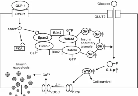
FIG. 7. Proposed mechanism to explain the effects of GLP-1 on GK activity in pancreatic-cells. The cAMP-dependent interactions of Epac2, Rim2, and Piccolo based on an unknown stoichiometry are depicted (30). The Epac2/Rim2/Piccolo macromolecular complex interacts with the GTP-bound form of Rab3A to potentiate the activity of GK, which is an integral component of insulin secretory granules (31).⌬, Membrane depolarization; VDCC, voltage-dependent calcium channels.
cAMP-dependent and cAMP-independent pathways by exploiting plasmalemmal GLP-1R and epidermal growth factor receptors (15). Cell survival and GSIS require ade- quate cellular response to glucose. In addition to previous studies, the potentiating action of GLP-1 on GK activity shown in the present study may also contribute to pro- moting-cell survival and GSIS by enhancing cellular glu- cose sensitivity (22). GK with a low affinity for glucose is selectively expressed in glucose-sensing and hormone (transmitter)-secreting neuronal/neuroendocrine cells, gut endocrine cells, pancreatic-cells, and hepatocytes (42).
In a motor neuron cell line, we have recently found that GLP-1 selectively potentiates GK activity through cAMP/
Epac signaling (43), which is a similar mechanism to the mechanism found in-cells in the present study. In con- trast, acute glucose uptake regulation by GLP-1 is likely to be mediated by GLUT4 trafficking to the plasma mem- brane via phosphoinositide 3-kinase and/or protein kinase B in skeletal muscles and adipocytes that possess only the other high-affinity hexokinases for glucose (44, 45).
The action of GLP-1 on-cell that activates adenylate cyclase (46) to increase cytosolic cAMP levels is generally thought to activate the downstream signaling of PKA (15, 20, 47) and Epac (19, 25, 48). The present results sug- gested that cAMP/Epac signaling was more critical than cAMP/PKA signaling for GK activation by GLP-1. Epac proteins are novel cAMP sensors that regulate several piv- otal cellular processes (49 –52). Although two variants of Epac (Epac1 and Epac2) are expressed in-cells, Epac2 mRNA levels are approximately 125-fold higher than Epac1 mRNA levels in -cells (29). Thus, Epac2 most likely constitutes the predominant effector protein acti- vated by GLP-1 in-cells. For Epac2-mediated enhance- ment of insulin exocytosis, it has been recently reported that Rim2 and the Ca2⫹sensor Piccolo form a macromo- lecular complex with Epac2 in the presence of Ca2⫹, but the stoichiometry of this complex has yet to be determined (30). This complex then interacts with the GTP-bound form of Rab3A located on the cytoplasmic surface of in- sulin secretory granules. Although some GK can be found in the mitochondrial-rich fraction, the majority of GK has been reported to be an integral component of insulin se- cretory granules (31) where Rab3A is embedded. The present findings support that both Rim2 and Rab3A are needed for Epac2 to enhance GK activity at least in INS-1 cells. Therefore, it may be suggested that GLP-1 could stimulate both GK activity and insulin exocytosis with a common mechanism via association of Epac2 with Rim2 and Rab3A downstream of GLP-1R and cAMP. By acti- vating GK, GLP-1 facilitates the glucose supply available for mitochondrial glucose metabolism. In addition, GK associated with insulin granules may also participate in
glucose-induced changes in granule movement or mem- brane fusion during insulin secretion (31). The GLP-1- mediated modulation of GK activity is glucose dependent, which is similar to the other roles of GLP-1 in GSIS, and the GLP-1-mediated modulation of GK activity is not ob- served at low glucose concentrations, which is a finding relevant to clinical applications in treating T2D.
Acknowledgments
Address all correspondence and requests for reprints to: Dae-Kyu Song, M.D., Ph.D., 2800 Dalgubeoldae-Ro, Dalseo-Gu, Daegu 704-701, Korea. E-mail: [email protected].
This research was supported by Technology Development Program for Agriculture and Forestry (110135-3), Ministry for Food, Agriculture, Forestry, and Fisheries, Republic of Korea, and by the KRF Grant funded by the Korean Government (MOEHRD) (KRF-2006-00008).
Disclosure Summary: The authors have nothing to disclose.
References
1. Tripathy D, Chavez AO 2010 Defects in insulin secretion and action in the pathogenesis of type 2 diabetes mellitus. Curr Diab Rep 10:
184 –191
2. Wajcberg E, Amarah A 2010 Liraglutide in the management of type 2 diabetes. Drug Des Devel Ther 4:279 –290
3. Affourtit C, Brand MD 2008 On the role of uncoupling protein-2 in pancreatic-cells. Biochim Biophys Acta 1777:973–979
4. Jitrapakdee S, Wutthisathapornchai A, Wallace JC, MacDonald MJ 2010 Regulation of insulin secretion: role of mitochondrial signal- ling. Diabetologia 53:1019 –1032
5. Liang Y, Bonner-Weir S, Wu YJ, Berdanier CD, Berner DK, Efrat S, Matschinsky FM 1994 In situ glucose uptake and glucokinase ac- tivity of pancreatic islets in diabetic and obese rodents. J Clin Invest 93:2473–2481
6. Jo¨rns A, Tiedge M, Ziv E, Shafrir E, Lenzen S 2002 Gradual loss of pancreatic -cell insulin, glucokinase and GLUT2 glucose trans- porter immunoreactivities during the time course of nutritionally induced type-2 diabetes in Psammomys obesus (sand rat). Virchows Arch 440:63– 69
7. Matschinsky FM 2009 Assessing the potential of glucokinase acti- vators in diabetes therapy. Nat Rev Drug Discov 8:399 – 416 8. Matschinsky FM 2002 Regulation of pancreatic-cell glucokinase:
from basics to therapeutics. Diabetes 51(Suppl 3):S394 –S404 9. Pal M 2009 Recent advances in glucokinase activators for the treat-
ment of type 2 diabetes. Drug Discov Today 14:784 –792 10. Ahre´n B 2009 Islet G protein-coupled receptors as potential targets
for treatment of type 2 diabetes. Nat Rev Drug Discov 8:369 –385 11. Baggio LL, Drucker DJ 2007 Biology of incretins: GLP-1 and GIP.
Gastroenterology 132:2131–2157
12. Byrne MM, Gliem K, Wank U, Arnold R, Katschinski M, Polonsky KS, Go¨ke B 1998 Glucagon-like peptide 1 improves the ability of the
-cell to sense and respond to glucose in subjects with impaired glucose tolerance. Diabetes 47:1259 –1265
13. Nauck MA 2009 Unraveling the science of incretin biology. Am J Med 122:S3–S10
14. Holz 4th GG, Ku¨htreiber WM, Habener JF 1993 Pancreatic-cells
are rendered glucose-competent by the insulinotropic hormone glu- cagon-like peptide-1(7–37). Nature 361:362–365
15. MacDonald PE, Wang X, Xia F, El-kholy W, Targonsky ED, Tsu- shima RG, Wheeler MB 2003 Antagonism of rat-cell voltage- dependent K⫹currents by exendin 4 requires dual activation of the cAMP/protein kinase A and phosphatidylinositol 3-kinase signaling pathways. J Biol Chem 278:52446 –52453
16. Suga S, Kanno T, Nakano K, Takeo T, Dobashi Y, Wakui M 1997 GLP-I(7–36) amide augments Ba2⫹current through L-type Ca2⫹
channel of rat pancreatic-cell in a cAMP-dependent manner. Di- abetes 46:1755–1760
17. Tsuboi T, da Silva Xavier G, Holz GG, Jouaville LS, Thomas AP, Rutter GA 2003 Glucagon-like peptide-1 mobilizes intracellular Ca2⫹and stimulates mitochondrial ATP synthesis in pancreatic MIN6-cells. Biochem J 369:287–299
18. Eliasson L, Ma X, Renstro¨m E, Barg S, Berggren PO, Galvanovskis J, Gromada J, Jing X, Lundquist I, Salehi A, Sewing S, Rorsman P 2003 SUR1 regulates PKA-independent cAMP-induced granule priming in mouse pancreatic B-cells. J Gen Physiol 121:181–197 19. Kashima Y, Miki T, Shibasaki T, Ozaki N, Miyazaki M, Yano H,
Seino S 2001 Critical role of cAMP-GEFII-Rim2 complex in incre- tin-potentiated insulin secretion. J Biol Chem 276:46046 – 46053 20. Renstrom E, Eliasson L, Rorsman P 1997 Protein kinase A-depen-
dent and -independent stimulation of exocytosis by cAMP in mouse pancreatic B-cells. J Physiol 502(Pt 1):105–118
21. Murao K, Li J, Imachi H, Muraoka T, Masugata H, Zhang GX, Kobayashi R, Ishida T, Tokumitsu H 2009 Exendin-4 regulates glucokinase expression by CaMKK/CaMKIV pathway in pancreatic
-cell line. Diabetes Obes Metab 11:939–946
22. Kim YK, Park JH, Park SH, Lim B, Baek WK, Suh SI, Lim JG, Ryu GR, Song DK 2010 Protective role of glucagon-like peptide-1 against glucosamine-induced cytotoxicity in pancreatic-cells. Cell Physiol Biochem 25:211–220
23. Balkan B, Dunning BE 1994 Glucosamine inhibits glucokinase in vitro and produces a glucose-specific impairment of in vivo insulin secretion in rats. Diabetes 43:1173–1179
24. Uldry M, Ibberson M, Hosokawa M, Thorens B 2002 GLUT2 is a high affinity glucosamine transporter. FEBS Lett 524:199 –203 25. Kang G, Chepurny OG, Holz GG 2001 cAMP-regulated guanine
nucleotide exchange factor II (Epac2) mediates Ca2⫹-induced Ca2⫹
release in INS-1 pancreatic-cells. J Physiol 536:375–385 26. O’Reilly CM, Fogarty KE, Drummond RM, Tuft RA, Walsh Jr JV
2003 Quantitative analysis of spontaneous mitochondrial depolar- izations. Biophys J 85:3350 –3357.
27. Ster J, de Bock F, Bertaso F, Abitbol K, Daniel H, Bockaert J, Fagni L 2009 Epac mediates PACAP-dependent long-term depression in the hippocampus. J Physiol 587:101–113
28. Zhong N, Zucker RS 2005 cAMP acts on exchange protein activated by cAMP/cAMP-regulated guanine nucleotide exchange protein to regulate transmitter release at the crayfish neuromuscular junction.
J Neurosci 25:208 –214
29. Leech CA, Dzhura I, Chepurny OG, Schwede F, Genieser HG, Holz GG 2010 Facilitation of-cell KATPchannel sulfonylurea sensitivity by a cAMP analog selective for the cAMP-regulated guanine nucle- otide exchange factor Epac. Islets 2:72– 81
30. Fujimoto K, Shibasaki T, Yokoi N, Kashima Y, Matsumoto M, Sasaki T, Tajima N, Iwanaga T, Seino S 2002 Piccolo, a Ca2⫹sensor in pancreatic
-cells. Involvement of cAMP-GEFII. Rim2. Piccolo complex in cAMP- dependent exocytosis. J Biol Chem 277:50497–50502
31. Arden C, Harbottle A, Baltrusch S, Tiedge M, Agius L 2004 Glu- cokinase is an integral component of the insulin granules in glucose- responsive insulin secretory cells and does not translocate during glucose stimulation. Diabetes 53:2346 –2352
32. Matschinsky F, Liang Y, Kesavan P, Wang L, Froguel P, Velho G, Cohen D, Permutt MA, Tanizawa Y, Jetton TL, Niswender K, Mag- nuson MA 1993 Glucokinase as pancreatic-cell glucose sensor and diabetes gene. J Clin Invest 92:2092–2098
33. Matschinsky FM 1990 Glucokinase as glucose sensor and metabolic signal generator in pancreatic-cells and hepatocytes. Diabetes 39:
647– 652
34. Matschinsky FM, Glaser B, Magnuson MA 1998 Pancreatic-cell glucokinase: closing the gap between theoretical concepts and ex- perimental realities. Diabetes 47:307–315
35. Sweet IR, Li G, Najafi H, Berner D, Matschinsky FM 1996 Effect of a glucokinase inhibitor on energy production and insulin release in pancreatic islets. Am J Physiol 271:E606 –E625
36. Kamata K, Mitsuya M, Nishimura T, Eiki J, Nagata Y 2004 Struc- tural basis for allosteric regulation of the monomeric allosteric en- zyme human glucokinase. Structure 12:429 – 438
37. Lenzen S, Panten U 1988 Signal recognition by pancreatic B-cells.
Biochem Pharmacol 37:371–378
38. Meglasson MD, Matschinsky FM 1986 Pancreatic islet glucose me- tabolism and regulation of insulin secretion. Diabetes Metab Rev 2:163–214
39. Thorens B, De´riaz N, Bosco D, DeVos A, Pipeleers D, Schuit F, Meda P, Porret A 1996 Protein kinase A-dependent phosphoryla- tion of GLUT2 in pancreatic-cells. J Biol Chem 271:8075–8081 40. Valera A, Solanes G, Ferna´ndez-Alvarez J, Pujol A, Ferrer J, Asins G, Go- mis R, Bosch F 1994 Expression of GLUT-2 antisense RNA in-cells of transgenic mice leads to diabetes. J Biol Chem 269:28543–28546 41. Mukai E, Fujimoto S, Sato H, Oneyama C, Kominato R, Sato Y,
Sasaki M, Nishi Y, Okada M, Inagaki N 2011 Exendin-4 suppresses SRC activation and reactive oxygen species production in diabetic Goto-Kakizaki rat islets in an Epac-dependent manner. Diabetes 60:218 –226
42. Matschinsky FM, Magnuson MA, Zelent D, Jetton TL, Doliba N, Han Y, Taub R, Grimsby J 2006 The network of glucokinase-ex- pressing cells in glucose homeostasis and the potential of glucoki- nase activators for diabetes therapy. Diabetes 55:1–12
43. Lim JG, Lee JJ, Park SH, Park JH, Kim SJ, Cho HC, Baek WK, Kim DK, Song DK 2010 Glucagon-like peptide-1 protects NSC-34 motor neurons against glucosamine through Epac-mediated glucose up- take enhancement. Neurosci Lett 479:13–17
44. Arne´s L, Gonza´lez N, Tornero-Esteban P, Sancho V, Acitores A, Valverde I, Delgado E, Villanueva-Pen˜acarrillo ML 2008 Charac- teristics of GLP-1 and exendins action upon glucose transport and metabolism in type 2 diabetic rat skeletal muscle. Int J Mol Med 22:127–132
45. Sancho V, Nuche B, Arne´s L, Cancelas J, Gonza´lez N, Díaz-Miguel M, Martín-Duce A, Valverde I, Villanueva-Pen˜acarrillo ML 2007 The action of GLP-1 and exendins upon glucose transport in normal human adipocytes, and on kinase activity as compared to morbidly obese patients. Int J Mol Med 19:961–966
46. MacDonald PE, El-Kholy W, Riedel MJ, Salapatek AM, Light PE, Wheeler MB 2002 The multiple actions of GLP-1 on the process of glucose-stimulated insulin secretion. Diabetes 51(Suppl 3):S434–S442 47. Drucker DJ 2002 Biological actions and therapeutic potential of the
glucagon-like peptides. Gastroenterology 122:531–544
48. Kang G, Joseph JW, Chepurny OG, Monaco M, Wheeler MB, Bos JL, Schwede F, Genieser HG, Holz GG 2003 Epac-selective cAMP analog 8-pCPT-2⬘-O-Me-cAMP as a stimulus for Ca2⫹-induced Ca2⫹release and exocytosis in pancreatic-cells. J Biol Chem 278:8279–8285 49. Holz GG 2004 Epac: A new cAMP-binding protein in support of
glucagon-like peptide-1 receptor-mediated signal transduction in the pancreatic-cell. Diabetes 53:5–13
50. Holz GG, Chepurny OG, Schwede F 2008 Epac-selective cAMP analogs: new tools with which to evaluate the signal transduction properties of cAMP-regulated guanine nucleotide exchange factors.
Cell Signal 20:10 –20
51. Holz GG, Kang G, Harbeck M, Roe MW, Chepurny OG 2006 Cell physiology of cAMP sensor Epac. J Physiol 577:5–15
52. Roscioni SS, Elzinga CR, Schmidt M 2008 Epac: effectors and bi- ological functions. Naunyn Schmiedebergs Arch Pharmacol 377:
345–357
![FIG. 1. Effect of GLP-1 on 2-deoxy-[ 3 H]glucose uptake, cellular ATP levels and ⌬m in INS-1 cells](https://thumb-ap.123doks.com/thumbv2/123dokinfo/5012468.306684/2.877.72.821.102.654/fig-effect-deoxy-glucose-uptake-cellular-levels-cells.webp)
![FIG. 6. Effect of GLP-1 on GK activity, 2-deoxy-[ 3 H]glucose uptake, and cellular ATP levels in Epac2-, Rim2-, or Rab3A-knockdown INS-1 cells](https://thumb-ap.123doks.com/thumbv2/123dokinfo/5012468.306684/6.877.118.390.413.865/effect-activity-deoxy-glucose-uptake-cellular-levels-knockdown.webp)