www.earticle.net
Bisphenol S가 돼지정자와 난소내 과립막세포의 생존성과 활성산소에 미치는 영향
이유섭․이승형․양부근* 강원대학교 동물생명과학대학
Effects of Bisphenol S on Viability and Reactive Oxygen Species of the Sperm and Ovarian Granulosa Cells in Pigs
Yu-Sub Lee, Seunghyung Lee and Boo-Keun Yang*
College of Animal Life Sciences, Kangwon National University, Chuncheon 24341, Korea
ABSTRACT1)
The effect of bisphenol S (BPS) on the viability and production of reactive oxygen species (ROS) was studied in boar sperm and ovarian granulosa cells. Boar semen was incubated in Beltsville thawing solution with either 0 or 5 μM BPS for 3 and 6 h. The viability of sperm was analyzed by SYBR14/PI doubling staining, and production of ROS was detected. Ovarian granulosa cells were also treated with BPS for 24, 48, and 72 h. Then, cell viability (0, 5, 10, and 20 μM) and ROS production (only 0 and 5 μM BPS) were assessed.
The results showed that, BPS decreased sperm viability at 3 and 6 h, and that BPS increased ROS production (p<0.05). Also, BPS reduced the viability of ovarian granulosa cells (p<0.05), and stimulated ROS production (p<0.05). These results suggest that BPS damages sperm activation and ovarian granulosa cells in the reproductive system.
(Key words: Bisphenol S, Boar sperm, Ovarian granulosa cells, Viability, Reactive oxygen species)
* Corresponding author: Boo-Keun Yang, College of Animal Life Sciences, Kangwon National University, Chuncheon 24341, Korea.
Tel: +82-33-250-8623, E-mail: [email protected]
This is an Open Access article distributed under the terms of the Creative Commons Attribution Non-Commercial License (http://creativecommons.org/licenses/by-nc-nd/3.0/deed.ko), which permits unrestricted non-commercial use, distribution, and reproduction in any medium, provided the original work is properly cited. The moral rights of the named author(s) have been asserted.
Ⅰ. 서론
현대사회의 산업이 급속하게 발달함에 따라 합성화학물 질의 개발과 이용이 증가 되었다. 하지만 유해성이 충분히 검증되지 않은 합성화학물질은 우리 일상생활에서 자주 쓰 이고 접촉되는 가정용품 또는 공산품의 원료로써 사용되고 있으며, 이러한 화학물질 중 천연호르몬을 모방하여 호르 몬 수용체와 결합하여 내분비계의 이상을 나타내는 물질을 내분비교란물질(endocrine disrupting chemical, EDC) 이 라고 한다. 세계보건기구(WHO)에 규정된 내분비교란물질 에는 poly-chlorinated biphenyls(PCBs), dioxins,
phthalates, vinclozolin, di-ethyl-stilbestrol(DES), dichloro- diphenyl-trichloroethane(DDT), bisphenol A(BPA) 등이 있 으며, BPA가 내분비교란물질로 규정됨에 따라 화학구조가 비슷한 여러 종류의 Bisphenol 화합물이 BPA의 대체물질 로서 사용되고 있다.
BPA의 대체 물질로서 가장 많이 사용되고 있는 Bisphenol S(Bis-hydroxyphenyl sulfone, BPS)는 폴리카보네이트 플 라스틱, 에폭시 레진, 식품포장재, 개인위생 용품, 영수증, 감열지 등의 원료로 사용되고 있으며, BPA 보다 고온과 내 광성에 대한 안정성이 우수한 것으로 보고되었다(Liao and Kannan, 2013; Liao and Kannan, 2014; Bjornsdotter et al.,
[Provider:earticle] Download by IP 118.70.52.165 at Monday, December 20, 2021 7:49 PM
www.earticle.net
2017). 그러나, BPS의 독성영향과 관련한 동물실험에서 BPS에 노출시킨 zebra fish의 난자의 생성 및 수정율의 감 소와 배아의 기형율이 증가하였고, 생식기관 발달을 저해 하는 것으로 나타났다(Ji et al., 2013). 또한, 피하주사를 통 해 BPS에 노출시킨 암컷 쥐의 자궁 발달에 유해한 영향을 미치며(Yamasaki et al., 2004), BPS에 노출시킨 수컷 쥐에 서 testosterone의 감소와 생식기관의 발달을 저해하며, 정 자 형성의 감소와 산화스트레스를 유발하는 것으로 나타 났다(Ullah et al., 2016).
BPA의 대체물질인 BPS가 생식세포에 미치는 영향에 관한 연구는 미비한 실정이다. 따라서, 본 연구는 BPS가 BPA의 대체물로서 적합한지 여부와 생식세포에 미치는 영향을 알아보기 위한 기초자료로 활용하고자, BPS가 돼 지의 정자와 난소내 과립막세포에 미치는 영향을 검토하 고자 실시하였다.
Ⅱ. 재료 및 방법
1. 시약
Bisphenol S(BPS)는 Tokyo Chemical(Tokyo, Japan) 사 에서 구입하여 사용하였으며, 그 외 시약 및 소모품은 Sigma-Aldrich(St. Louis, MO, USA)에서 구입하였다.
2. 정액의 처리
돼지정액은 원주 인공수정센터(금보유전자)에서 공급받 았으며, 정자 운동성이 85% 이상인 것을 실험에 사용하였 으며, 정자의 농도는 2.0×10⁶마리/mL로 희석하여 실험을 실시하였다. 희석 용액은 beltsville thawing solution(BTS;
37mg/mL glucose, 1.25mg/mL EDTA, 6mg/mL sodium citrate, 1.25mg/mL sodium bicarbonate, 0.75mg/mL potassium chloride, 0.6mg/mL penicillin 및 1mg/mL streptomycin)을 사용하였다(Pursel and Johnson, 1975).
정자는 BPS(0, 5μM)를 처리한 후, 37℃의 5% CO2환경에 서 3, 6 시간 동안 배양하고 정자의 생존성과 활성산소를 측정하였다.
3. 정자의 생존성
SYBR-14(Invitrogen, Eugene, OR, USA)와 propidium
iodide(PI; Invitrogen)로 염색하여 정자의 생존성을 측정하 였다(Lee and Park, 2015). 100μL 정액에 5μL SYBR-14(10μ L/mL in DMSO)를 10분 동안 암실에서 처리한 후, 5μL 2.4mM PI와 10분 동안 처리하였다. 처리 후, 정액은 슬라 이드 글라스에 분주하고, 형광현미경(Axioskop, Zeiss, Germany)을 사용하여 정자의 생존성을 분석하였다. 녹색 형광을 띄는 생존정자와 붉은색 형광을 띄는 사멸정자로 구분하였으며, 최소 500개 이상의 정자를 분석하여 생존율 을 평가하였다.
4. 정자의 활성산소 생산
정자의 활성산소 생산은 2',7'-dichlorofluorescein diacetate (DCF-DA; Sigma) 형광염색방법으로 측정하였다. 1mL 정 자를 원심분리(1500✕g)하고, 200μL 상층액을 회수하여 96-well black plate에 분주하였다. Kit 매뉴얼에 따라 20mM DCF-DA 20μL를 첨가하고 37℃, 5% CO2암실에서 30분 동안 배양하였다. Micro fluorescent plate reader (Spectra max M2e, Molecular Devices, USA)를 이용하여 485nm에서 활성산소의 생산량을 측정하였다.
5. 과립막세포의 처리
과립막세포는 포천 소재의 제일 산업에서 도축한 3원 교 잡종의 돼지난소에서 26–gage의 주사바늘을 이용하여 채 취하였으며, 채취한 과립막세포는 DMEM(Dulbecco’s modified Eagle’s medium, Gibco) 용액과 혼합 하여 원심 분리를 실시하였다(1500✕g, 5분). DMEM 용액으로 2회 세 척 후, DMEM 용액과 5% Fetal bovine serum(FBS), 1%(v/v) Penicillin/Streptomycin mix(Sigma-Aldrich)을 혼합하여 배양하였다. 3일 동안 배양 후(37℃, 5% CO2), 0.25% trypsin-EDTA(Gibco)를 처리하여 회수한 세포를 사 용하여 생존성 및 실험에 사용하였다.
6. 과립막세포의 생존성
생존성은 MTT(Sigma) 방법을 이용하여 측정하였으며, 회수한 과립막세포는 96-well clear plate(Nunc, USA)에 분 주하여 24시간동안 배양하였다. Monolayer가 형성된 과립 막세포는 PBS 용액으로 2회 세척하고 , 배양액에 0, 5, 10, 20μM BPS를 첨가하여 24, 48, 72시간 동안 배양하였다(3 7℃, 5% CO2). 각각의 시간 별로 처리된 과립막세포은 상 층액을 제거하고, PBS 용액으로 2회 세척하였다. 그 후,
[Provider:earticle] Download by IP 118.70.52.165 at Monday, December 20, 2021 7:49 PM
www.earticle.net
10μL MTT(5mg MTT/mL)와 90μL PBS 용액을 분주하고 4시간 동안 암실에서 배양하였다. 상층액을 제거하고 100μ L DMSO 용액을 분주하고 1시간 동안 2차 배양하였다.
Micro plate reader(Spectra max M2e)를 사용하여 570nm 에서 생존성을 측정하였으며, optical density(O.D) 값을 백 분율로 환산하여 과립막세포의 생존성을 나타냈다. 7. 과립막세포의 활성산소 생산
과립막세포의 활성산소 생산은 제시된 방법과 동일하게 실시하였고, 세포에 5μM BPS를 첨가하여 24, 48, 72시간 동안 배양하였다. 각각의 배양시간 별로 배양된 세포에서 활성산소를 측정하였다.
8. 통계처리
실험의 결과는 평균과 표준 오차(mean±SEM, n=3)로 나 타냈으며, 통계처리는 statistical analysis system software version 9.3(SAS Institute Inc. USA)을 사용하였다. 실험구별 유의성 검증은 Duncan 다중검정법을 사용하여 나타냈다.
Ⅲ. 결과 및 고찰
내분비교란물질은 신경전달물질 및 호르몬의 운반과 합 성 및 대사를 모방하여 정상적인 매커니즘에 혼란을 야기하 고 핵 수용체의 결합 및 발현의 변이를 나타내며, estrogen,
testosterone, luteinizing hormone(LH), follicle stimulation hormone(FSH) 와 같은 생식호르몬의 대사 및 합성에 관 여하여 불임, 난임, 유산, 생식기관의 발달 및 형태적 이상 등의 번식관련 질병을 야기한다(Gallet and Vanacker, 2010; Kiyama et al., 2015; Sifakis et al., 2017). 본 실험은 내분비교란물질인 BPA의 대체물질로 사용되고 있는 BPS 가 돼지 정자와 돼지 난소내 과립막세포 특성에 미치는 영 향을 검토하여 포유동물의 번식건강에 미치는 영향을 간 접적으로 알아보고, BPS가 BPA의 대체물로서 적합한지 알 아보기 위하여 실시하였다.
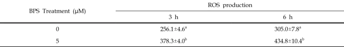
본 실험에서 정자배양액에 5μM BPS를 첨가하고 3, 6시 간 처리하여 정자의 생존성과 활성산소를 측정한 결과 (Table 1, 2), 대조구와 비교하여 BPS 처리구에서 시간과 관계없이 정자 생존성이 모두 유의적으로 감소하였으며 (p<0.05), 정자의 활성산소량은 대조구와 비교하여 BPS 처 리구에서 시관과 관계없이 활성산소가 증가하는 것을 나 타냈다(p<0.05).
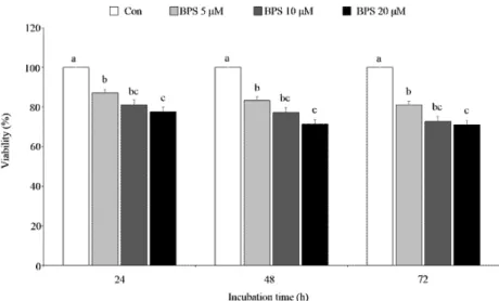
또한, 난소내 과립막세포 배양액에 0, 5, 10, 20μM의 BPS를 24, 48, 72시간동안 처리한 결과(Fig. 1), 대조구와 비교하여 모든 처리농도와 시간에서 생존성이 유의적으로 감소하였고(p<0.05), 농도 의존적으로 생존성이 더욱 감소 하는 것으로 나타냈으며, 난소내 과립막세포에 5μM BPS 를 24, 48, 72시간 처리하여 활성산소를 측정한 결과(Fig.
2), 대조구와 비교하여 BPS 처리구에서 활성산소량이 모든 처리시간에서 유의적으로 증가하였으며, 시간이 경과할수 록 활성산소의 증가하는 경향을 나타냈다(p<0.05).
Table 1. Effect of BPS on sperm viability in the pigs
BPS Treatment (μM) Viability (%)
3 h 6 h
0 70.3±1.5a 64.1±1.2a
5 61.1±0.8b 50.2±1.1b
a,bValues with different superscripts within same column are significantly different, p<0.05.
Table 2. Effect of BPS on ROS production in boar sperm
BPS Treatment (μM) ROS production
3 h 6 h
0 256.1±4.6a 305.0±7.8a
5 378.3±4.0b 434.8±10.4b
a,bValues with different superscripts within same column are significantly different, p<0.05.
[Provider:earticle] Download by IP 118.70.52.165 at Monday, December 20, 2021 7:49 PM
www.earticle.net
Fig. 1. Effect of BPS on viability of ovarian granulosa cells.
a~dValues with different superscripts within same incubation time are significantly different, p<0.05.
Fig. 2. Effect of BPS on ROS production in ovarian granulosa cells.
*Values are significantly different, p<0.05.
본 실험의 결과와 비교하여 Shi 등(2017)의 연구에서 BPS 에 노출시킨 CD-1 mice에서 정자수와 운동성이 감소하였 으며 정원세포의 발달을 방해한다는 연구결과와 유사하게 나타났다. 또한, 인간 기관지 상피세포에 산화스트레스를 유발하여 활성산소가 유의적으로 증가하여 세포의 DNA 손상과 세포 사멸의 증가하였다는 연구(George and Rupasinghe, 2018) 결과를 비추어 보아, 본 실험에서 돼지 의 정자 및 과립막세포에 BPS가 산화스트레스를 유발하여 세포사멸을 유도한 것으로 판단된다. 또한, 경구 투여로 BPS에 노출시킨 rat의 정자에서 산화스트레스를 유발하여 활성산소를 증가시켜 정자의 DNA 손상을 나타내어 정자
의 유해한 영향을 나타냈지만, 정자의 운동성에는 영향을 미치지 않았다는 연구(Ullah et al., 2017) 결과와 비교하여 본 실험의 결과에서 정자의 활성산소가 증가하는 것을 확 인하였고, 이에 따른 정자의 생존성에도 유해한 영향을 미 쳐 정자의 질을 감소시킨 것으로 생각된다. 또한, BPS는 BPA와 유사한 estrogen 활성을 나타내고, BPS에 노출된 닭의 배아의 성장률의 감소와 BPA와 유사한 독성영향을 나타낸다고 보고하였으며(Hashimoto and Nakamura, 2000; Crump et al., 2016), BPS에 노출된 암컷 mice에서 수정률의 감소 및 배반포기의 성장을 저해하고(Nourian et al., 2017), 300nM BPS 저농도에 노출된 돼지난모세포와
[Provider:earticle] Download by IP 118.70.52.165 at Monday, December 20, 2021 7:49 PM
www.earticle.net
난구세포에서 내분비교란물질로서 유해한 영향을 줄 뿐만 아니라, 돼지난모세포의 발달과 관련하여 BPA와 유사하게 유해한 영향을 미치는 것으로 나타났다(Žalmanová et al., 2017). BPS에 노출된 rat의 정소와 난소의 기능의 저해와 생식기관의 발달과 호르몬 분비의 유해한 영향을 미치며, 이는 불임을 야기할 것이라고 보고하였다(Ullah et al., 2016; Ahsan et al., 2018). 위의 연구결과와 유사하게, 본 실험에서 BPS는 과립막세포의 생존성의 감소와 활성산소 의 증가로 인한 자성생식세포에 유해한 영향을 미치는 것 으로 나타났다.
결론적으로, BPS 는 돼지정자의 생존성의 감소, 활성산소의 증가로 인한 돼지정자 특성에 유해한 영향을 나타냈으며, 난소내 과립막세포의 생존성의 감소와 활성산소가 유의적으로 증가하는 것이 나타났다. 따라서, BPS 는 웅성 및 자성 생식세포에 유해한 영향을 미치는 것으로 판단되며, 생식세포의 수정능력의 저하로 인한 인간과 동물의 불임 또는 난임 등의 번식 질병을 유도할 것으로 생각된다. 또한, BPS 는 BPA 의 대체물질로서 적합하지 않다고 생각되며, 아직까지 BPA 의 많은 연구와 비교하여 BPS 의 관한 연구는 미흡한 실정이기 때문에 BPS 에 관한 메커니즘과 BPS 의 독성 영향에 대한 더 많은 연구가 진행되어야 할 것으로 판단된다.
Ⅳ. 요약
본 연구는 돼지의 정자와 난소내 과립막세포에서 bisphenol S(BPS)가 생존성과 활성산소 생산에 미치는 영향을 알아보 고자 연구하였다. 돼지정액은 0, 5μM BPS를 처리하여 3, 6 시간동안 배양하였다. 정자의 생존성은 SYBR14/PI를 이중 염색하여 분석하였으며, 활성산소의 생산을 측정하였다.
또한, BPS(0, 5, 10, 20μM)를 과립막세포에 처리하여 24, 48, 72시간동안 처리하였다. 처리 후, 세포의 생존율과 활 성산소 생산(단, 5μM BPS)을 측정하였다. 그 결과, 돼지에 서 정자의 생존율은 BPS에 의해 감소하였고, 활성산소의 생산은 모든 처리시간에서 증가하였다(p<0.05). 또한 과립 막세포의 생존은 BPS에 의해 억제되었고, 활성산소는 유 의적으로 증가하였다(p<0.05). 이상의 결과를 토대로, BPS 의 노출은 정자의 활성과 번식과 관련된 세포에 나쁜 영향 을 미칠 것이다.
사사
2017년도 강원대학교 대학회계 학술연구조성비로 연구 하였음(관리번호-520170244).
Ⅴ. 참고문헌
1. Amaral, A., Lourenço, B., Marques, M. and Ramalho- Santos, J. 2013. Mitochondria functionality and sperm quality. Reproduction. 146:163-174.
2. Ahsan, N., Ullah, H., Ullah, W. and Jahan, S.
Comparative effects of Bisphenol S and Bisphenol A on the development of female reproductive system in rats; a neonatal exposure study. Chemosphere. 197:
336-343.
3. Crump, D., Chiu, S. and Williams, K. L. 2016.
Bisphenol S alters embryonic viability, development, gallbladder size, and messenger RNA expression in chicken embryos exposed via egg injection. Environ.
Toxicol. Chem. 35:1541-1549.
4. Gallet, M. and Vanacker, J. M. 2010. ERR receptors as potential targets in osteoporosis. Trends Endocrinol.
Metab. 21:637-641.
5. George, V. C. and Rupasinghe, H. P. V. DNA damaging and apoptotic potentials of Bisphenol A and Bisphenol S in human bronchial epithelial cells.
Environ. Toxicol. Pharmacol. 60:52-57.
6. Hashimoto, Y. and Nakamura, M. 2000. Estrogenic activity of dental materials and bisphenol-A related chemicals in vitro. Dent. Mater. J. 19:245-62.
7. Ji, K., Hong, S., Kho, Y. and Choi, K. 2013. Effects of bisphenol s exposure on endocrine functions and reproduction of zebrafish. Environ. Sci. Technol.
47:8793-800.
8. Kiyama, R. and Wada-Kiyama, Y. 2015. Estrogenic endocrine disruptors: Molecular mechanisms of action.
Environ. Int. 83:11-40.
9. Lee, S. H. and Park, C. K. 2015. Effect of magnetized extender on sperm membrane integrity and development of oocytes in vitro fertilized with liquid storage boar semen. Anim. Reprod. Sci. 154:86-94.
[Provider:earticle] Download by IP 118.70.52.165 at Monday, December 20, 2021 7:49 PM
www.earticle.net
10. Liao, C. and Kannan, K. 2013. Concentrations and profiles of Bisphenol A and other Bisphenol Analogues in foodstuffs from the United States and their implications for Human exposure. J. Agric.
Food Chem. 61:4655–4662.
11. Liao, C. and Kannan, K. 2014. A survey of bisphenol A and other bisphenol analogues in foodstuffs from nine cities in China. Arch. Environ. Contam. Toxicol.
67:50-59.
12. Mahfouz, R. Z., du Plessis, S. S., Aziz, N. Sharma, R.
Sabanegh, E. and Agarwal, A. 2010. Sperm viability, apoptosis, and intracellular reactive oxygen species levels in human spermatozoa before and after induction of oxidative stress. Fertil. Steril. 93:814-21.
13. Nourian, A., Soleimanzadeh, A., Shalizar Jalali, A.
and Najafi, G. Effects of bisphenol-S low concentrations on oxidative stress status and in vitro fertilization potential in mature female mice. Vet.
Res. Forum. 4:341-345.
14. Pursel, V. G. and Johnson, L. A. 1975. Freezing of boar spermatozoa: fertilizing capacity with concentrated semen and a new thawing procedure. J.
Anim. Sci. 40:99-102.
15. Shi, M., Sekulovski, N., MacLean, J. A. and Hayashi, K. 2017. Effects of bisphenol A analogues on
reproductive functions in mice. Reprod. Toxicol.
73:280-291.
16. Sifakis, S., Androutsopoulos, V. P., Tsatsakis, A. M.
and Spandidos, D. A. 2017. Human exposure to endocrine disrupting chemicals: Effects on the male and female reproductive systems. Environ. Toxicol.
Pharmacol. 51:56-70.
17. Ullah, H., Jahan, S., Ain, Q. U., Shaheen, G. and Ahsan, N. 2016. Effect of bisphenol S exposure on male reproductive system of rats: A histological and biochemical study. Chemosphere. 52:383-391
18. Ullah, H., Ambreen, A., Ahsan, N. and Jahan, S.
2017. Bisphenol S induces oxidative stress and DNA damage in rat spermatozoa in vitro and disrupts daily sperm production in vivo. Toxicol. Environ.
Chem. 99:953-965.
19. Žalmanová, T., Hošková, K., Nevoral, J., Adámková, K., Kott, T., Šulc, M., Kotíková, Z., Prokešová, Š., Jílek, F., Králíčková, M. and Petr, J. 2017. Bisphenol S negatively affects the meotic maturation of pig oocytes. Sci. Rep. 1:485.
(Received 04 July 2018, Revised 12 December 2018, Accepted 17 December 2018)
[Provider:earticle] Download by IP 118.70.52.165 at Monday, December 20, 2021 7:49 PM