에탄올 투여 랫드에서 곰취 열수 추출 발효물이 간 독성에 미치는 영향
유근형1․이선엽1․양현모1․함영안1․이수응1․채승완2․이용진1
1(재)춘천바이오산업진흥원 기술개발실
2성균관대학교 의과대학 강북삼성병원 병리과
Effects of Fermented Water Extracts from Ligularia fischeri on Hepatotoxicity in Ethanol-Induced Rats
Keun-Hyung Yu1, Sun-Yeop Lee1, Hyun-Mo Yang1, Young-Ahn Ham1, Soo-Ung Lee1, Seoung-Wan Chae2, and Yong-Jin Lee1
1Department of Technical Development Chuncheon Bioindustry Foundation
2Department of Pathology, Kangbuk Samsung Hospital, Sungkyunkwan University School of Medicine
ABSTRACT This study was conducted to determine the effects of fermented water extracts from Ligularia fischeri (LAF) on reduction of hepatotoxicity induced by ethanol in rats. Ethanol-treated Sprague-Dawley rats were divided into the following eight groups: ethanol-treated group (control), ethanol and ursodeoxycholic acid-treated group (positive control), ethanol and non-fermented water extracts from Ligularia fischeri (LA)-treated groups [100, 200, and 400 mg/kg BW (body weight)], ethanol and LAF-treated groups (100, 200, and 400 mg/kg BW). γ-Glutamyl transferase activities of the ethanol+LA-treated (100, 200, and 400 mg/kg BW) groups and ethanol+LAF-treated (400 mg/kg BW) group decreased significantly compared to those in the control group (P<0.05). Aspartate aminotransferase activities of the ethanol+LAF-treated (100, 200, and 400 mg/kg BW) groups and ethanol+LA-treated (200 and 400 mg/kg BW) groups decreased significantly compared to those in the control group (P<0.05). Alanine aminotransferase and lactate dehydrogenase activities of all groups significantly decreased compared to those in the control group (P<0.05). The total cholesterol, low density lipoprotein-cholesterol, and triglyceride levels of all groups tended to decrease compared to those in the control group, but the differences were not significant. Superoxide dismutase activity of liver tissues was enhanced in the ethanol+LAF-treated (400 mg/kg BW) group (P<0.05). The contents of malondialdehyde in liver tissues decreased in the ethanol+LAF-treated groups (P<0.05). All treated groups showed well preserved lobular archi- tectures with no evidence of steatosis or liver damage compared to the control group. As the results of this study, LAF may improve the plasma lipid profile and alleviate hepatic damage by ethanol.
Key words: Ligularia fischeri, fermented water extracts, ethanol, hepatoprotective effect
Received 29 June 2015; Accepted 25 July 2015
Corresponding author: Yong-Jin Lee, Department of Technical Development Chuncheon Bioindustry Foundation, Chuncheon, Gangwon-do 24232, Korea
E-mail: [email protected], Phone: +82-33-258-6950
서 론
최근 사회가 복잡해지고 급변함에 따라 스트레스 해소를 위해 술의 소비가 급증하고 있다. 지나친 음주는 개인, 가정 및 사회에 부정적인 영향을 초래함으로써 사회경제적 손해 를 초래하고 있으며, 만성적인 음주로 인해 췌장염, 심근경 색, 신경장애, 결핵 등의 장애요소가 되고 간 조직의 구조 및 기능에 치명적 손상을 초래하고 있다(1-7). 만성적인 알코 올의 섭취는 간세포 내 NADH/NAD+의 비율을 증가시켜 지 방의 합성을 증가시키고 reactive oxygen species(ROS)를 생성시켜 간세포에 손상을 유발하며, 각종 질환의 발생 및
노화를 촉진하는 것으로 보고되고 있다(8-13). 산화적 스트 레스로 인해 생성된 ROS를 제거하기 위해서는 인체 내 su- peroxide dismutase(SOD), catalase, glutathione S-trans- ferase(GST), glutathione peroxidase(GSH-px) 등과 같 은 항산화 효소의 활성을 증가시키고 식이를 통해 비타민이 나 polyphenol류와 같은 항산화제를 공급함으로써 세포의 산화적 손상을 막을 수 있다(14,15).
에탄올 투여 동물모델 연구에서 알코올성 간 손상은 지질 과산화의 증가, protein carbonyl 생성, 지질 라디칼 생성, 간 내 항산화 효소의 활성 감소 등을 유발하지만(16-18) 식이 중 불포화지방산 및 항산화제를 투여하면 지질과산화 억제, 국소적 괴사 및 염증반응을 감소시켜 지방 축적을 개 선하는 것으로 알려져 있다(19).
곰취(Ligularia fischeri)는 전국의 산림이나 습기가 많은 초생지에서 자생하는 다년생 풀로, 봄에 어린잎을 채취하여 생채, 나물, 쌈 등으로 식용하고 있으나 이용방법이 제한적
Table 1. Chemical compositions of diet
Compositions Concentration (%) Protein
Lipid Fiber Ash
Ca P
≥20.0
≥4.5
≤6.0
≤7.0
≥0.5
≤1.0 이고 출하시기가 한정되는 등 활용범위가 크게 제한될 수밖
에 없는 실정이다. 곰취는 예로부터 주로 기침, 가래, 다리 아픔, 요통, 두통, 백일해, 천식에 효험을 나타내고 혈액순환 을 활발하게 한다고 알려져 있으며(20,21), 다양한 방법을 통해 항염증 효과(20), 항돌연변이성 및 유전독성 억제 효과 (21), 항산화 효과(22-28) 및 angiotensin converting en- zyme(ACE) 저해 활성(29) 등이 과학적으로 입증되었으며, Choi 등(30)은 곰취로부터 분리한 3,4-di-O-caffeoylquin- ic acid(3,4-DCQA)가 간 지질과산화를 억제한다고 보고한 바 있다.
따라서 본 연구에서는 유산균을 이용하여 발효한 곰취 열 수 추출 발효물의 간 기능 개선 효과를 알아보고자 연구하였 으며, 이를 통해 향후 곰취의 활용 가치를 높이고 건강기능 식품으로서의 가능성을 알아보고자 하였다.
재료 및 방법
곰취 열수 추출물 제조
강원도 양구군 소재 (주)영동기능식품으로부터 제공받은 곰취 분말 시료를 속슬렛 추출장치(MS-EAM, Misung, Yangju, Korea)에 넣고 10배의 증류수를 가하여 80°C에서 8시간 환류냉각 추출하였다. 추출 후 4,000×g에서 30분간 원심분리 및 여과를 실시하였으며, 감압 농축(N-1110, EYELA, Tokyo, Japan)하고 동결 건조(PVTF20R, ilShin- BioBase, Dongducheon, Korea)하여 곰취 열수 추출물을 제조하였다.
곰취 열수 추출 발효물 제조
곰취 분말 시료를 속슬렛 추출장치에 넣고 10배의 증류수 를 가하여 80°C에서 8시간 환류냉각 추출한 후 4,000×g에 서 30분간 원심분리 및 여과를 실시하였다. 여과 후 4 N NaOH를 이용하여 pH를 7.0으로 조정하고 121°C에서 15 분간 멸균하였으며, 2회 이상 계대 배양한 Lactobacillus plantarum(KCTC 10887BP)을 1% 접종하여 25°C에서 72 시간 발효하였다. 발효한 시료를 4,000×g에서 30분간 원심 분리 및 여과를 실시하였으며, 감압 농축(N-1110, EYELA) 후 동결 건조(PVTF20R, ilShinBioBase)하여 곰취 열수 추 출 발효물을 제조하였다.
실험동물 사육 및 식이
생후 6주령의 Sprague Dawley계 수컷 랫드를 (주)대한 바이오링크(Chungbuk, Korea)로부터 분양받아 일정한 조 건(온도 22±1°C, 습도 55±3%, 명암주기 12시간) 하에서 일주일간 적응 기간을 거친 후 각 군당 5마리씩 정상식이군 (normal), 대조군(control), 양성대조군(UDCA; ursodeox- ycholic acid, 600 μg/kg BW(body weight)), 곰취 열수 추출물(100, 200, 400 mg/kg BW) 및 발효물(100, 200, 400 mg/kg BW) 투여군으로 구분하여 6주간 사육하였다.
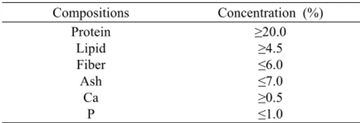
시작일부터 6주간 매일 오전에 UDCA 600 μg/kg BW, 곰취 열수 추출물 및 발효물을 100, 200 및 400 mg/kg BW 농도 가 되도록 생리식염수에 현탁 후 zonde를 이용하여 경구 투여하였다. 시료 경구 투여 4시간 후에 25% 에탄올을 2 mL/kg BW 용량이 되도록 zonde를 이용하여 경구 투여하 였다. 물과 식이는 무제한 급이하였으며(ad libitum), 식이 의 조성은 Table 1과 같다. 본 동물실험은 (재)춘천바이오산 업진흥원 동물실험윤리위원회의 승인을 거쳐 진행하였다 (CBF IACUC No. 2013-004).
시료 채취
실험 마지막 날 시료와 에탄올 경구 투여 후 사료를 제거 하여 18시간 동안 절식시켰다. 마취제로 zoletil(25 mg/kg) 과 xylazine(5 mg/kg)을 혼합하여 사용하였으며, 마취 후 개복하여 심장에서 채혈 및 간 조직 적출을 실시하였다. 채 취한 혈액은 원심분리기(centrifuge 5810R, Eppendorf, Hamburg, Germany)를 이용하여 4°C, 3,200×g에서 20분 간 원심분리 하고 혈청을 분리하여 -80°C deep-freezer에 보관 후 사용하였다. 간 조직은 멸균된 생리식염수를 이용하 여 3회 세척하고 여과지로 표면의 수분을 제거한 후 간의 좌우엽에서 각각 2개의 절편을 떼어냈다. 각각 1개의 절편 은 10% formalin에 고정시켰으며, 나머지 절편은 효소 활성 측정을 위해 액체질소에 급랭시키고 -80°C deep-freezer 에 보관하여 실험에 사용하였다.
체중 및 간 무게 변화 측정
실험기간 동안 체중 측정은 2일 간격으로 일정 시간에 측정하였으며, 식이섭취로 인한 체중 변화 오차를 줄이고자 체중 측정 1시간 전에 사료를 제거하여 측정하였다. 간 조직 은 멸균된 생리식염수로 3회 세척하고 여과지로 표면의 수 분을 제거한 후 무게를 측정하였다.
혈청 분석
분리한 혈청은 생화학분석기(Konelab 20B, Thermo, Vantaa, Finland)를 이용하여 GGT(γ-glutamyl trans- ferase), AST(aspartate aminotransferase), ALT(alanine aminotransferase), LDH(lactate dehydrogenase), tri- glyceride, total cholesterol, HDL(high density lipopro- tein) cholesterol, LDL(low density lipoprotein) choles- terol과 같은 간 주요지표 효소와 지질을 분석하였다. 또한
Table 2. Changes of body weight gain and liver weight in etha- nol-induced rats
Group1) Body weight gain (g/d) Liver weight (g) Normal
Control UDCA LA-100 LA-200 LA-400 LAF-100 LAF-200 LAF-400
3.07±0.40NS2)3) 3.08±0.39 3.36±0.71 3.35±0.42 3.13±0.51 3.08±0.58 3.78±0.46 3.36±0.44 3.61±0.62
8.0±0.5NS 8.5±0.9 8.4±0.9 8.9±0.8 7.9±0.7 8.6±0.5 8.9±0.8 8.6±0.8 8.3±0.9
1)Normal, non-treated; Control, 25% ethanol (2 mL/kg BW) treated; UDCA, ursodeoxycholic acid (positive control, 600 μg/
kg BW and 25% ethanol (2 mL/kg BW)); LA-100, non-fer- mented water extracts from Ligularia fischeri (100 mg/kg BW and 25% ethanol (2 mL/kg BW)); LA-200, non-fermented wa- ter extracts from Ligularia fischeri (200 mg/kg BW and 25%
ethanol (2 mL/kg BW)); LA-400, non-fermented water extracts from Ligularia fischeri (400 mg/kg BW and 25% ethanol (2 mL/kg BW)); LAF-100, fermented water extracts from Ligularia fischeri (100 mg/kg BW and 25% ethanol (2 mL/kg BW));
LAF-200, fermented water extracts from Ligularia fischeri (200 mg/kg BW and 25% ethanol (2 mL/kg BW)); LAF-400, fermented water extracts from Ligularia fischeri (400 mg/kg BW and 25% ethanol (2 mL/kg BW)).
2)Mean±SD.
3)Not significant.
심혈관계 질환의 위험도 판정에 사용되는 동맥경화지수(AI:
atherogenic index) 및 HDL-콜레스테롤 함량 비율(HTR:
high density lipoprotein cholesterol by total cholesterol ratio)은 Han 등(6)의 방법에 근거하여 계산하였다.
간 조직의 항산화 효소 활성 및 과산화지질 함량 측정 혈액을 제거한 간 조직 1 g에 5배의 RIPA buffer[20 mM Tris-HCl(pH 7.5), 150 mM NaCl, 1 mM Na2EDTA, 1 mM EGTA, 1% NP-40, 1% sodium deoxycholate, 2.5 mM sodium pyrophosphate, 1 mM beta-glycerophosphate, 1 mM Na3VO4, 1 µg/mL leupeptin, Cell Signaling, Dan- vers, CO, USA]를 가하여 간 조직을 homogenizer(HS- 30E, DAIHAN, Seoul, Korea)로 4°C에서 10분간 균질화 한 후 10,000×g에서 10분간 원심분리 하여 상등액을 회수 하였다. 회수한 상등액을 4°C에서 105,000×g에서 20분간 원심분리 하여 cytosol을 분리하였다.
Cytosol에 존재하는 SOD의 활성도는 McCord 및 Fri- dovich(31)의 방법을 변형하여 측정하였다. 2 mL의 혼합용 액(5 μmol xanthine, 2 μmol cytochrome c, 0.1 mM EDTA, 50 mM Tris-HCl buffer, pH 7.8)을 cuvette에 넣 은 후 20 μL의 cytosol을 첨가하고 0.2 U/mL의 xanthine oxidase를 첨가하여 550 nm에서 흡광도를 측정하였다. 과 산화지질 함량은 Troncoso Brindeiro 등(32)의 방법에 따 라 균질한 간 조직에 TBA reagent(Cell Biolabs, San Diego, CA, USA)를 첨가하고 95°C에서 60분간 열처리하였다. 열 처리 후 얼음에서 냉각하고 3,200×g에서 15분간 원심분리 하여 상등액을 회수하였으며, 회수한 상등액을 micro-tube 에 300 μL씩 분주하고 동량의 n-butanol을 첨가하여 교반 하였다. 교반 후 20,000×g에서 5분간 원심분리 하여 n- butanol 층을 회수하고 회수한 n-butanol 층을 532 nm에서 흡광도를 측정하였다.
간 조직 병리학적 분석 방법
10% formalin에 고정시킨 간 조직은 함수-탈수 처리를 거쳐 파라핀 블록을 제작하였으며, 4 μm 두께의 미세절편을 만들어 슬라이드 글라스에 부착시켰다. 제작된 슬라이드는 hematoxylin and eosin(H&E) 염색과 Masson's trichrome (MT) 염색을 시행하여 광학현미경으로 200배율로 관찰하 였다(33).
통계 처리
실험결과는 실험군당 평균(mean)과 표준편차(SD)로 나 타내었고, GraphPad prism 4(GraphPad software, La Jolla, CA, USA)를 이용하여 일원배치 분산분석(one-way anal- ysis of variance)을 실시한 후 P<0.05 수준에서 Tukey's multiple comparison test에 의해 각 실험군 평균치 간의 유의성을 검정하였으며, 염색한 간 조직은 3집단 이상을 비 교하는 비모수법(Kruskal-Wallis test)을 이용하여 처리군
간의 유의성을 검정하였다.
결과 및 고찰
체중 증가 및 간 무게 변화에 미치는 영향
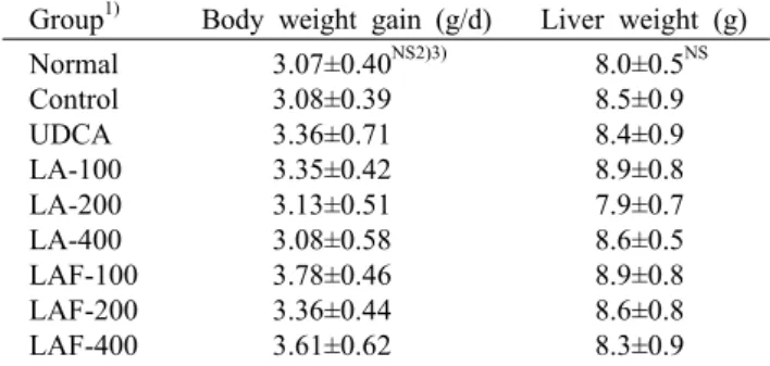
실험기간 동안 실험동물의 체중 증가량 및 간 무게의 경우 Table 2에서 보는 바와 같이 처리군 간 유의적인 차이가 없 는 것으로 나타났다.
간 기능 효소에 미치는 영향
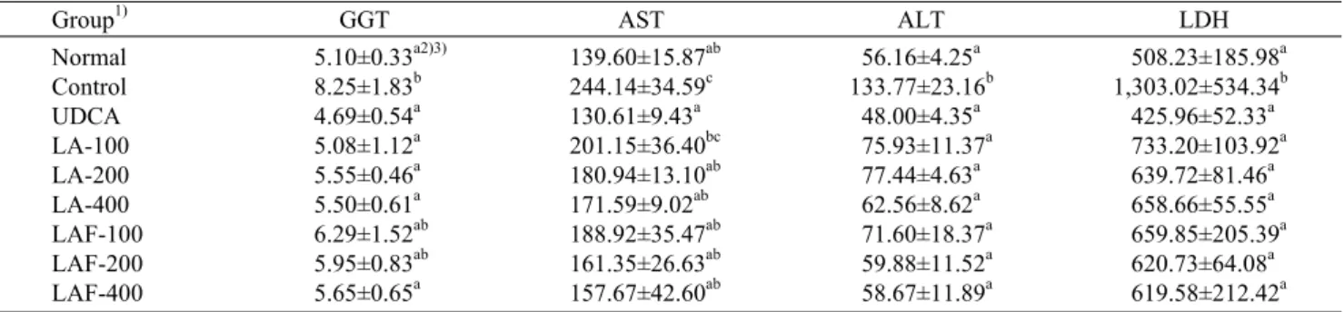
에탄올과 각각의 시료 투여 후 혈청 GGT, AST, ALT 및 LDH 활성을 분석한 결과는 아래 Table 3과 같다. GGT는 일반적으로 간, 신장 및 췌장에 분포하며, 질병 방어기능을 하여 간질환이나 알코올성 간질환 지표로 널리 사용되고, AST는 간과 심장에 고농도로 존재하며 간염, 간 경변 등의 지표로 널리 사용되고 있다. ALT는 여러 조직에 광범위하 게 존재하나 특히 간에서 가장 높은 활성도를 나타내며 간염 과 간 괴사, 간 경변 등으로 인해 활성이 증가하는 것으로 알려져 간 손상 지표로 사용하고 있고, LDH의 경우 심장질 환, 간질환, 악성종양 및 백혈병, 악성빈혈 및 급성간염 등에 서 증가하는 것으로 알려져 있다(6).
혈청 GGT 측정 결과 정상식이군과 비교하여 대조군에서 GGT가 유의하게 증가하였으며(P<0.05), 곰취 열수 추출물 (100, 200, 400 mg/kg BW) 및 발효물(400 mg/kg BW) 투여군에서 대조군과 비교하여 유의하게 감소하였다(P<
0.05). AST는 정상식이군과 비교하여 대조군의 경우 AST
Table 3. Effects of non-fermented and fermented water extracts from Ligularia fischeri on the GGT, AST, ALT, and LDH activity in serum of ethanol-induced rats (U/L)
Group1) GGT AST ALT LDH
Normal Control UDCA LA-100 LA-200 LA-400 LAF-100 LAF-200 LAF-400
5.10±0.33a2)3) 8.25±1.83b 4.69±0.54a 5.08±1.12a 5.55±0.46a 5.50±0.61a 6.29±1.52ab 5.95±0.83ab 5.65±0.65a
139.60±15.87ab 244.14±34.59c 130.61±9.43a 201.15±36.40bc 180.94±13.10ab 171.59±9.02ab 188.92±35.47ab 161.35±26.63ab 157.67±42.60ab
56.16±4.25a 133.77±23.16b 48.00±4.35a 75.93±11.37a 77.44±4.63a 62.56±8.62a 71.60±18.37a 59.88±11.52a 58.67±11.89a
508.23±185.98a 1,303.02±534.34b 425.96±52.33a
733.20±103.92a 639.72±81.46a 658.66±55.55a 659.85±205.39a 620.73±64.08a 619.58±212.42a
1)Refer to Table 2. 2)Mean±SD.
3)Values with the different letters within the same column are significantly different (P<0.05).
Table 4. Effects of non-fermented and fermented water extracts from Ligularia fischeri on the concentration of serum lipids in serum of ethanol-induced rats (mg/dL)
Group1) Triglyceride Total cholesterol HDL cholesterol LDL cholesterol AI2) HTR3) Normal
Control UDCA LA-100 LA-200 LA-400 LAF-100 LAF-200 LAF-400
61.34±4.40NS4)5) 72.88±9.81 57.54±5.86 71.54±8.44 71.13±11.04 63.92±6.71 70.67±10.70 62.36±7.16 58.65±13.33
74.09±10.48bcd6) 88.36±4.89d 83.72±2.25cd 74.56±5.58bcd 72.73±5.28bcd 83.96±10.11d 78.79±9.24cd 58.30±9.34ab 65.22±6.18abc
58.96±7.55bcd 69.56±3.30d 68.47±2.34d 59.70±5.55bcd 58.81±4.21bcd 66.92±7.79d 61.21±6.38cd 46.86±7.30ab 52.75±3.95abc
14.50±2.96NS 17.32±2.78 15.31±1.80 13.94±1.07 14.24±1.55 14.48±2.11 14.00±5.52 12.96±1.81 11.47±1.32
0.25±0.03NS 0.27±0.03 0.22±0.03 0.25±0.03 0.24±0.02 0.25±0.02 0.29±0.02 0.25±0.03 0.23±0.03
0.80±0.02NS 0.79±0.02 0.82±0.02 0.80±0.01 0.81±0.01 0.80±0.01 0.78±0.01 0.80±0.01 0.81±0.02
1)Refer to Table 2.
2)Atherogenic index (AI)=(total cholesterol-HDL cholesterol)/ HDL cholesterol.
3)HTR=HDL cholesterol/ total cholesterol.
4)Mean±SD. 5)Not significant.
6)Values with the different letters within the same column are significantly different (P<0.05).
수치가 유의하게 증가하였으며(P<0.05), 곰취 열수 추출물 (200, 400 mg/kg BW) 및 발효물(100, 200, 400 mg/kg BW) 투여군에서 대조군과 비교하여 유의하게 감소하였다 (P<0.05). ALT 및 LDH 측정 결과 대조군에서 정상식이군 과 비교하여 ALT 및 LDH 수치가 유의하게 증가하였으며 (P<0.05), 곰취 열수 추출물 및 발효물 모든 투여군에서 대 조군과 비교하여 유의하게 감소하였다(P<0.05). 본 연구 결 과 곰취 열수 추출물 및 발효물 투여군에서 대조군과 비교하 여 혈청 중 간 기능 효소의 활성이 유의하게 감소하는 것으 로 나타났다(P<0.05). 담즙산의 핵심 성분인 UDCA를 처리 한 양성대조군의 경우 대조군과 비교하여 GGT, AST, ALT 및 LDH 수치가 현저하게 감소하는 것으로 나타났으나 (P<0.05) 곰취 열수 추출물 및 발효물 투여군 간 유의적인 차이는 없는 것으로 나타났다.
Kim 등(20)은 온라인 항산화 장치를 이용하여 곰취 추출 물의 주요 항산화 성분이 5-O-caffeoylquinic acid(5- CQA), 3,4-di-O-caffeoylquinic acid(3,4-DCQA), 3,5- di-O-caffeoylquinic acid(3,5-DCQA) 및 4,5-di-O-caf- feoylquinic acid(4,5-DCQA)로 구성된 caffeoylquinic acid(CQA)류의 화합물임을 확인하였으며, 곰취 추출물이 에탄올에 의해 간 손상이 유발된 랫드의 혈청 GGT, AST
및 ALT 수치를 유의하게 감소시키고 간 조직 내에 존재하는 효소의 활성을 증가시켜 간을 보호한다고 보고한 바 있다 (34). Choi 등(30)은 carbon tetrachloride(CCl4) 및 D- galactosamine(D-GalN) 투여로 간 손상이 유발된 랫드의 혈청을 분석한 결과 곰취로부터 분리한 3,4-di-O-caf- feoylquinic acid(3,4-DCQA) 투여군에서 혈청 중 GGT, AST, ALT, LDH, sorbitol dehydrogenase(SDH) 및 al- kaline phosphatase(ALP) 수치가 유의하게 감소하는 것을 확인하였다. 따라서 곰취 열수 추출물 및 발효물 투여군의 혈청 GGT, AST, ALT 및 LDH 수치 감소는 곰취 추출물 내에 존재하는 CQA에 의한 것으로 사료되나 이와 관련된 추가적인 연구가 필요할 것으로 판단된다.
혈청지질 성분에 미치는 영향
혈청 내 총 콜레스테롤(total cholesterol), HDL-콜레스 테롤, LDL-콜레스테롤 및 중성지방(triglyceride)을 분석 한 결과는 Table 4와 같다. 혈청 내 총 콜레스테롤 및 HDL- 콜레스테롤 측정 결과 정상식이군과 대조군 간의 유의적인 차이는 나타나지 않았으나 발효물 투여군(200, 400 mg/kg BW)의 경우 대조군과 비교하여 유의하게 감소하였다(P<
0.05). LDL-콜레스테롤 측정 결과 대조군의 경우 정상식이
0.00 2.00 4.00 6.00 8.00 10.00 12.00 14.00
Normal Control UDCA LA- 100
LA- 200
LA- 400
LAF- 100
LAF- 200
LAF- 400
SOD (U/mg protein) .
a
b
ab a a a
a a a
25% ethanol (2 mL/kg BW) UDCA1) (600 µg/kg BW) LA2) (mg/kg)
LAF3) (mg/kg)
-
-
-
- +
-
-
- + +
-
- +
- 100
- +
- 200
- +
- 400
- +
-
- 100
+
-
- 200
+
-
- 400
Fig. 1. Effects of non-fermented and fermented water extracts from Ligularia fischeri on the superoxide dis- mutase (SOD) activity in liver tissue of ethanol-induced rats. Values with the different letters above bar graphs are significantly different (P<0.05). 1)Ursodeoxycholic acid. 2)Non-fermented water extracts from Ligularia fischeri. 3)Fermented water extracts from Ligularia fischeri.
0.00 5.00 10.00 15.00 20.00 25.00 30.00 35.00 40.00 45.00 50.00
Normal Control UDCA LA- 100
LA- 200
LA- 400
LAF- 100
LAF- 200
LAF- 400
MDA (nM/mg protein) .
abc
abc ab abc bcd bcd
cd
bcd d
25% ethanol (2 mL/kg BW) UDCA1) (600 µg/kg BW) LA2) (mg/kg)
LAF3) (mg/kg)
-
-
-
- +
-
-
- + +
-
- +
- 100
- +
- 200
- +
- 400
- +
-
- 100
+
-
- 200
+
-
- 400
Fig. 2. Effects of non-fermented and fermented water extracts from Ligularia fischeri on malondialdehyde (MDA) contents in liver tissue of ethanol-induced rats.
Values with the different letters above bar graphs are significantly different (P<0.05). 1)Ursodeoxycholic acid.
2)Non-fermented water extracts from Ligularia fischeri.
3)Fermented water extracts from Ligularia fischeri.
군과 비교하여 LDL-콜레스테롤이 증가하였으나 유의적인 차이는 없었으며, 곰취 열수 추출물 및 발효물의 경우 대조 군과 비교하여 LDL-콜레스테롤이 감소하였으나 유의적인 차이는 없었다. 중성지방의 경우 정상식이군과 비교하여 대 조군에서 증가하였으나 유의적인 차이는 없었으며, UDCA 투여군과 곰취 열수 추출물(400 mg/kg BW) 및 발효물(200, 400 mg/kg BW) 투여군에서 감소하는 것으로 나타났으나 유의성은 없었다. 총 콜레스테롤 함량 중 HDL-콜레스테롤 함량 비율 및 동맥경화지수는 모든 처리군에서 유의적인 차 이는 없는 것으로 나타났다.
항산화 효소 및 과산화지질에 미치는 영향
만성적인 알코올 섭취는 대사과정 중 산화적 스트레스를 유발하여 ROS를 생성하며, NADH의 전자전달체계 방해로 인해 미토콘드리아 내에 생성된 ROS는 지질산화를 일으켜 MDA(malondialdehyde)를 생성하고 간세포 내 염증반응을 유발하여 간 손상을 초래하는 것으로 알려져 있다(35,36).
산화적 스트레스로 인해 생성되는 ROS는 생체 내 항산화 효소들에 의해 대부분 소거되지만, 과량 생성된 ROS는 항산
화 방어계의 균형을 깨트리며 각종 질환의 발생 및 노화를 촉진시킨다. 따라서 항산화 효소의 활성이 증가하게 되면 세포손상을 막아 질환 발생과 노화 촉진을 억제할 수 있다고 보고되어 왔다(7,37-39).
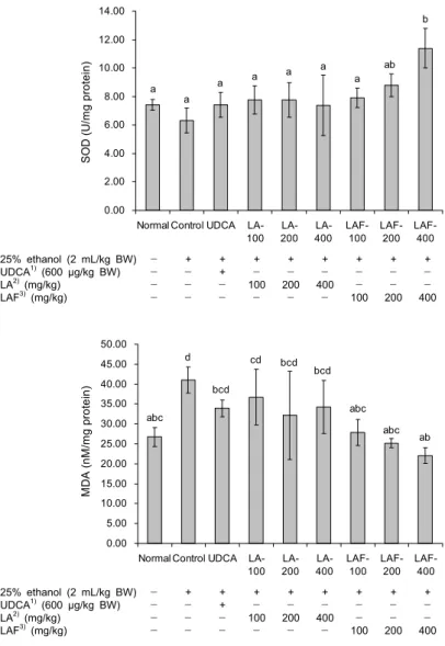
간 조직 내 SOD 활성 변화 결과는 Fig. 1과 같다. 에탄올 을 단독 투여한 대조군의 경우 정상식이군과 비교하여 SOD 활성이 감소하였으며, 곰취 열수 추출물 투여군에서 SOD 활성이 증가하였으나 유의적인 차이가 없는 것으로 나타났 고, 발효물 투여군(400 mg/kg BW)에서 대조군과 비교하여 유의하게 SOD 활성이 증가하였다(P<0.05). Fig. 2에서 보 는 바와 같이 간 조직 내 과산화지질의 분해산물인 MDA 함량을 측정한 결과 에탄올을 단독 투여한 대조군에서 MDA 함량이 가장 높게 나타났으며(P<0.05), 곰취 열수 추출물을 투여한 처리군에서는 대조군과 비교하여 MDA 함량이 감소 하는 것으로 나타났고, 발효물 투여군의 경우 모든 농도에서 MDA 함량이 유의하게 감소하였다(P<0.05). 알코올은 주로 간에서 alcohol dehydrogenase(ADH), cytochrome P450 2E1 및 catalase 등 3가지 효소계에 의해 acetaldehyde로 대사되지만 과량의 알코올 섭취 시 microsomal ethanol
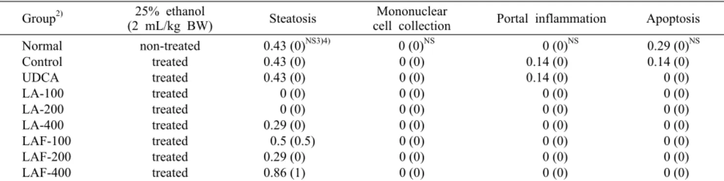
Table 5. A histological score1) on the ethanol-induced liver damage in H&E and MT staining patterns on SD rats (n=5) Group2) 25% ethanol
(2 mL/kg BW) Steatosis Mononuclear
cell collection Portal inflammation Apoptosis Normal
Control UDCA LA-100 LA-200 LA-400 LAF-100 LAF-200 LAF-400
non-treated treated treated treated treated treated treated treated treated
0.43 (0)NS3)4) 0.43 (0) 0.43 (0) 0 (0) 0 (0) 0.29 (0) 0.5 (0.5) 0.29 (0) 0.86 (1)
0 (0)NS 0 (0) 0 (0) 0 (0) 0 (0) 0 (0) 0 (0) 0 (0) 0 (0)
0 (0)NS 0.14 (0) 0.14 (0) 0 (0) 0 (0) 0 (0) 0 (0) 0 (0) 0 (0)
0.29 (0)NS 0.14 (0) 0 (0) 0 (0) 0 (0) 0 (0) 0 (0) 0 (0) 0 (0)
1)A histological score was blindly calculated on liver sections as follows: 0∼3 for steatosis; 0∼3 for mononuclear cell collection;
0∼3 for extent of portal inflammation; 0∼4 for extent of apoptosis; and 0=no involvement; 1=mild; 2=moderate; 3=severe for each of the four parameters.
2)Refer to Table 2. 3)Not significant. 4)Mean (median).
Fig. 3. Representative histopathological sections of normal, control and exper- imental groups (H&E, ×200). There is occasionally minimal hepatocytic va- cuolization around porta tract in UDCA (B)and Control (C) groups. No signifi- cant pathologic findings are noted among the groups. A, Normal; B, UDCA; C, Control; D, LA-100; E, LA-200; F, LA- 400; G, LAF-100; H, LAF-20; and I, LAF-400. Groups are the same as in Table 2. Bar=0.1 mm.
oxidizing system(MEOS)에 의해 acetaldehyde로 대사되 고 만성적인 알코올 섭취는 지방 합성 증가 및 ROS를 생성 하여 간세포 손상을 유발한다(6-8). 랫드에 에탄올을 지속 적으로 투여할 경우 간에서 활성산소흡수능력(ORAC; ox- ygen radical absorbance capacity) 감소, MDA 증가, 총 glutathione 감소, SOD 및 GSH-px 효소 활성이 감소하지 만 곰취 추출물을 투여할 경우 ORAC 증가, MDA 감소, 총 glutathione 증가, SOD 및 GSH-px 효소 활성이 증가하고 SOD, catalase(CAT), GSH-px, hemeoxygenase(HO)의 단백질 발현량이 증가하는 것으로 알려졌다(34). 이와 같이 곰취 추출물의 항산화 및 간 보호 효과는 곰취 추출물의 주
요 성분인 CQA에 의한 것으로 보고되고 있다(20,30). 곰취 열수 추출물 및 발효물 투여군에서의 MDA 감소는 SOD와 같은 항산화 효소의 활성 증가로 인해 지질과산화 반응을 억제하였기 때문으로 사료되나 추가적으로 CAT 및 GSH- px와 같은 항산화 효소 활성 검증이 필요할 것으로 판단된 다.
간 조직 병리학적 분석 결과
적출한 간을 H&E 염색과 MT 염색을 실시하여 지방증 (steatosis; fatty change or hepatocytic vacuolization), 간문맥 염증(portal inflammation), 단핵구 침윤(mononu-
clear cell collection) 및 세포사멸(apoptosis)을 관찰하였 다(Table 5). 대조군의 간문맥(portal tract) 주위에서 약간 의 지방증이 일부 관찰되었으며, 곰취 열수 추출물 및 발효 물 투여군에서도 간문맥 주위에 미약한 지방증이 관찰되었 으나 특이소견은 관찰되지 않았다(Fig. 3).
요 약
본 연구는 항산화 활성이 우수한 것으로 알려진 곰취의 활용 가치를 높이고 향후 건강기능식품 소재로서의 가능성을 확 인하고자 에탄올 동물모델에서의 간 기능 개선 효과를 연구 하였다. 실험기간 동안 실험동물의 체중 증가량 및 간 무게 의 경우 처리군 간 유의적인 차이는 없었다. 혈청 중 GGT (γ-glutamyl transferase), AST(aspartate aminotrans- ferase), ALT(alanine aminotransferase) 및 LDH(lactate dehydrogenase) 측정 결과 대조군에서 정상식이군과 비교 하여 유의하게 증가하였다(P<0.05). 곰취 열수 추출물(100, 200, 400 mg/kg BW) 및 발효물(400 mg/kg BW) 투여군에 서 대조군과 비교하여 GGT 수치가 유의하게 감소하였으며 (P<0.05), 곰취 열수 추출물(200, 400 mg/kg BW) 및 발효 물(100, 200, 400 mg/kg BW) 투여군에서 대조군과 비교하 여 AST 수치가 유의하게 감소하였다(P<0.05). 곰취 열수 추 출물 및 발효물 모든 투여군에서 대조군과 비교하여 ALT 및 LDH 수치가 유의하게 감소하였다(P<0.05). 혈청 중 총 콜레스테롤 측정 결과 정상식이군과 대조군 간의 유의적인 차이는 없었으나 발효물 투여군(200, 400 mg/kg BW)에서 대조군과 비교하여 유의하게 감소하였다(P<0.05). HDL(high density lipoprotein)-콜레스테롤 측정 결과 정상식이군과 대조군 간 유의적인 차이는 없었으나, 발효물 투여군(200, 400 mg/kg BW)에서 대조군과 비교하여 유의하게 감소하 였다(P<0.05). 곰취 열수 추출물 및 발효물의 경우 대조군 과 비교하여 LDL(low density lipoprotein)-콜레스테롤이 감소하는 것으로 나타났으나 유의적인 차이는 없었다. 중성 지방의 경우 정상식이군과 비교하여 대조군에서 증가하는 것으로 나타났으나 유의적인 차이는 없는 것으로 나타났으 며, 곰취 열수 추출물(400 mg/kg BW) 및 발효물(200, 400 mg/kg BW) 투여군에서 감소하는 것으로 나타났으나 유의 적인 차이는 없었다. 총 콜레스테롤 함량 중 HDL-콜레스테 롤 함량 비율 및 동맥경화지수를 조사한 결과 모든 처리군 간에 유의적인 차이는 없었다. 간 조직 내 superoxide dis- mutase(SOD) 활성을 측정한 결과 발효물 투여군(400 mg/
kg BW)에서 대조군과 비교하여 유의하게 증가하였다(P<
0.05). 간 조직 내 과산화지질의 분해산물인 malondialde- hyde(MDA) 측정 결과 에탄올을 단독 투여한 대조군에서 MDA 함량이 가장 높게 나타났으며, 발효물을 투여한 모든 농도에서 MDA 함량이 유의적으로 감소하였다(P<0.05). 간 조직의 병리학적 분석 결과 모든 처리군에서 간문맥에 염증 이 거의 관찰되지 않았고 곰취 열수 추출물 및 발효물 처리
군에서 대조군과 비교하여 지방증 등이 개선되는 경향을 보 였으나 유의적인 차이는 없는 것으로 나타났다. 곰취 열수 추출 발효물의 투여가 혈청 GGT, AST, ALT 및 LDH 활성 을 유의적으로 감소시켜 간 기능 개선 효과가 있음을 확인할 수 있었으며, 간 조직 내 SOD 활성을 증가시키고 지질과산 화 반응을 억제시킴으로써 알코올로 인한 간 손상을 완화시 키는 데 효과적일 것으로 사료되며, 향후 건강기능식품 소재 로 이용 가능할 것으로 사료된다.
REFERENCES
1. Tsukamoto S, Kanegae T, Uchigasaki S, Kitazawa M, Fujioka T, Fujioka S, Imamura Y, Nagoya T, Shimamura M, Mieda Y. 1993. Changes in free and bound alcohol me- tabolites in the urine during ethanol oxidation. Arukoru Ken- kyuto Yakubutsu Ison 28: 441-452.
2. Lieber CS, Garro A, Leo MA, Mak KM, Worner T. 1986.
Alcohol and cancer. Hepatology 6: 1005-1019.
3. Lieber CS. 1994. Alcohol and liver update: 1994 update.
Gastroenterology 106: 1085-1105.
4. Lieber CS. 1995. Medical disorders of alcoholism. N Engl J Med 333: 1058-1065.
5. Lieber CS. 2004. The unexpected outcomes of medical re- search: serendipity and the microsomal ethanol oxidizing system. J Hepatol 40: 198-202.
6. Han EK, Jin YX, Yoo YS, Jung EJ, Lee JY, Chung CK.
2009. Effect of Artemisia capillaris and Paecilomyces ja- ponica on the reduction of hepatotoxicity and lipid metabo- lism induced by ethanol. J Korean Soc Food Sci Nutr 38:
1016-1023.
7. Yu KH, Lee SY, Yang HM, Ham YA, Lee SU, Chae SW, Lee YJ. 2013. Effects of the Artemisia capillaris extract on the hepatotoxicity in ethanol-induced rats. J Korean Soc Food Sci Nutr 42: 1560-1566.
8. Zakhari S. 2006. Overview: how is alcohol metabolized by the body? Alcohol Res & Health 29: 245-254.
9. Lieber CS. 1970. New pathway of ethanol metabolism in the liver. Gastroenterology 59: 930-937.
10. Bleich HL, Boro ES. 1977. Metabolic and hepatic effects of alcohol. N Engl J Med 296: 612-616.
11. Gramenzi A, Caputo F, Biselli M, Kuria F, Loggi E, Andreone P, Bernardi M. 2006. Review article: alcoholic liver disease-pathophysiological aspects and risk factors.
Aliment Pharmacol Ther 24: 1151-1161.
12. Das SK, Vasudevan DM. 2007. Alcohol-induced oxidative stress. Life Sci 81: 177-187.
13. Nagata K, Suzuki H, Sakaguchi S. 2007. Common patho- genic mechanism in development progression of liver injury caused by non-alcoholic or alcoholic steatohepatitis. J Toxi- col Sci 32: 453-468.
14. Byers T, Perry G. 1992. Dietary carotenes, vitamin C, and vitamin E as protective antioxidants in human cancers. Annu Rev Nutr 12: 135-159.
15. Lawrence RA, Burk RF. 1976. Glutathione peroxidase ac- tivity in selenium-deficient rat liver. Biochem Biophys Res Commun 71: 952-958.
16. Polavarapu R, Spitz DR, Sim JE, Follansbee MH, Oberley LW, Rahemtulla A, Nanji AA. 1998. Increased lipid perox- idation and impaired antioxidant enzyme function is asso- ciated with pathological liver injury in experimental alco- holic liver disease in rats fed diets high in corn oil and
fish oil. Hepatology 27: 1317-1323.
17. Rouach H, Fataccioli V, Gentil M, French SW, Morimoto M, Nordmann R. 1997. Effect of chronic ethanol feeding on lipid peroxidation and protein oxidation in relation to liver pathology. Hepatology 25: 351-355.
18. French SW, Wong K, Jui L, Albano E, Hagbjork AL, Ingel- man-Sundberg M. 1993. Effect of ethanol on cytochrome P450 2E1 (CYP2E1), lipid peroxidation, and serum protein adduct formation in relation to liver pathology pathogenesis.
Exp Mol Pathol 58: 61-75.
19. Nanji AA. 2004. Role of different dietary fatty acids in the pathogenesis of experimental alcoholic liver disease. Alco- hol 34: 21-25.
20. Kim SM, Kang SW, Um BH. 2010. Extraction condition of radical scavenging caffeoylquinic acids from Gomchui (Ligu- laria fischeri) tea. J Korean Soc Food Sci Nutr 39: 399-405.
21. Bae JH, Yu SO, Kim YM, Chon SU, Kim BW, Heo BG.
2009. Physiological activity of methanol extracts from Ligu- laria fischeri and their hyperplasia inhibition activity of can- cer cell. J Bio-Environ Control 18: 67-73.
22. Kim DW, Son KH, Chang HW, Bae K, Kang SS, Kim HP.
2004. Anti-inflammatory activity of Sedum kamtschaticum.
J Ethnopharmacol 90: 409-414.
23. Ham SS, Lee SY, Oh DH, Jung SW, Kim SH, Chung CK, Kang IJ. 1998. Antimutagenic and antigenotoxic effects of Ligularia fischeri extracts. J Korean Soc Food Sci Nutr 27:
745-750.
24. Jeong S, Kim E, Hwangbo H, Ham S. 1998. Effects of Ligu- laria fischeri extracts on oxidation of low density lipopro- tein. Korean J Food Sci Technol 30: 1214-1221.
25. Choi EM, Kim YH. 2008. A preliminary study of the effects of an extract of Ligularia fischeri leaves on type Ⅱ colla- gen-induced arthritis in DBA/1J mice. Food Chem Toxicol 46: 375-379.
26. Choi EM, Suh KS. 2009. Ligularia fischeri leaf extract sup- presses proinflammatory mediators in SW982 human syno- vial cells. Phytother Res 23: 1575-1580.
27. Choi EM, Ding Y, Nguyen HT, Park SH, Kim YH. 2007.
Antioxidant activity of Gomchi (Ligularia fischeri) leaves.
Food Sci Biotechnol 16: 710-714.
28. Choi EM. 2007. Ligularia fischeri leaf extract prevents the oxidative stress in DBA/1J mice with type Ⅱ collagen-in- duced arthritis. J Appl Toxicol 27: 176-182.
29. Choi GP, Chung BH, Lee DI, Lee HY, Lee JH, Kim JD.
2002. Screening of inhibitory activities on angiotensin con- verting enzyme from medicinal plants. Korean J Medicinal Crop Sci 10: 399-402.
30. Choi J, Park JK, Lee KT, Park KK, Kim WB, Lee JH, Jung HJ, Park HJ. 2005. In vivo antihepatotoxic effects of Ligu- laria fischeri var. spiciformis and the identification of the active component, 3,4-dicaffeoylquinic acid. J Med Food 3: 348-352.
31. McCord JM, Fridovich I. 1969. Superoxide dismutase. An enzymatic function for erythrocuprein (hemocuprein). J Biol Chem 244: 6049-6055.
32. Troncoso Brindeiro CM, Lane PH, Carmines PK. 2012.
Tempol prevents altered K+ channel regulation of afferent arteriolar tone in diabetic rat kidney. Hypertension 59: 657- 664.
33. Gove ME, Rhodes DH, Pini M, van Baal JW, Sennello JA, Fayad R, Cabay RJ, Myers MG Jr, Fantuzzi G. 2009. Role of leptin receptor-induced STAT3 signaling in modulation of intestinal and hepatic inflammation in mice. J Leukoc Biol 85: 491-496.
34. Yoo JH, Oidovsambuu S, Kim SM, Jeon NR, Yun JH, Kang YK, Jho EH, Lee SB, Nho CW. 2011. Hepatoprotective ef- fect of Handaeri-gomchi (Ligularia fischeri var. spiciformis Nakai) extract against chronic alcohol-induced liver damage in rats. Food Sci Biotechnol 20: 1655-1661.
35. Thurman RG, Bradford BU, Iimuro Y, Knecht KT, Connor HD, Adachi Y, Wall C, Arteel GE, Raleigh JA, Forman DT, Mason RP. 1997. Role of Kupffer cells, endotoxin and free radicals in hepatotoxicity due to prolonged alcohol con- sumption: studies in female and male rats. J Nutr 127: 903S- 906S.
36. Nordmann R, Ribière C, Rouach H. 1992. Implication of free radical mechanisms in ethanol-induced cellular injury.
Free Radic Biol Med 12: 219-240.
37. Jeon SM, Bok SH, Jang MK, Lee MK, Nam KT, Park YB, Rhee SJ, Choi MS. 2001. Antioxidative activity of naringin and lovastatin in high cholesterol-fed rabbits. Life Sci 69:
2855-2866.
38. Moon SH, Lee MK, Chae KS. 2001. Inhibitory effects of the solvent fractions from persimmon leaves on xanthine oxidase activity. Korean J Food & Nutr 14: 120-125.
39. de Haan JB, Cristiano F, Iannello RC, Kola I. 1995. Cu/
Zn-superoxide dismutase and glutathione peroxidase during aging. Biochem Mol Biol Int 35: 1281-1297.