Pro-apoptotic and Anti-adipogenic Effects of Proso Millet ( Panicum miliaceum ) Grains on 3T3-L1 Preadipocytes
Do Youn Jun
1, Ji Young Lee
1, Cho Rong Han
1, Kwan-Pil Kim
2, Myung Chul Seo
3, Min Hee Nam
4and Young Ho Kim
1*
1
School of Life Science and Biotechnology, College of Natural Sciences, Kyungpook National University, Daegu 702-701, Korea
2
Lotte Confectionary Co., LTD, Yangsan 626-120, Korea
3
Crop Environment Division, NICS, RDA, Suwon 441-857, Korea
4
Functional Cereal Crop Research Division, NICS, RDA, Miryang 627-803, Korea
Received May 20, 2014 /Revised May 24, 2014 /Accepted May 26, 2014To examine the anti-obese activity of miscellaneous cereal grains, 80% ethanol extracts from eight se- lected miscellaneous cereal grains were compared for their cytotoxic effects on 3T3-L1 murine pre- adipocytes. The ethanol extract of proso millet exhibited the highest cytotoxicity. Further fractionation of the ethanol extract with methylene chloride, ethyl acetate, and n-butanol showed that the cytotox- icity of the ethanol extract was mainly partitioned into the butanol fraction. As compared with differ- entiated mature adipocytes, 3T3-L1 preadipocytes were more susceptible to the cyctotoxicity of the bu- tanol fraction. When each organic solvent fraction (25 μg/ml) was added during the differentiation period for 6 days, the cell viability was not affected significantly except for the butanol fraction, but the intracellular lipid accumulation declined to a level of 81.5%~50.3% of the control. The Oil Red O staining data also demonstrated that the ethanol extract as well as the butanol fraction could inhibit the differentiation of 3T3-L1 preadipocytes into mature adipocytes. The presence of the butanol extract during the induced adipocytic differentiation also resulted in a significant reduction in the expression levels of critical adipogenesis mediators (C/EBPα, PPARγ, aP2, and LPL) to a barely detectable or un- detectable level and the cells retained the fibroblast-like morphology of 3T3-L1. In 3T3-L1 cells, the cytotoxicity of the butanol fraction (50-100 μg/ml) was accompanied by mitochondrial membrane po- tential (Δψm) loss, caspase-3 activation, and PARP degradation. Taken together, these results indicate that proso millet grains possess pro-apoptotic and anti-adipocytic activities toward adipocytes, which can be applicable to prevention of obesity.
Key words : 3T3-L1 preadipocyte, anti-adipogenesis, anti-obesity, Panicum miliaceum, proso millet
*Corresponding author
*Tel : +82-53-950-5378, Fax : +82-53-955-5522
*E-mail : [email protected]
This is an Open-Access article distributed under the terms of the Creative Commons Attribution Non-Commercial License (http://creativecommons.org/licenses/by-nc/3.0) which permits unrestricted non-commercial use, distribution, and reproduction in any medium, provided the original work is properly cited.
Journal of Life Science 2014 Vol. 24. No. 5. 505~514 DOI : http://dx.doi.org/10.5352/JLS.2014.24.5.505
Introduction
Obesity, which is known to be induced by the imbalance between energy intake and energy expenditure, has been im- plicated in development of several metabolic diseases in- cluding type 2 diabetes, hyperlipidemia, osteoarthritis, sleep apnea, and cardiovascular disease, and cancers [11, 23, 42].
Obesity is mediated by increased adipose tissue mass result- ing from increased fat-cell numbers as well as by increased fat-cell size [7]. The number of adipocytes present in an or- ganism is determined by the capacity of the adipogenic proc-
ess involving the preadipocyte proliferation (hyperplasia) and their differentiation into mature adipocytes (hypertro- phy), whereas the increase of fat-cell size is mainly affected by the amount of lipids accumulated in the adipocytes. The adipose tissue mass can be reduced by the inhibition of adi- pogenesis from preadipocytes to mature adipocytes, pre- vention of lipid accumulation in the adipocytes, and in- duction of apoptosis in adipose cells [33].
In this regard, several naturally occurring phytochemicals
from various plant sources, which have potential for inhibit-
ing adipogenesis of preadipocytes and inducing apoptosis
of preadipocyte and/or adipocytes, have been reported to
possess anti-obesity effects. Polyphenol, coumarin de-
rivatives, carotenoids, and phytoalexins are among the phy-
tochemicals that appear to possess the potential for inhibit-
ing adipogenesis and inducing apoptosis of adipocytes [1,
2, 12, 13, 25, 41]. Because these phytochemicals are also
found in plant-derived foods such as fruits, vegetables,
beans, and grains, much attention has been paid to the health benefits of these foods with respect to their function to lower the incidence of obesity. Furthermore, many studies have reported that a diet rich in whole-grain cereals and their products can exert protective effects against develop- ment of obesity, diabetes, cardiovascular disease, and can- cers [3, 4, 23, 34, 37].
After treatment of 3T3-L1 preadipocytes, derived from a mouse embryo, with a mixture of isobutylmethylxanthine (IBMX), insulin and dexamethasone, the cells can be differ- entiated into adipocytes along with acquisition of expression of functional differentiation markers, and specific cellular morphology along with an accumulation of lipid droplets within the cells [10]. In this context, 3T3-L1 cells have been used as the in vitro experimental model to mimic adipocyte hyperplasia, to elucidate the mechanism of adipocytic differ- entiation, and to assess suppressive effects of anti-adipocytic agents [5, 8, 33].
Agriculture in Korea has traditionally focused on pro- duction of the major cereal grains such as rice, barley and wheat, whereas the cultivation and harvesting of other mis- cellaneous cereal grains has remained in a low level. In re- cent years, however, due to increased demand for well-being foods, the interest in miscellaneous cereal grains as crude fibers and bioactive phytochemical sources that benefit hu- man health and thus the consumption of miscellaneous ce- real grains are also increasing in the developed countries.
Although several studies have been performed to extend our understanding on nutritional importance, antioxidant, anti- microbial, antimutagenic, and anticarcinogenic, and anti- diabetic properties of miscellaneous cereal grains harvested in Korea [9, 15, 17, 18, 29], the systematic study on their bioactive components associated with the anti-obese efficacy is still rare. If miscellaneous cereal grains are proven as a proper source of anti-obese phytochemicals, it is likely that these grains become highly effective in obesity-related chronic diseases by consuming as diet [22, 34, 38].
As an attempt to compare anti-obese effects of eight se- lected miscellaneous cereal grains (proso millet, hwang- geumchal sorghum, glutinous sorghum, yellow glutinous foxtail millet, green glutinous foxtail millet, golden foxtail millet, barnyard millet, and adlay) harvested in Korea, in the present study, we intended to investigate the anti-adipo- genic effects of 80% ethanol extracts of the individual grains using 3T3-L1 murine preadipocytes. The 80% ethanol extract of proso millet grains, which appeared to possess the highest
cytotoxicity toward 3T3-L1 cells among the selected grains, was sequentially fractionated by methylene chloride, ethyl acetate, and n-butanol. Since the cytotoxicity toward 3T3-L1 cells was mainly detected in the butanol fraction, the an- ti-adipogenic activity of the butanol fraction has been exam- ined further by investigating not only its inhibitory effect on adipocytic differentiation of 3T3-L1 preadipocytes, but al- so its pro-apoptotic effect on 3T3-L1 preadipocytes.
Materials and Methods
Reagents, chemicals, antibodies and culture medium The ECL Western blotting kit was purchased from Amersham (Arlington Height, IL, USA), and Immobilon-P membrane was obtained from Millipore Corporation (Bedford, MA, USA). Anti-caspase-3 and anti-PARP were purchased from Cell Signaling Technology (Beverly, MA, USA), and anti-β-actin was obtained from Santa Cruz Biotechnology (Santa Cruz, CA, USA). The cell viability as- say kit (CellTiter 96® Aqueous One Solution Cell Prolifera- tion assay) was purchased from Promega (Madison, WI, USA). 3T3-L1 preadipocytes were purchased from ATCC (Manassas, VA, USA), and cultured in Dulbecco's modified Eagles medium (DMEM) (Hyclone, Gaithersburg, MD, USA) containing 10% bovine calf serum (BCS) and 100 μg/ml gentamycin. Insulin, IBMX, and dexamethasone were ob- tained from Sigma Chemical (St. Louis, MO, USA). Cells were cultured at 37ºC in a humidified 5% CO
2atmosphere.
Preparation of sample extracts
Eight miscellaneous cereal grains, including proso millet
(polished grains), hwanggeumchal sorghum (unpolished
grains), glutinous sorghum (polished grains), yellow gluti-
nous foxtail millet (polished grains), green glutinous foxtail
millet (polished grains), golden foxtail millet (unpolished
grains), barnyard millet (unpolished grains), and adlay
(polished grains) were provided by National Institute of
Crop Science, Rural Development Administration, Miryang,
Gyeongnam 627-803, Korea. Individual dried grains were
milled on a Blender 7012 (Dynamics Corporation, USA), and
then extracted with 80% ethanol for 3 hr at 80°C as described
elsewhere [28]. The ethanol extract was evaporated, dis-
solved in water, and then sequentially extracted with meth-
ylene chloride, ethyl acetate, and n-butanol. Each organic
solvent fractionation was repeated three times. The organic
solvent fraction was concentrated by rotary vacuum evapo-
rator (Heidolph LR 4000, Germany).
Cell viability assay
Cytotoxic effect of grain extracts on 3T3-L1 cell was ana- lyzed using the cell viability assay kit (CellTiter 96®
Aqueous One Solution Cell Proliferation Assay) containing 3-(4,5-dimethylthiazol-2-yl)-5-(3-carboxymethoxyphenyl)- 2-(4-sulfophenyl)-2H-tetrazolium (MTS) reagent. Briefly, 3T3-L1 cells (1~3 × 10
4/well) were cultured with serial dilu- tions of proso millet extract in 96-well plate. At 48 hr after incubation, the medium was removed and replaced with 100 μ l fresh culture media and 10 μl MTS solution (Promega, Mdadison, WI, USA). After incubation for an additional 2 h, 25 μl 10% sodium dodecyl sulfate (SDS) was added as a stop solution. The absorbance was measured at 490 nm by a plate reader to determine the formazan concentration, which reflects the cell viability.
Quantification of lipid content
Lipid (triglyceride) content was determined using the AdipoRed
™assay reagent (Cambrex Bio Science Walkers- ville, Walkersville, MD, USA) according to manufacturer's instructions. Briefly, 3T3-L1 cells (1 × 10
4/well) were seeded in 96-well microtiter plates and treated for induction of adi- pocytic differentiation. On day 6 after induction, the cells were washed with PBS and 100 μl of PBS was added to the wells. Five microliter of AdipoRed
™assay reagent was add- ed to each well and incubated for 10 min at RT. The amount of triglyceride was determined by measuring with an ex- citation wave/length of 485 nm and emission wave/length of 572 nm using PerkinElmer
™Walac Victor 3.
Induction of adipocytic differentiation and oil droplet staining
The 3T3-L1 murine preadipose cell line was purchased from American Type Culture Collection (Manassas, VA, USA). Adipocytic differentiation of 3T3-L1 preadipocytes was induced as previously described [13]. Briefly, cells were cultured in Dulbecco's modified Eagles medium (DMEM) containing 10% bovine calf serum (BCS) and 100 µg/ml of gentamycin until confluent. Two days after confluence (day 0, D0), the differentiation of cells were stimulated with DMEM containing 10% fetal bovine serum (FBS), 167 nM insulin, 0.5 mM IBMX, and 1 μM dexamethasone for 2 days (day 2, D0~D2). On day 2, the media was changed with DMEM/10% FBS containing 100 nM insulin and was in-
cubated for two days (day 4, D2~D4), followed by culturing with DMEM/10% FBS for 2 additional days (day 6, D4~D6).
Grain extracts were administered during indicated time peri- ods of the adipocytic differentiation. For Oil Red O staining, on day 6, the accumulated lipid droplet within the cells was stained with Oil Red O Dye using the Adipogenesis assay kit (Cayman Chemical Company, Ann Arbor, MI, USA) ac- cording to manufacturer’s instructions. 3T3-L1 preadipo- cytes were used at passage 3 through 9 of cells for all experiments.
Total RNA isolation and RT-PCR
Cells were washed twice in PBS, then total RNA was iso- lated using the Trizol reagent from Invigtrogen (Carlsbad, CA, USA) according to the manufacturer’s instructions and DNase I treatment. After RNA quantification by GE NanoVue Spectrophotometer (GE healthcare, Buckingham- shire, UK), 1 μg RNA was reverse transcribed using First strand cDNA synthesis kit (Thermo scientific, Logan, UT, USA) for cDNA synthesis. GAPDH was amplified with for- ward (5’-ATCCTGCGTCTGGACCTGGCT-3’) and reverse (5’-CTGATCCACATCTGCTGGAAG-3’) primers. Primers used in the PCR were as follows: CCAAT/enhancer binding protein-alpha (CEBP/α) forward, (5’-GTGGACAAGAAC- AGCAACGA-3’) and reverse, (5’-GGTCAACTCCAGCACC- TTCT-3’); peroxisome proliferator-activated receptor gam- ma (PPARγ) forward, (5’-GAACCTGCATCTCCACCTTA-3’) and reverse, (5’-TTCAGCTTGAGCTGCAGTTC–3’); adipo- cyte protein 2 (aP2) forward, (5’-TCACCTGGAAGA- CAGCTCCT-3’) and reverse, (5’-ACTCTCTGACCGG- ATGGTGA-3’); lipoprotein lipase (LPL) forward, (5’- GACTGAGGATGGCAAGCAAC-3’) and reverse, (5’-CAGTT- CTCCGATGTCCACCT -3’).
Preparation of cell lysate and western blot analysis Cellular lysates were prepared by suspending cells (5 × 10
6 )in 300 μl of lysis buffer (137 mM NaCl, 15 mM EGTA, 1 mM sodium orthovanadate, 15 mM MgCl
2, 25 mM MOPS, 1 mM PMSF, and 2.5 g/ml proteinase inhibitor E-64, 0.1%
Triton X-100, pH 7.2). The cells were disrupted by sonication and extracted at 4ºC for 30 min. An equivalent amount of protein lysate (25 μg) was electrophoresed on 4~12%
NuPAGE gradient gel (Invitrogen/Novex, Carlsbad, CA,
USA) with MOPS buffer and then electrotransferred to
Immobilon-P membranes. Detection of each protein was per-
formed using the ECL Western blotting kit following the
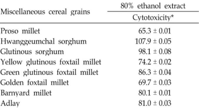
Table 1. Cytotoxic effect of 80% ethanol extract of eight miscella- neous cereal grains on 3T3-L1 preadipocytes Miscellaneous cereal grains 80% ethanol extract
Cytotoxicity*
Proso millet
Hwanggeumchal sorghum Glutinous sorghum
Yellow glutinous foxtail millet Green glutinous foxtail millet Golden foxtail millet Barnyard millet Adlay
65.3 ± 0.01 107.9 ± 0.05 98.1 ± 0.08 74.2 ± 0.02 86.3 ± 0.04 69.7 ± 0.03 80.1 ± 0.01 81.0 ± 0.03
* After 3T3-L1 cells (1 × 10⁴/well) were treated with each sam- ple in 96-well plate for 48 hr, cell viability was determined by the MTS coloimetric assay. Each value is expressed as mean
± SD (n=3 with six replicates per independent experiment).
manufacturer's instructions.
Statistical analysis
Unless otherwise indicated, each result in this paper is representative of at least three separate experiments. Values represent the mean ± standard deviation (SD) of these experiments. The statistical significance was calculated with Student’s t-test. P values less than 0.05 were considered significant.
Results and Discussion
Cytotoxic effect of the 80% ethanol extracts and their solvent fractions obtained from eight selected miscellaneous cereal grains on viability of 3T3 preadipocytes
To evaluate anti-adipogenic properties of miscellaneous cereal grains, which were harvested in Korea, including pro- so millet, hwanggeumchal sorghum, glutinous sorghum, yellow glutinous foxtail millet, green glutinous foxtail millet, golden foxtail millet, barnyard millet, and adlay, the in- hibitory effect of 80% ethanol extracts prepared from the in- dividual grains on the viability of 3T3-L1 preadipocytes was measured using MTS assay. As shown in Table 1, the pres- ence of 80% ethanol extract (100 μg/ml) of proso millet, yel- low glutinous foxtail millet, barnyard grass millet, or adlay appeared to reduce the viability of 3T3-L1 cells to a level of 65.3 ± 0.01%, 74.2 ± 0.02%, 69.7 ± 0.03%, 80.1 ± 0.01%, or 81.0 ± 0.03%, respectively. These results show that the 80%
ethanol extract of proso millet grains exhibited the highest cytotoxicity followed by those of golden foxtail millet grains,
and yellow glutinous foxtail millet grains.
Although proso millet (Panicum miliaceum L.) is one of the oldest cereal crop used for humans in Asia and Africa [21], pharmaceutical studies on the health benefits of proso millet grains are not well-established. In the nutritional aspects, the proso millet grains are known to be relatively rich in protein, mineral substances, and vitamins, when compared with common cereal grains [14]. In the physiological and func- tional aspects, it has been reported that the extract of proso millet possesses anti-oxidant activity [6], hepatic protective activity [26], anti-diabetic and anti-hyperlipidemic effects [29, 31], anti-inflammatory activity [30], and a beneficial in- fluence on metabolism of cholesterol and lipid [27, 36]. With respect to anti-obese effect of proso millet grains, the aque- ous extract of proso millet grains has recently been shown to down-regulate the expression level of adipogenic tran- scription factors [32]. However, little information has been known regarding the involvement of apoptotic cell death in the anti-obesity effect of proso millet grains.
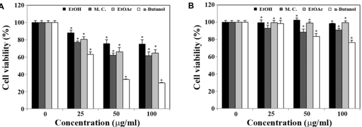
To examine further the anti-adipogenic property of proso millet grains, the 80% ethanol extract of proso millet grains was sequentially fractionated with methylene chloride, ethyl acetate and n-butanol, and then individual fractions were tested for their inhibitory effects on the viability of 3T3-L1 preadipocytes or mature adipocytes that could be obtained from induced adipocytic differentiation of 3T3-L1 cells by exposure to the mixture of 167 nM insulin, 0.5 mM IBMX, and 1 μM dexamethasone for 6 days. When 3T3-L1 pre- adipocytes were treated with the ethanol extract at a concen- tration of 25 μg/ml, 50 μg/ml, or 100 μg/ml, the cell viability appeared to decline to a level of 80.2%, 77.5% or 68.0%, re- spectively (Fig. 1A). In addition, the cytotoxicity of the 80%
ethanol extract of proso millet grains toward 3T3-L1 pre-
adipocytes appeared to be mainly partitioned into the buta-
nol fraction. Treatment of 3T3-L1 cells with the butanol frac-
tion at a concentration of 25 μg/ml, 50 μg/ml, or 100 μg/ml
resulted in a reduction in cell viability to a level of 63.2%,
34.3% or 30.0%, respectively. As shown in Fig. 1B, the ma-
ture adipocytes appeared to be less sensitive to the cytotox-
icity of the butanol fraction compared with 3T3-L1 pre-
adipocytes, and the viability of the mature adipocytes was
sustained up to a level of 80.0% in the presence of the buta-
nol fraction (100 μg/ml). Consequently, these results show
that among the eight selected miscellaneous cereal grains,
the proso millet grains possessed the highest cytotoxicity to-
ward 3T3-L1 preadipocytes, and the cytotoxicity was more
A B
Fig. 1. Effect of 80% ethanol extract and its organic solvent fractions of proso millet grains on cell viability of 3T3-L1 preadipocyes (A) and mature adipocytes (B). 3T3-L1 preadipocytes and mature adipocytes obtained from induced differentiation of 3T3-L1 cells by standard procedures as described in Materials and Methods were treated with 80% ethanol extract, or subsequent four organic solvent fraction (methylene chloride, ethyl acetate, or n-butanol) of proso millet grains at various concentrations (vehicle, 25 μg/ml , 50 μg/ml, and 100 μg/ml). After individual cells (1 × 10⁴/well) were treated with each sample in 96-well plate for 48 hr, cell viability was determined by the MTS coloimetric assay. Each value is expressed as mean ± SD (n=3 with six replicates per independent experiment). *
p
<0.05 compared to control.effective on 3T3-L1 preadipocytes than on the mature adipo- cytes which could be obtained from induced differentiation of 3T3-L1 cells.
Inhibitory effect of the butanol fraction of proso millet grains on adipocytic differentiation of 3T3-L1 preadipocytes
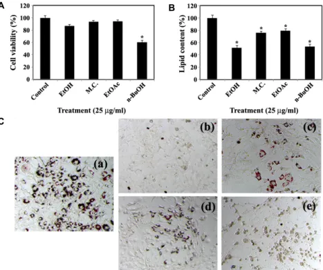
Previously, it has been reported that a reduction in the adipose tissue mass in the body can be mediated by various approaches, such as inhibiting adipogenesis from pre- adipocytes to mature adipocytes, preventing lipid accumu- lation in the adipocytes, and inducing apoptosis in adipose cells [33]. Because current results showed that the ethanol extract or the butanol fraction of proso millet grains could exert a potent cytotoxicity toward 3T3-L1 preadipocytes, it was likely that that proso millet grains might possess a po- tential benefit to reduce the number of adipocytes.
We next decided to examine whether the ethanol extract and organic solvent fractions from proso millet grains can suppress the induced differentiation of 3T3-L1 preadipocytes to adipocytes at a low dosage, which is insufficient to exert cytotoxicity. In this regard, 3T3-L1 cells were continuously exposed to the ethanol extract, methylene chloride fraction, ethyl acetate fraction, or n-butanol fraction at a concentration of 25 μg/ml during the differentiation period for 6 days, and then cell viability and intracellular lipid accumulation were measured. As shown in Fig. 2A and 2B, although the presence of each fraction at a concentration of 25 μg/ml, except for the butanol fraction, during the differentiation pe-
riod for 6 days did not influence significantly the cell via- bility, it could reduce the intracellular lipid accumulation to a level of 81.5%~50.3% of the control. Under these con- ditions, the Oil Red O staining, which was performed to vis- ualize intracellular lipid accumulation during the induced adipocytic differentiation of 3T3-L1 cells [5, 13], showed that the presence of the butanol fraction during the 6-day differ- entiation period caused not only almost complete prevention of intracellular lipid accumulation, but also retaining of fi- broblast-like morphology of 3T3-L1 preadipocytes (Fig. 2C).
These Oil Red O staining data confirmed the inhibitory effect of the butanol fraction on differentiation of 3T3-L1 pre- adipocytes into mature adipocytes.
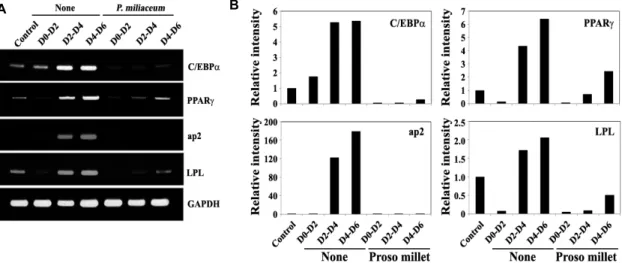
To examine the mechanism underlying the inhibitory ef-
fect of the butanol fraction on the induced differentiation
of 3T3-L1 preadipocytes to mature adipocytes, 3T3-L1 pre-
adipocytes were stimulated to induce differentiation for 6
days, during which periods the cells were exposed to the
butanol extract (25 μg/ml) for 2 days at various time points
(D0-D2, D2-D4 or D4-D6). Under these conditions, the ex-
pression levels of CCAAT/enhancer binding protein-alpha
(C/EBPα) and peroxisome proliferator-activated receptor-
gamma (PPARγ), which are known to be crucial tran-
scription factors required for adipocytic differentiation [5, 19,
33], and the endogenous gene expression of transcriptional
targets of PPARγ, such as aP2 and LPL [40], were inves-
tigated by RT-PCR method. During the induced adipocytic
differentiation of 3T3-L1 preadipocytes for 6 days, the ex-
pression level of C/EBPα and PPARγ became gradually
A B
C
Fig. 2. Effect of 80% ethanol extract and its organic solvent fractions of proso millet grains on cell viability (A) and intracellular lipid accumulation (B and C) during adipocytic differentiation of 3T3 L1. Inhibitory effect of presence of 25 μg/ml of various extracts of proso millet grains during induced adipocytic differentiation of 3T3-L1 preadipocytes for 6 days on cell viability (A), intracellular lipid accumulation (B and C). Cell viability was determined by the MTS colorimetric assay, and intracellular lipid content was measured by using the AdipoRed Assay™ reagent as described in Materials and Methods. Each value is expressed as mean SD (n=3 with six replicates per independent experiment). *
p
<0.05 compared with control. On day 6, cells were stained with Oil Red O dye to visualize the accumulated lipid droplet. A representative study is shown and two additional experiments yielded similar results. Control cells (a). Cells treated with 80% ethanol extract (b), methylene chloride fraction (c), ethyl acetate fraction (d), or n-butanol fraction (e).up-regulated (Fig. 3A). At the same time, however, the pres- ence of the butanol extract during the induced adipocytic differentiation resulted in a significant reduction in the ex- pression levels of C/EBPα, PPARγ, aP2, and LPL to a barely detectable or undetectable level (Fig. 3B). In particular, the suppressive effect of the butanol extract (25 μg/ml) on the expression level of C/EBPα appeared to be more significant if the butanol fraction was added at the early stage (D0-D2) of the differentiation period. These results indicated that the butanol fraction of proso millet could suppress differ- entiation of 3T3-L1 preadipocytes into mature adipocytes and suggest that down-regulation of expression levels of the transcription factors such as C/EBPα, and PPARγ was asso- ciated with the butanol fraction-induced suppression of adi- pocytic differentiation.
Apoptogenic effect of the butanol fraction of proso millet grains on 3T3-L1 preadipocytes
Because the induction of apoptosis in adipose cells can
lead to their own destruction into apoptotic bodies which are cleared by surrounding phagocytic cells without induc- ing a local damaging inflammatory response [16, 35], apop- tosis is considered as an efficient mechanism by which adi- pose cells can be removed upon treatment with anti-obesity drugs.
In order to examine whether the cytotoxic effect of the butanol fraction on 3T3-L1 cells was responsible to induced apoptotic cell death, several apoptotic events including in- duction of apoptotic sub-G
1peak, mitochondrial membrane potential (Δψm) loss, activation of caspase-3, and cleavage of poly (ADP-ribose) polymerase (PARP) into two fragments were investigated. When 3T3-L1 cells were treated with 0.1
% DMSO (vehicle) or the butanol fraction (25 μg/ml, 50 μg/
ml, and 100 μg/ml) for 24 hr, the ratio of negative fluo-
rescence at concentrations of 25 μg/ml, 50 μg/ml, and 100
μ g/ml of the butanol fraction were 26.7%, 41.8% and 44.6%,
respectively (Fig. 4A), suggesting that mitochondrial damage
causing Δψm loss was induced in a dose-dependent manner.
A B
Fig. 3. Effect of the butanol extract of proso millet grains on the expression level of transcription factors such as C/EBPα and PPARγ during the early (D0–D2), intermediate (D2–D4) and late stage (D4–D6) of adipocytes differentiation. 3T3L1 pre- adipocytes were induced to differentiate according to the standard procedures in Material and Methods. The butanol fraction (25 μg/ml) was added during the days 0–2, 2–4, or 4–6 of the 6-day differentiation periods. On day 6, mRNAs were prepared and cDNAs were synthesized as described in Materials and Methods. The expression level of C/EBPα, PPARγ, aP2 and Lpl was determined by RT-PCR. The synthesized cDNA was subjected to RT-PCR with specific primers to amplify the target genes and the PCR products were resolved on a 1.5% agarose gel electrophoresis. A representative study is shown and two additional experiments yielded similar results.
A
B
Fig. 4. Effect of the butanol extract of proso millet grains on the mitochondrial membrane po- tential (A), and kinetic analysis of activation of caspases cascade and PARP degradation (B) in 3T3-L1. To measure the changes in the mitochondrial membrane potential, 3T3-L1 cells were treated with vehicle, 25 μg/ml, 50 μg/ml, and 100 μg/ml of the butanol extract for 24 hr, stained with DiOC6and subjected to flow cytometric analysis. After cells were treated with 50 μg/ml of the butanol extract for 48 hr, cell lysates were prepared as de- scribed in Materials and Methods. For west- ern blot analysis, 4-12% SDS gradient poly- acrlyamide gels and electrotransferred to Immobilon-p membrane. Western blot analy- sis was performed using ECL western blot- ting detection system. A representative study is shown and two additional experiments yielded similar results.
In addition, western blot analysis revealed that while there was no detectable active caspase-3 (17 kDa) in 3T3-L1 cells treated with vehicle, the cleavage of procaspase-3 (32 kDa) into active form (17 kDa) was detected in 3T3-L1 cells after treatment with the butanol fraction (50 μg/ml) in a time-de- pendent manner (Fig. 4B).
Previously, it has been reported that mitochondrial dam- age causing Δψm loss and cytochrome c release into cyto-
plasm are frequently involved in chemical induced apoptotic
signaling pathways as early pro-apoptotic events [24, 39],
and resultant activation of caspase cascade including ca-
pase-3, leading to cleavage of PARP. As the downstream
events of Δψm loss, the activation of caspase-3 and PARP
cleavage were provoked in 3T3-L1 cells treated with the bu-
tanol extract (50 μg/ml) in a time-dependent manner. The
cleavage of PARP into two fragments by active caspse-3 has
been proposed as a marker of apoptosis in many ex- perimental models [20]. Consequently, current results dem- onstrated that the cytotoxic effect of the butanol fraction to- ward 3T3-L1 was attributable to apoptotic cell death medi- ated by Δψm loss and subsequent activation of caspase-3 and cleavage of PARP.
In conclusion, this is the first report to demonstrate that the butanol fraction obtained from 80% ethanol extract of proso millet (Panicum miliaceum) grains exerts cytotoxicity, which is attributable to inducing apoptotic cell death, in 3T3-L1 preadipocytes and inhibits terminal differentiation of 3T3-L1 preadipocytes into mature adipocytes. The results show that the cytotoxic effect of the butanol extract was more dominant on 3T3-L1 preadipocytes than on mature adipocytes. The induced apoptotic cell death appeared to be mediated by mitochondria-dependent activation of cas- pase-3 and subsequent cleavage of PARP. In addition to in- duction of apoptosis, the butanol extract (25 μg/ml) could effectively suppress adipocytic differentiation of 3T3-L1 cells, without exerting a significant cytotoxic effect, via down-regulation of the expression level of transcription fac- tors, such as C/EBPα and PPARγ1, which are known to be crucial for adipogenesis. These results provide an insight in- to the mechanism underlying the anti-adipogenic effect of proso millet grains.
Acknowledgements
This research was carried out with the support of
“Cooperative Research Program for Agriculture Science &
Technology Development (Project No. PJ006638)”, Rural Development Administration, Republic of Korea.
References
1. Ambati, S., Yang, J. Y., Rayalam, S., Park, H. J., Della-Fera, M. A. and Baile, C. A. 2009. Ajoene exerts potent effects in 3T3-L1 adipocytes by inhibiting adipogenesis and induc- ing apoptosis.
Phytother Res
23, 513-518.2. Ahn, J., Lee, H., Kim, S. and Ha, T. 2010. Curcumin-induced suppression of adipogenic differentiation is accompanied by activation of Wnt/beta-catenin signaling.
Am J Physiol Cell Physiol
298, 1510-1516.3. Anderson, J. W. 2003. Whole grains protect against athero- sclerotic cardiovascular disease.
Proc Nutr Soc
62, 135-142.4. Awika, J. M. and Rooney, L. W. 2004. Sorghum phytochem- icals and their potential impact on human health.
Phytochemistry
65, 1199-1221.5. Camp, H. S., Ren, D. and Leff, T. 2002. Adipogenesis and
fat-cell function in obesity and diabetes.
Trends Mol Med
8, 442-447.6. Chandrasekara, A. and Shahidi, F. 2010. Content of in- soluble bound phenolics in millets and their contribution to antioxidant capacity.
J Agric Food Chem
58, 6706-6714.7. Couillard, C., Mauriege, P., Imbeault, P., Prudhomme, D., Nadeau, A., Tremblay, A., Bouchard, C. and Despres, J. P.
2000. Hyperleptinemia is more closely associated with adi- pose cell hypertrophy than with adipose tissue hyperplasia.
Int J Obes Relat Metab Disord
24, 782–788.8. Gregoire, F. M. 2001. Adipocyte differentiation: from fibro- blast to endocrine cell.
Exp Biol Med
226, 997-1002.9. Ha, Y. D. and Lee, S. P. 2001. Characteristics of proteins in Italian millet, sorghum and common millet.
Korean J Postharvest Sci Technol
8, 187-192.10. Hemati, N., Ross, S. E., Erickson, R. L., Groblewski, G. E.
and MacDougald, O. A. 1997. Signaling pathways through which insulin regulates ccaat/enhancer binding protein al- pha (C/EBPα) phosphorylation and gene expression in 3T3-L1 adipocytes. Correlation with GLUT4 gene expression.
J Biol Chem
272, 25913-25919.11. Hirahatake, K. M., Slavin, J. L., Maki, K. C. and Adams, S. H. 2014. Associations between dairy foods, diabetes, and metabolic health: potential mechanisms and future directions.
Metabolism
63, 618-627.12. Hirata, T., Fujii, M., Akita, K., Yanaka, N., Ogawa, K., Kuroyanagi, M. and Hongo, D. 2009. Identification and physiological evaluation of the components from citrus fruits as potential drugs for anti-corpulence and anticancer.
Bioorg Med Chem
17, 25-28.13. Jun, D. Y., Han, C. R., Choi, M. S., Bae, M. A., Woo, M.
H. and Kim, Y. H. 2011. Effect of mollugin on apoptosis and adipogenesis of 3T3-L1 preadipocytes.
Phytother Res
25, 724-731.14. Kalinova, J. and Moudry, J. 2006. Content and quality of protein in Proso millet (
Panicum miliaceum
L.) varieties.Plant Foods for Human Nutrition
61, 45-49.15. Kil, H. Y., Seong, E. S., Ghimire, B. K., Chung, I. M., Kwon, S. S., Goh, E. J., Heo, K., Kim, M. J., Lim, J. D., Lee, D.
and Yu, C. Y. 2009. Antioxidant and antimicrobial activities of crude sorghum extract.
Food Chem
115, 1234-1239.16. Kurosaka, K., Takahashi, M., Watanabe, N. and Kobayashi Y. 2003. Silent cleanup of very early apoptotic cells by macrophages.
J Immunol
171, 4672-4679.17. Kwak, C. S., Lim, S. J., Kim, S. A., Park, S. C. and Lee, M.
S. 2004. Antioxidative and antimutagenicity effects of Korean buckwheat, sorghum, millet, and Job's tears
. J Korean Soc Food Sci Nutr
33, 921-929.18. Kweon, Y. M. and Park, K. Y. 1998. Antimutagenic and anti- carcinogenic effect of sorghum. J
Cancer Prev
3, 128-135.19. Lazar, M. A. 2002. Becoming fat.
Gene Dev
16, 1-5.20. Lazebnik, Y. A., Kaufmann, S. H., Desnoyers, S., Poirier, G.
G. and Earnshaw, W. C. 1994. Cleavage of poly (ADP-ri- bose) polymerase by a proteinase with properties like ICE.
Nature
371, 346-347.21. Lu, H., Zhang, J., Liu, K. B., Wu, N., Li. Y., Zhou, K., Ye.,
M., Zhang, T., Zhang, H., Yang, X., Shen, L., Xu, D. and Li, Q. 2009. Earliest domestication of common millet (
Panicum miliaceum
) in East Asia extended to 10,000 years ago.Proc Natl Acad Sci USA
106, 7367-7372.22. Marquart, L., Jacobs, D. R. Jr. and Slavin, J. L. 2000. Whole grains and health.
J Am Coll Nutr
19, 289-290.23. McKeown, N. M., Meigs, J. B., Liu, S., Wilson, P. W and Jacques, P. F. 2002. Whole-grain intake is favorably asso- ciated with metabolic risk factors for type 2 diabetes and cardiovascular disease in the framingham offspring study.
Am J Clin Nutr
76, 390-398.24. Nagata, S. 1997. Apoptosis by death factor.
Cell
88, 355-365.25. Nakazato, K., Song, H. and Waga, T. 2006. Effects of dietary apple polyphenol on adipose tissues weights in Wistar rats.
Exp Anim
55, 383-389.26. Nishizawa, N., Sato, D., Ito, Y., Nagasawa, T., Hatakeyama, Y., Choi, M. R., Choi, Y. Y. and Wei, Y. M. 2002. Effects of dietary protein of proso millet on liver injury induced by D-galactosamine in rats.
Biosci Biotechnol Biochem
66, 92-96.27. Nishizawa, N., Shimanuki, S., Fujihashi, H., Watanab, H., Fudamoto, Y. and Nagasawa, T. 1996. Proso millet protein elevates plasma level of high-density lipoprotein: a new food function of proso millet.
Biomed Environ Sci
9, 209-212.28. Park, D. H., Lee, S. T., Jun, D. Y., Lee, J. Y., Woo, M. H., Kim, K. Y., Seo, M. C., Ko, J. Y., Woo, K. S., Jung, T. W., Kwak, D. Y., Nam, M. H. and Kim, Y. H. 2014. Comparative evaluation of antioxidant activities of ethanol extracts and their solvent fractions obtained from selected miscellaneous cereal grains.
J Life Sci
24, 26-38.29. Park, K. O., Ito, Y., Nagasawa, T., Choi, M. R. and Nishizawa, N. 2008. Effects of dietary Korean proso-millet protein on plasma adiponectin, HDL cholesterol, insulin lev- els, and gene expression in obese type 2 diabetic mice.
Biosci Biotechnol Biochem
72, 2918-2925.30. Park, M. Y., Kim, J. H. and Park, D. S. 2011. Anti-in- flammatory activities of hog millet (
Panicum miliaceum
L.) in murine macrophages through IRAK-4 signaling.Korean J Food Nutr
24, 268-272.31. Park, M. Y., Jang, H. H, Kim, J. B., Yoon, H. N., Lee, J.
Y., Lee, Y. M., Kim, J. H, and Park, D. S. 2011. Hog millet (
Panicum miliaceum
L.)-supplemented diet ameliorates hy- perlipidemia and hepatic lipid accumulation in C57BL/6J-ob/ob mice.Nutr Res Pract
5, 511-519.32. Park, M. Y., Seo, D. W., Lee, J. Y., Sung, M. K., Lee, Y. M., Jang, H. H., Choi, H. Y, Kim, J. H. and Park, D. S. 2011.
Effects of
Panicum miliaceum
L. extract on adipogenic tran- scription factors and fatty acid accumulation in 3T3-L1 adipocytes.Nutr Res Pract
5, 192-197.33. Rosen, E. D. and MacDougald, O. A. 2006. Adipocyte differ- entiation from the inside out.
Nat Rev Mol Cell Biol
7, 885-896.34. Sahyoun, N. R., Jacques, P. F., Zhang, X. L., Juan, W. and McKeown, N. M. 2006. Whole-grain intake is inversely asso- ciated with the metabolic syndrome and mortality in older adults.
Am J Clin
Nutr 83, 124-131.35. Savill, J. and Fadok, V. 2000. Corpse clearance defines the meaning of cell death.
Nature
407, 784-788.36. Shimanuki, S., Nagasawa, T. and Nishizawa, N. 2006.
Plasma HDL subfraction levels increase in rats fed pro- so-millet protein concentrate.
Med Sci Monit
12, 221-226.37. Slavin, J. 2003. Why whole grains are protective: biological mechanisms.
Proc Nutr Soc
62, 129-134.38. Slavin, J. L., Martini, M. C., Jacobs, D. R. Jr. and Marquart, L. 1999. Plausible mechanisms for the protectiveness of whole grains.
Am J Clin Nutr
70, 459-463.39. Sun, X. M., MacFarlane, M., Zhuang, J., Wolf, B. B., Green, D. R. and Cohen, G. M. 1999. Distinct caspase cascades are initiated in receptor-mediated and chemical-induced apoptosis.
J Biol Chem
274, 5053-5040.40. Tamori, Y., Masugi, J., Nishino, N. and Kasuga, M. 2002.
Role of peroxisome proliferator-activated receptor-gamma in maintenance of the characteristics of mature 3T3-L1 adipocytes.
Diabetes
51, 2045-2055.41. Urizar, N. L. and Moore, D. D. 2003. GUGULIPID: a natural cholesterol-lowering agent.
Annu Rev Nutr
23, 303-313.42. Visscher, T. L. and Seidell, J. C. 2001. The public health im- pact of obesity.