Anti-inflammatory Effect of Flavonoids Kaempferol and Biochanin A-enriched Extract of Barnyard Millet (Echinochloa crus-galli var. frumentacea) Grains in LPS-stimulated RAW264.7 Cells
Ji Young Lee
1, Do Youn Jun
2, Young Ho Yoon
3, Jee Youn Ko
3, Koan Sik Woo
3, Mi Hee Woo
4and Young Ho Kim
1*
1Laboratory of Immunobiology, School of Life Science and Biotechnology, College of Natural Sciences, Kyungpook National University, Daegu 702-701, Korea
2Institute of Life Science and Biotechnology, Kyungpook National University, Daegu 702-701, Korea
3Functional Cereal Crop research Division. Department of Functional Crop, NICS, RDA, 627-803, Korea
4Department of Pharmacology, College of Pharmacology, Daegu Catholic University, Daegu 712-702, Korea Received November 12, 2014 /Revised November 13, 2014 /Accepted November 14, 2014
In order to compare the anti-inflammatory effects of five selected cereal grains-proso millet, hwang- geumchal sorghum, foxtail millet, barnyard millet, and adlay-the inhibitory activities of 80% ethanol (EtOH) extracts obtained from the individual grains on lipopolysaccharide (LPS)-induced nitric oxide (NO) generation were investigated in RAW264.7 cells. The EtOH extract of barnyard millet (Echinochloa crus-galli var. frumentacea) grains exhibited more potent anti-inflammatory activity than that of the other grains. When the EtOH extract of barnyard millet grains was sequentially fractio- nated with n-hexane, methylene chloride (MC), ethyl acetate (EtOAc), and n-butanol, the majority of the anti-inflammatory activity was detected in the MC fraction, followed by the EtOAc fraction.
Pretreatment with the MC fraction caused downregulation of the expression levels of iNOS- and COX-2-specific transcripts and proteins, as well as proinflammatory cytokine gene transcripts (IL-1β, IL-6, and TNF-α) in LPS-stimulated RAW264.7 cells. Additionally, the MC fraction could suppress not only the LPS-induced nuclear translocation of cytosolic NF-kB, but also the LPS-induced activation of MAPKs, such as ERK, JNK, and p38MAPK. Further analysis of the MC fraction by HPLC identified kaempferol, biochanin A, and formononetin as the major phenolic components. Both kaempferol and biochanin A, but not formononetin, could exert anti-inflammatory effect at the same concentrations as those of the MC fraction. Consequently, these results indicate that kaempferol and biochanin A are among the most effective anti-inflammatory phenolic components in barnyard millet grains. This find- ing suggests that barnyard millet grains and the MC extract enriched in kaempferol and biochanin A could be beneficial functional food sources that have an anti-inflammatory effect.
Key words :
Anti-inflammation, barnyard millet grains, cytokines, MAPKs, NF-κB
*Corresponding author
*Tel : +82-53-950-5378, Fax : +82-53-955-5522
*E-mail : [email protected]
This is an Open-Access article distributed under the terms of the Creative Commons Attribution Non-Commercial License (http://creativecommons.org/licenses/by-nc/3.0) which permits unrestricted non-commercial use, distribution, and reproduction in any medium, provided the original work is properly cited.
Journal of Life Science 2014 Vol. 24. No. 11. 1157~1167 DOI : http://dx.doi.org/10.5352/JLS.2014.24.11.1157
Introduction
Lipopolysaccharide (LPS) is an endotoxic component of the outer membranes of gram-negative bacteria and stim- ulates several pathological inflammatory responses, such as systemic inflammatory response syndrome, septic shock, disseminated intravascular coagulation and multiple organ dysfunctions [5]. These LPS-induced inflammatory diseases
are known to be mediated primarily via deregulated over-
production of pro-inflammatory mediators, such as nitric ox-
ide (NO), prostaglandin E
2(PGE
2), interleukin-1β (IL-1β), in-
terleukin-6 (IL-6), and tumor necrosis factor-α (TNF-α), by
activated macrophages [21, 38]. The LPS-induced production
of these pro-inflammatory mediators by macrophages is
tightly regulated by two principal transcription factors, NF-κ
B and AP-1. These factors are activated by the trans-
membrane signaling pathway, which is triggered by the in-
teraction of the cell surface CD14/toll-like receptor 4 (TLR4)
complex with the hydrophobic lipid A portion of LPS, and
then relayed by sequential activation of protein kinases [14,
22]. Among the critical protein kinases required for the sig-
naling pathway are IL-1 receptor-associated kinase 4
(IRAK4) and IRAK1, TGF-β-activated kinase 1 (TAK1), IκB
kinase (IKK), and MAPKs (p38MAPK, JNK and ERK) [2, 11].
In relation to LPS-induced TLR4-dependent activation of NF-κB in macrophages, the redox signaling mediated by re- active oxygen species (ROS) has also been implicated [9, 17, 20]. Supplementation of antioxidants in early inflammation stage has been shown to attenuate the oxidative stress by acting as ROS scavenger, and could prevent developing chronic inflammation [3]. In this context, it is likely that in- hibition of oxidative stress might be one of the reliable strat- egies, which is beneficial for ameliorating metabolic diseases being accompanied by inflammation.
Much attention has been paid to the physiological func- tionality of foods, due to the increasing interest in human health, and research into the health benefits of foods has been increasing last years. Traditionally, miscellaneous ce- real grains have been considered as a beneficial functional food source, which can improve the metabolic diseases [7, 8, 30]. Barnyard millet is one of the hardy crops that have adapted to sterile environments throughout the world [28].
The cultivation area of barnyard millet was gradually re- duced due to various changes in current diet and cereal breeding. However, in recent years, barnyard millet grains are re-evaluated because of their high nutritional value in preventing metabolic diseases. The nutritive and biological studies about barnyard millet grains have demonstrated that the grains are rich in protein, lipid, vitamin B complex and nicotinic acid, when compared with common cereal grains.
Previously, it has been reported that the extract of barnyard millet grains possess anti-oxidant activity [12, 13], a benefi- cial influence on metabolism of cholesterol and lipid [27], immunosuppressive activity [15], anti-microbial activity [29]
and lowering glycemic index and anti-diabetes effects [23, 27, 36]. Several bioactive components have been found from barnyard millet grains including serotonin, luteolin, tricin, deoxynojirimycin and coumaric acid derivatives [33, 37], but studies on the health benefits of barnyard millet grains with respect to anti-inflammatory activity are not well-established.
In the present study, as an attempt to compare anti-in- flammatory activities of five selected miscellaneous cereal grains, such as proso millet (Panicum miliaceum), hwang- geumchal sorghum (Sorghum bicolor (L.) Moench var. hwang-
geumchal), yellow glutinous foxtail millet (Setaria italica),adlay (Coix lacryma-jobi), and barnyard millet (Echinochloa
crus-galli var. frumentacea) harvested in Korea, we inves-tigated the anti-NO production activity of the 80% ethanol (EtOH) extracts from the individual grains in LPS-stimulated
murine macrophage RAW264.7 cells. The EtOH extract of barnyard millet grains, which exhibited more potent anti-in- flammatory effect compared with other grain extracts, were sequentially fractionated by n-hexane, MC, EtOAc, and BuOH. Because the inhibitory effect on LPS-induced NO production in RAW264.7 cells were mainly detected in the MC fraction followed by the EtOAc fraction, the anti-in- flammatory activity of the MC fraction was further exam- ined by investigating its inhibitory action against LPS-in- duced inflammatory events and by detecting the active phe- nolic components in the MC fraction.
Materials and Methods
Reagents, chemicals, antibodies and culture medium The ECL Western blotting kit was purchased from PerkinElmer (Boston, MA, USA), and Immobilon-P mem- brane was obtained from Millipore Corporation (Bedford, MA, USA). The anti-COX-2, anti-β-actin, anti-NF-κB p65, an- ti-p-JNK, anti-JNK, anti-Sp1 and anti-p38 MAPK antibodies were purchased from Santa Cruz Biotechnology (Santa Cruz, CA, USA), and the anti-p-p38 MAPK, anti-IκBα, anti-p-IκBα and p-c-jun antibodies were obtained from Cell Signaling Technology (Beverly, MA, USA). The anti-p-ERK was ob- tained from Millipore Corporation, and anti-ERK antibody was obtained from Zymed Laboratories (South San Francisco, CA, USA). Anti-iNOS was purchased from BD Biosciences (Chicago, IL, USA). Horse radish peroxide (HRP)-conjugated anti-mouse IgG and anti-rabbit IgG were obtained from Cell Signaling, and HRP-conjugated anti-goat IgG was obtained from Santa Cruz Biotechnology. The mur- ine macrophage cell line RAW264.7 was purchased from ATCC (Manassas, VA, USA) and cultured in Dulbecco's modified Eagle's medium (DMEM) which was supple- mented with 10% fetal bovine serum (FBS) and 100 μg/ml gentamycin at 37°C in a humidified 5% CO
2atmosphere.
For the experiments, the cells were grown to 80-90% con- fluences, and were subjected to no more than 15 cell passages.
Sample extraction
Barnyard millet grains were provided by National
Institute of Crop Science of Miryang, Korea, and a voucher
specimen has been deposited in Laboratory of Immunology,
College of Natural Sciences, Kyungpook National
University, Daegu, Korea. The dried grains (250 g) were mil-
led on a Blender 7012 (Dynamics Corporation, USA) for 10 min, and then extracted with 80% ethanol (EtOH) for 3 hr at 80°C. The ethanol extract was evaporated, dissolved in water, and then sequentially extracted with n-hexane, meth- ylene chloride (MC), ethyl acetate (EtOAc) and n-butanol (BuOH). Each organic solvent fractionation was repeated three times. Each organic solvent fraction as well as the rem- nant aqueous fraction was centrifuged at 7,500 rpm for 15 min to remove insoluble substances. The recovered super- natant of each fraction was then concentrated by rotary vac- uum evaporator (Heidolph LR 4000, Germany). The yields of hexane fraction, MC fraction, EtOAc fraction, BuOH frac- tion, remnant aqueous fraction were 9.4 g, 2.5 g, 1.1 g, 0.7 g, 2.3 g and 3.1 g, respectively.
Cell viability assay
The Cytotoxic effect of samples on RAW264.7 cells was analyzed by 3-(4,5-dimethylthiazol-2-yl)-2,5-diphenylte- trazolium bromide (MTT) assay. Briefly, cells (0.5×10
5/well) were cultured with serial dilutions of samples in 96-well plates. After incubation for 18 hr, 50 μl of MTT solution (1.1 mg/ml) was added to each well and incubated for an addi- tional 2 hr. The colored formazan crystal produced from MTT was dissolved in DMSO. The absorbance was meas- ured at 540 nm by a plate reader (Molecular Devices, Thermo Max, USA) to determine the formazan concen- tration, which reflects the cell viability.
Nitric oxide assay
As an indicator of NO production, the concentration of nitrite, a stable metabolite of NO, in the culture medium was assessed by Griess reagent [10]. Briefly, RAW264.7 cells (2×10
5cells/well) were cultured overnight in 96-well plates, and then treated with LPS (0.1 μg/ml) in the absence or in the presence of various concentrations of sample for 16 hr.
The culture supernatant (100 μl) was mixed with an equal volume of Griess reagent for 15 min at room temperature in dark condition, and then the absorbance of the chromo- phoric azo-derivative molecule was measured using a micro- plate reader at 540 nm. To ensure the validity of the results, experiments were done in three independent experiments with three replicates per independent experiment.
Total RNA isolation and RT-PCR
Cells were washed twice in PBS, then total RNA was iso-
lated using the Trizol reagent from Invitrogen (Carlsbad, CA, USA) according to the manufacturer’s instructions and DNase I treatment. After RNA quantification by GE NanoVue Spectrophotometer (GE healthcare, Buckingham- shire, UK), 1 μg RNA was reversely transcribed using First strand cDNA synthesis kit (Thermo scientific, Logan, UT, USA) for cDNA synthesis. Gene expression values were nor- malized to housekeeping GAPDH gene. GAPDH was ampli- fied with forward (5’-ATCCTGCGTCTGGACCTGGCT-3’) and reverse (5’-CTGATCCACATCTGCTGGAAG-3’) primers.
PCR amplification was done using AccuPower
™PCR PreMix (Bioneer, Seoul, Korea) and specific primers. The following primers were used for PCR: iNOS-forward, 5’- ATGTCCGAAGCAAACATCAC-3’; iNOS-reverse, 5’-TAA- TGTCCAGGAAGTAGGTG-3’; COX-2-forward, 5-CAGCA- AATCCTTGCTGTTCC-3; COX-2-reverse, 5’-TGGGCAAA- GAATGCAAACATC-3’; TNF-α-forward, 5’-TACTGAACTTC- GGGGTGATCGGTCC-3’; TNF-α-reverse, 5’-CAGCCTTGT- CCCTTGAAGAGAACC-3’; IL-6-forward, 5’-GAAATGA- TGGATGCTTCCAAACTGG-3’; IL-1β-forward, 5’-CAAGG- AGAACCAAGCAAC-3’; IL-1β-reverse, 5’-GGGGAAGGC-
AATAGAAAC-3’. To ensure that the same amount of RNA was being used, the concentration of the total RNA for each sample was confirmed by spectrophotometry and normal- ized with GAPDH as the message of a housekeeping gene.
The PCR products were electrophoresed using 1.2% agarose gel and visualized under UV light after ethidium bromide staining.
Preparation of cell lysate and western blot analysis Cellular lysates were prepared by suspending cells (5×10
6) in 300 μl of lysis buffer (137 mM NaCl, 15 mM EGTA, 1 mM sodium orthovanadate, 15 mM MgCl
2, 25 mM MOPS, 1 mM PMSF, and 5.0 μg/ml proteinase inhibitor E-64, 0.1%
Triton X-100, pH 7.2). The cells were disrupted by sonication and extracted at 4
oC for 30 min. An equivalent amount of protein lysate (25 μg) was electrophorersed on 4~12%
NuPAGE gradient gel (Invitrogen/Novex, Carlsbad, CA,
USA) with MOPS buffer and then electrotransferred to
Immobilon-P membranes. Detection of each protein was per-
formed utilizing the ECL Western blotting kit following the
manufacturer's instructions. Densitometry was performed
using ImageQuant TL software (Amersham, Arlington
Heights, IL, USA). Arbitrary densitometric units for the pro-
tein of interest were normalized to the densitometric units
of β-actin.
A
B
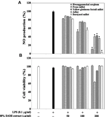
Fig. 1. Effect of the 80% EtOH extracts of five selected different miscellaneous cereal grains on LPS-induced NO pro- duction (A) and viability (B) in RAW264.7 cells. After RAW 264.7 cells (2×10⁵ cells/well) were incubated in 96 well plates for 16 hr, the cells were treated with the in- dividual 80% ethanol extracts (50, 100, and 200 μg/ml) for 1 hr and then continuously incubated with LPS (0.1 μg/ml) for 20 hr. The nitrite concentration as an in- dicator of NO production in culture medium was meas- ured using Griess reagent. The cell viability was de- termined by the MTT assay as described in Materials and Methods. Each value is expressed as mean ± SD (n=3 with six replicates per independent experiment).
*p<0.05, significant compared with vehicle- treated control.
High-performance liquid chromatography (HPLC) analysis of 80% ethanol extract and its organic solvent fractions of barnyard millet grains
The contents of phenolic compounds were analyzed as previously described [32]. Samples were filtered through a 0.45 μm syringe filter (Millipore, Billerica, MA, USA) and analyzed by HPLC (Agilent 1200, Agilent Technologies, Waldbronn, Germany). The analytical column was a ZORBAX ODS (4.6×250 mm, Agilent Technologies) with a guard column (Phenomenex, Torrance, CA, USA). The de- tection wavelength was set at 280 nm and the solvent flow rate was held constant at 1.0 ml/min. The mobile phase used for the separation consisted of solvent A (distilled water in- cluded 0.1% acetic acid) and solvent B (acetonitrile included 0.1% acetic acid). A gradient elution procedure was used as 0 min 92% A, 2-27 min 90% A, 27-50 min 70% A, 50-51 min 10% A, 51-60 min 0% A, and 60-62 min 92% A. The injection volume was 20 μl for analysis. The standards used were biochanin A, caffeic acid, (±)-catechin hydrate, chloro- genic acid, trans-cinnamic acid, formononetin, gallic acid, hesperidin, homogentisic acid, isoorientin, kaempferol, nar- ingin, orientin, protocatechuic acid, pyrogallol, quercetin, re- sveratrol, rutin hydrate, syringic acid, vanillic acid, vanillin, veratric acid and all samples were analyzed in triplicate.
Statistical analysis
Unless indicated otherwise, each result in this paper is representative of at least three separate experiments. Values represent the mean standard deviation (SD) of these experiments. The statistical significance was calculated with Student’s t-test. P values less than 0.05 were considered significant.
Results and Discussion
Anti-NO production activity of the 80% EtOH extract and its organic solvent fractions obtained from barn yard millet grains in LPS-stimulated RAW264.7 cells
In order to compare the anti-inflammatory effects of five selected miscellaneous cereal grains, including proso millet (Panicum miliaceum), hwanggeumchal sorghum (Sorghum bi-
color (L.) Moench var. hwanggeumchal), yellow glutinous fox-tail millet (Setaria italica), adlay (Coix lacryma-jobi), and barn- yard millet (Echinochloa crus-galli var. frumentacea) harvested in Korea, the inhibitory activities of the 80% ethanol extracts of the individual cereal grains on LPS-induced NO pro-
duction were investigated in RAW264.7 cells. As shown in Fig. 1A, the EtOH extracts of barnyard millet grains, hwang- geumchal sorghum grains, and proso millet grains appeared to exhibit more potent anti-NO production activities com- pared with other cereal grains tested. The LPS-induced NO productions in the presence of the EtOH extracts of barnyard millet grains and hwanggeumchal sorghum grains at con- centrations of 100 μg/ml were reduced to the levels of 56.4%
and 52.3%, respectively, whereas those at concentrations of
200 μg/ml were reduced to the levels of 3.4% and 10.8%,
respectively. However, under the same conditions, none of
these EtOH extracts could affect the cell viability of
RAW264.7 cells (Fig. 1B). These results indicated that anti-in-
flammatory effect of barnyard millet grains was the most
A
B
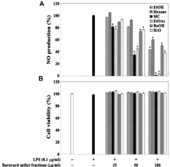
Fig. 2. Effect of the 80% EtOH extract and its organic solvent fraction of barnyard millet grains on LPS-induced NO production (A) and viability (B) in RAW264.7 cells. After RAW 264.7 cells (2×10⁵ cells/well) were incubated in 96 well plates for 16 hr, the cells were treated with the in- dividual 80% ethanol extracts (25, 50, and 100 μg/ml) for 1 hr and then continuously incubated with LPS (0.1 μg/ml) for 20 hr. The nitrite concentration as an in- dicator of NO production in culture medium was meas- ured using Griess reagent. The cell viability was de- termined by the MTT assay as described in Materials and Methods. Each value is expressed as mean ± SD (n=3 with six replicates per independent experiment).
*p<0.05, significant compared with vehicle-treated control.
potent among the five selected miscellaneous cereal grains tested, and that the EtOH extract of barnyard millet grains at concentrations of 100-200 μg/ml could significantly re- duce the LPS-induced production of NO without affecting cell viability in RAW264.7 cells.
In order to examine further the anti-NO production prop- erty of barnyard millet grains, the 80% EtOH extract of barn- yard millet grains was sequentially fractionated with n-hex- ane, MC, EtOAc, and BuOH, and then individual organic solvent fractions at concentrations ranging from 25-100 μg/
ml were tested for the anti-NO production activity. As shown in Fig. 2A, the LPS-induced NO production was the most significantly suppressed in the presence of the MC frac- tion, followed by the EtOAc fraction. In addition, the LPS-in- duced NO productions in the presence of the MC fraction at concentrations of 25 μg/ml, 50 μg/ml and 100 μg/ml were
reduced to the levels of 81.7%, 35.2% and 1.4%, respectively.
At the same time, the MC fraction at concentrations of up to 100 μg/ml did not affect the cell viability (Fig. 2B).
Consequently, these results indicate that the MC fraction of barnyard millet grains at concentrations of 25-100 μg/ml could suppress the LPS-induced NO production in a dose-dependent manner, and that the IC
50value of the MC fraction was 44.8 μg/ml.
Inhibitory effect of the MC fraction of barnyard millet grains on LPS-induced expression of iNOS, COX-2, and pro-inflammatory cytokines in RAW264.7 cells
Because the production of pro-inflammatory mediators, such as NO and prostaglandin E
2(PGE
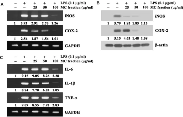
2), in RAW64.7 cells stimulated with LPS is governed by the enzymes iNOS and COX-2, RT-PCR and western blot analysis were performed to examine whether the MC fraction of barnyard millet grains could suppress the expression levels of iNOS and COX-2 in RAW264.7 cells stimulated with LPS. As shown in Fig. 3A, RT-PCR data revealed that the expression level of mRNAs specific for iNOS and COX-2, which were not detected in unstimulated RAW264.7 cells, were significantly enhanced following LPS-stimulation; however, the presence of the MC fraction at concentration of 25-100 μg/ml down-regulated the levels of iNOS and COX-2 mRNAs.
Under these conditions, western blot analysis data also re- vealed that although the proteins specific for iNOS and COX-2 were not detected in continuously growing RAW 264.7 cells, the levels of both proteins were enhanced by 5.8 folds and 5.2 folds, respectively, in RAW264.7 cells stimu- lated with LPS (Fig. 3B). However, the LPS-induced increase in the levels of iNOS and COX-2 proteins was markedly re- duced by the MC fraction in a dose-dependent manner. In particular, LPS-induced expression of both iNOS and COX-2 proteins was not detected in the presence of the MC fraction at a concentration of 100 μg/ml. These results demonstrated that the MC fraction of barnyard millet grains at concen- trations of 25‒100 μg/ml could reduce the expression levels of the pro-inflammatory proteins (iNOS and COX-2) in RAW264.7 cells stimulated with LPS via the down-regu- lation of mRNA levels.
To examine whether the MC fraction could suppress the
expression of pro-inflammatory cytokines such as IL-1β,
IL-6, and TNF-α, RAW264.7 cells were stimulated with LPS
for 4 hr following pretreatment with the MC fraction (25-100
A B
C
Fig. 3. Effect of the MC fraction of barnyard millet grains on the expression levels of iNOS, COX-2 and pro-inflammatory cytokines (IL-1β, IL-6 and TNF-α) in RAW264.7 cells stimulated with LPS. The cells were pretreated with the MC fraction at indicated concentrations, prior to stimulation with LPS (0.1 μg/ml) for 20 hr. RT-PCR analysis of transcripts of iNOS, COX-2 and GAPDH (A) and IL-6, IL-1β, TNF-α and GAPDH (C), and western blot analysis of iNOS, COX-2 and β-actin proteins (B) were performed as described in the Materials and Methods. The expression level of GAPDH mRNA or β-actin protein was used as control.
Arbitrary densitometric units for the individual transcripts and proteins of interest were normalized to the densitometric units of GAPDH transcript and β-actin protein, respectively.
A representative study is shown and two additional experiments yielded similar results.μg/ml) for 1 hr. The levels of mRNAs specific for IL-1β, IL-6, and TNF-α were assessed by RT-PCR. As shown in Fig.
3C, the mRNAs specific for IL-1β, IL-6, and TNF-α were not detected in unstimulated RAW264.7 cells, but the expression levels of these cytokine-specific mRNAs were markedly up-regulated in RAW264.7 cells following stimulation with LPS. Under the same conditions, the LPS-induced expression of IL-1β, IL-6, and TNF-α mRNA was reduced in the pres- ence of the MC fraction, more efficiently at a concentration of 100 μg/ml. These results suggested that the MC fraction at concentrations of 25-100 μg/ml could inhibit the LPS-in- duced up-regulation of the expression levels of pro-in- flammatory cytokines including IL-1β, IL-6, and TNF-α.
Inhibitory effect of the MC fraction of barnyard millet grains on the nuclear translocation of NF-κB complex in RAW264.7 cells
The LPS-induced expression of pro-inflammatory media- tors including iNOS, COX-2, IL-1β, IL-6 and TNF-α in mac-
rophages is known to be tightly regulated by two principal transcription factors, NF-κB and AP-1 [21, 34, 38]. Nuclear translocation of NF-κB and proteasomal degradation of IκBα have been considered as prominent markers that exhibit mo- lecular inflammation initiation for the expression of pro-in- flammatory mediators in activated macrophages.
To determine whether the MC fraction of barnyard millet
grains could inhibit LPS-induced activation of NF-κB in
RAW264.7 cells, LPS-induced phosphorylation of IκBα, alter-
ations in the level of IκBα, and nuclear translocation of NF-κ
B (p65), all of which are known to be critical for the activa-
tion of NF-kB, were compared by western blot analysis in
RAW264.7 cells stimulated with LPS with and without the
MC fraction (25-100 μg/ml). As shown in Fig. 4, LPS-stim-
ulation resulted in an increase in the phosphorylation level
of IκBα and a decrease in the protein level of IκBα in
RAW264.7 cells. At the same time, LPS-stimulation caused
nuclear translocation of cytosolic NF-κB (p65) so that the lev-
el of nuclear NF-κB could be enhanced by approximately
7 folds compared with that of untreated RAW264.7 cells.
Fig. 4. Inhibition of the MC fraction of barnyard millet grains on IκBα phosphorylation, IκBα protein levels, and nu- clear translocation of cytosolic NF-κB (p65) in RAW264.7 cells following LPS stimulation. The cells were treated by the MC fraction at the indicated concentrations for 1 hr, and then stimulated with LPS (0.1 μg/ml) for 4 hr to detect IκBα and its phosphorylation by western analysis. A representative study is shown and two addi- tional experiments yielded similar results.
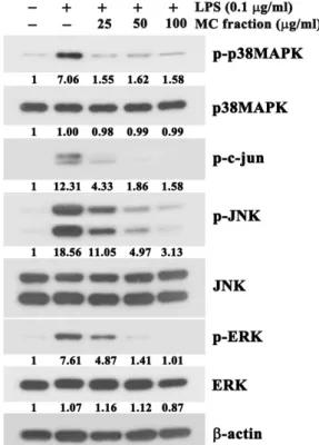
Fig. 5. Effect of the MC fraction of barnyard millet grains on LPS-induced activation of MAPKs (p38MAPK, JNK and ERK) in RAW264.7 cells. The cells were pre-treated with indicated concentrations of the MC fraction for 1 hr, and then with LPS (0.1 μg/ml) for 20 hr. The levels of the active phosphorylated forms of p38MAPK, c-Jun, JNK, and ERK were assessed by western blot analysis using antibodies specific for the phosphorylated forms of in- dividual kinases. A representative study is shown and two additional experiments yielded similar results.
However, the LPS-induced phosphorylation of IκBα, down-regulation of IκBα protein levels, and nuclear trans- location of cytosolic NF-κB (p65) appeared to be suppressed by the MC fraction in a dose-dependent manner. These re- sults suggested that the MC fraction of barnyard millet grains could inhibit LPS-induced activation of NF-κB by suppressing the LPS-induced phosphorylation and degrada- tion of IκBα, and subsequent nuclear translocation of NF-kB in RAW264.7 cells.
Inhibitory effect of the MC fraction of barnyard millet grains on LPS-induced phosphorylation of MAPKs in RAW264.7 cells
Several studies have reported that MAPKs (p38MAPK, JNK and ERK) are closely associated with the TLR4-mediat- ed proximal signaling events that lead to the activation of transcription factors NF-kB and AP-1 in LPS-stimulated mac- rophages [2, 11]. Because the MC fraction of barnyard millet grains appeared to suppress the LPS-induced phosphor- ylation of IκBα, alterations in the level of IκBα, and nuclear translocation of NF-κB (p65), all of which are critical for acti- vation of the transcription factor NF-kB, we decided to ex- amine whether the LPS-induced activation of p38MAPK, JNK, and ERK could be targeted by the inhibitory action of the MC fraction of barnyard millet grains by western blot analysis using individual antibodies specific for the phos-
phorylated active forms of these MAPKs. Although the total protein levels of p38MAPK, JNK, and ERK all of which were easily detected in unstimulated RAW264.7 cells, were not changed regardless of the presence of the MC fractions, the active phosphorylated forms of p38MAPK, JNK, and ERK were significantly enhanced after LPS treatment (Fig. 5).
Under these conditions, however, the LPS-induced phos-
phorylation of p38MAPK, JNK and ERK was commonly re-
duced by the MC fraction. In accordance with the observed
reduction in the level of LPS-induced phosphorylation of
JNK, the phosphorylation of c-Jun protein at Ser-63, which
is catalyzed by JNK [6], was markedly reduced in the pres-
ence of the MC fractions. This finding indicated that the
phosphorylated JNK that was detected in RAW264.7 cells
after treatment with LPS possessed enough enzymatic activ-
ity to phosphorylate c-Jun. Consequently, these results in-
dicated that the LPS-induced activation of p38MAPK, JNK
A
B
Fig. 6. Comparison of the anti-NO production activities (A) and cytotoxicity (B) of phenolic compounds (kaempferol, bio- chanin A, and formononetin) in LPS-stimulated RAW 264.7 cells. The cells were pre-incubated for 1 hr with the MC fraction (12.5 μg/ml, 20 μg/ml and 50 μg/ml), kaempferol (10 μM, 25 μM, and 50 μM), biochanin A (10 μM, 25 μM, and 50 μM), or formononetin (10 μM, 25 μM, and 50 μM) in triplicate and then treated with LPS (0.1 μg/ml) for 4 hr. The culture supernatants were saved and used to determine NO production. MTS were em- ployed to check the cell viability. Each value is expressed as mean ± SD (n=3 with six replicates per independent experiment). *p<0.05, significant compared with ve- hicle-treated control.
and ERK was influenced by the inhibitory action of the MC fraction. In addition, these results also suggested that in- hibitory action of the MC fraction against the LPS-induced nuclear translocation of NF-kB in RAW264.7 cells might be due to the decline in the level of activation of p38MAPK, JNK and ERK.
Identification of the major anti-inflammatory phenolic compounds in the MC fraction of barnyard millet grains
Among phytochemicals, phenolic compounds have been reported to have various ameliorating effects on neuro- degenerative diseases, multiple sclerosis, cardiovascular dis- eases, and metabolic syndrome from oxidative stress [16, 35].
In order to identify the major anti-inflammatory in- gredient(s) of the MC fraction of barnyard millet grains, we decided to analyze phenolic compounds contained in the MC fraction by HPLC. As the major phenolic components, kaempferol (9.17 μg/mg), biochanin A (2.22 μg/mg), and formononectin (1.52 μg/mg), which accounted for 85% of the total phenolic compounds, were detected in the MC fraction.
Because it has previously been reported that kaempferol [4, 18], biochanin A [19], and formononectin [26] possess an- ti-inflammatory activity, the anti-NO production activities of these phenolic compounds were examined in LPS-stimu- lated RAW264.7 cells. As shown in Fig. 6A, when the in- hibitory activities of kaempferol, biochanin A, and for- mononectin against LPS-induced NO production were ex- amined at concentrations of 10 μM, 25 μM, and 50 μM in RAW264.7 cells, kaempferol reduced the LPS-induced NO production to the levels of 85.3%, 69.1%, and 45.7%, re- spectively, whereas biochanin A reduced the NO production to the levels of 79.6%, 60.9%, and 39.7%, respectively. Under these conditions, both kaempferol and biochanin A did not show a significant cytotoxic effect on RAW264.7 cells.
Although formononetin has previously been reported to in- hibit the inflammation in mouse lung injury model [26], it failed to inhibit the LPS-induced NO production to a re- markable level. It is noteworthy that hesperidin (0.33 μg/
mg) [1, 31], naringin (0.56 μg/mg) [25], and protocatechuic acid (0.34 μg/mg) [24] as the minor components, which were reported to possess anti-inflammatory activity, were de- tected in the MC fraction. Consequently, these results in- dicated that kaempferol and biochanin A, but not for- mononetin, could inhibit the LPS-induced NO production
in a dose-dependent manner, and that both kaempferol and biochanin A were among the most effective anti-in- flammatory phenolic components in barnyard millet grains.
In conclusion, this study describes an anti-inflammatory activity of barnyard millet grains against LPS-induced in- flammatory events in mouse macrophage cell line RAW 264.7, and demonstrates that this anti-inflammatory action is attributable to suppression of LPS-induced up-regulation of pro-inflammatory modulators including iNOS, COX-2, IL-1β, IL-6, and TNF-α, via inhibition of nuclear trans- location of cytosolic NF-kB as well as inactivation of MAPKs.
As the active phenolic ingredient in the MC fraction respon-
sible for the inflammatory activities, kaempferol and bio-
chanin A, which are detected as the major phenolic com-
pounds in the MC fraction of barnyard millet grains, are
identified. Current results also suggest that barnyard millet
grains and the MC extract enriched in kaempferol and bio-
chanin A could be beneficial functional food sources appli- cable to improving inflammatory conditions.
Acknowledgement
This work was carried out with the support of
“Cooperative Research Program for Agriculture Science &
Technology Development (Project No. PJ009865)” Rural Development Administration, Republic of Korea.
References
1. Abuelsaad, A. S., Allam, G. and Al-Solumani, A. A. 2014.
Hesperidin inhibits inflammatory response induced by Aeromonas hydrophila infection and alters CD4+/CD8+ T cell ratio. Mediators Inflamm 2014, 393217.
2. Akira, S. and Takeda, K. 2004. Toll-like receptor signaling.
Nat Rev Immunol 4, 499-511.
3. Basu, S., Ghosh, A. and Hazra, B. 2005. Evaluation of the antibacterial activity of Ventilago madraspatana Gaertn., Rubia cordifolia Linn. and Lantana camara Linn.: isolation of emodin and physcion as active antibacterial agents. Phytother Res 19, 888-894.
4. Choi, I. S., Choi, E. Y., Jin, J. Y., Park, H. R., Choi, J. I. and Kim, S. J. 2013. Kaempferol inhibits P. intermedia lip- opolysaccharide-induced production of nitric oxide through translational regulation in murine macrophages: critical role of heme oxygenase-1-mediated ROS reduction. J Periodontol 84, 545-555.
5. Corriveau, C. C. and Danner, R. L. 1993. Endotoxin as a therapeutic target in septic shock. Infect Agents Dis 2, 35-43.
6. Davis, R. J. 2000. Signal transduction by the JNK group of MAP kinases. Cell 103, 239-252.
7. Esmaillzadeh, A., Mirmiran, P. and Azizi, F. 2005. Whole- grain intake and the prevalence of hypertriglyceridemic waist phenotype in Tehranian adults. Am J Clin Nutr 81, 55-63.
8. Fardet, A. 2010. New hypotheses for the health-protective mechanisms of whole-grain cereals: what is beyond fibre?
Nutr Res Rev 23, 65-134.
9. Flohe, L., Brigelius-Flohe, R., Saliou, C., Traber, M. G. and Packer, L. 1997. Redox regulation of NF-κB activation. Free Radic Biol Med 22, 1156-1126.
10. Green, L. C., Wanger, D. A. and Glogowski, J. 1982. Analysis of nitrate, nitrite, and [15N] nitrate in biological fluids. Anal Biochem 126, 131-138.
11. Guha, M. and Mackman, N. 2001. LPS induction of gene expression in human monocytes. Cell Signal 13, 85-94.
12. Hegde, P. S. and Chandra, T. S. 2005. ESR spectroscopic study reveals higher free radical quenching potential in ko- do millet (Paspalum scrobiculatum) compared to other millets.
Food Chem 92, 177-182.
13. Higashi-Okai, K., Ishida, E., Nakamura, Y., Fujiwara, S. and
Okai, Y. 2008. Potent antioxidant and radical-scavenging ac- tivities of traditional Japanese cereal grains. J UOEH 30, 375-389.
14. Hoebe, K., Janssen, E. and Beutler, B. 2004. The interface between innate and adaptive immunity. Nat Immunol 5, 971-974.
15. Hosoda, A., Okai, Y., Kasahara, E., Inoue, M., Snhimizu, M., Usui, Y., Sekiyama, A. and Higashi-Okai, K. 2012. Potent immunomodulating effects of bran extracts of traditional Japanese millets on nitric oxide and cytokine production of macrophages (RAW264.7) induced by lipopolysaccharide. J UOEH 34, 285-296.
16. Hur, S. J, Kang, S. H., Jung, H. S., Kim, S. C., Jeon, H. S., Kim, I. H. and Lee, J. D. 2012. Review of natural products actions on cytokines in inflammatory bowel disease. Nutr Res 32, 801-816.
17. Janessen-Heininger, Y. M., Poynter, M. E. and Baeuerle, P.
A. 2000. Recent advances towards understanding redox mechanisms in the activation of nuclear factor kappa B. Free Radic Biol Med 28, 1317-1327.
18. Kim, H. K., Park, A. H., Lee, J. S., Chung, T. S., Chung, H. Y. and Chung, A. J. 2007. Down-regulation of iNOS and TNF-a expression by kaempferol via NF-κB inactivation in aged rat gingival tissues. Biogerontology 8, 399-408.
19. Kole, L., Giri, B., Manna, S. K., Pal, B. and Ghosh, S. 2011.
Biochanin-A, an isoflavone, showed anti-proliferative and anti-inflammatory activities through the inhibition of iNOS expression, p38-MAPK and ATF-2 phosphorylation and blocking NF-κB nuclear translocation. Eur J Pharmacol 653, 8-15.
20. Kong, X., Thimmulappa, R., Kombairaju, P. and Biswal, S.
2010. NADPH oxidase-dependent reactive oxygen species mediate amplified TLR4 signaling and sepsis-induced mor- tality in Nrf2-deficient mice. J Immunol 185, 569-577.
21. Kubes, P. and McCafferty, D. M. 2000. Nitric oxide and in- testinal inflammation. Am J Med 109, 150-158.
22. Kurt-Jones, E. A., Popova, L., Kwinn, L., Haynes, L. M., Jones, L. P., Tripp, R. A., Walsh, E. E., Freeman, M. W., Golenbock, D. T., Anderson, L. J. and Finberg, R. W. 2000.
Pattern recognition receptors TLR4 and CD14 mediate re- sponse to respiratory syncytial virus. Nat Immunol 1, 398-401.
23. Lee, S. H., Chung, I. M., Cha, Y. S. and Park, Y. 2010. Millet consumption decreased serum concentration of triglyceride and C-reactive protein but not oxidative status in hyper- lipidemic rats. Nutr Res 30, 290-296.
24. Lende, A. B., Kshirsagar, A. D., Deshpande, A. D., Muley, M. M., Patil, R. R., Bafna, P. A. and Naik, S. R. 2011. Anti-in- flammatory and analgesic activity of protocatechuic acid in rats and mice. Inflammopharmacology 19, 255-263.
25. Liu, Y., Wu, H., Nie, Y. C., Chen, J. L., Su, W. W. and Li, P. B. 2011. Naringin attenuates acute lung injury in LPS-treated mice by inhibiting NF-κB pathway. Int Immunopharmacol 11, 1606-1612.
26. Ma, Z., Ji, W., Fu, Q. and Ma, S. 2013. Formononetin in- hibited the inflammation of LPS-induced acute lung injury
in mice associated with induction of PPAR gamma expression. Inflammation 36, 1560-1566.
27. Nishizawa, N., Togawa, T., Park, K. O., Sato, D., Miyakoshi, Y., Inagaki, K., Ohmori, N., Ito, Y. and Nagasawa, T. 2009.
Dietary Japanese millet protein ameliorates plasma levels of adiponectin, glucose, and lipids in type 2 diabetic mice.
Biosci Biotechnol Biochem 73, 351-360.
28. Odintsova, T., Rogozhin, E. A., Baranov, Y., Musolyamov, A. Kh., Yalpani, N., Egorov, T. A. and Grishin, E. V. 2008.
Seed defensins of barnyard grass Echinochloa crusgalli (L.) Beauv. Biochimie 90, 1667-1673.
29. Ryazantsev, D. Y., Rogozhin, E. A., Dimitrieva, T. V., Drobyazina, P. E., Khadeeva, N. V., Egorov, T. A., Grishin, E. V. and Zavriev, S. K. 2014. A novel hairpin-like anti- microbial peptide from barnyard grass (Echinochloa crusgalli L.) seeds: Structure functional and molecular-genetics characterization. Biochimie 99, 63-70
30. Sahyoun, N. R., Jacques, P. F., Zhang, X. L., Juan, W. and McKeown, N. M. 2006. Whole-grain intake is inversely asso- ciated with the metabolic syndrome and mortality in older adults. Am J Clin Nutr 83, 124-31.
31. Saiprasad, G., Chitra, P., Manikandan, R. and Sudhandiran, G. 2013. Hesperidin alleviates oxidative stress and down- regulates the expressions of proliferative and inflammatory markers in azoxymethane-induced experimental colon carci- nogenesis in mice. Inflamm Res 62, 425-440.
32. Seo, M. C., Ko, J. Y., Song, S. B., Lee, J. S., Kang, J. R., Kwak, D. Y., Oh, B. G., Yoon, Y. N., Nam, M. H., Jeong, H. S.
and Woo, K. S. 2011. Antioxidant compounds and activities
of foxtail millet, proso millet and sorghum with different pulverizing methods. J Korean Soc Food Sci Nutr 40, 790-797.
33. Seo, W. D., Kim, J. Y., Jang, K. C., Han, S. I., Ra, J. E., Oh, S. H., Lee, J. H., Kim, Y. G., Kang, H. J., Kim, B. J. and Nam, M. H. 2012. Anti-pigmentation effect of serotonin al- kaloid isolated from Korean barnyard millet (Echinochola uti- lis). J Korean Soc Appl Biol Chem 55, 579-586.
34. Surh, Y. J., Chun, K. S., Cha, H. H., Han, S. S., Keum, Y.
S., Park, K. K. and Lee, S. S. 2001. Molecular mechanisms underlying chemopreventive activities of anti-inflammatory phytochemicals: down-regulation of COX-2 and iNOS through suppression of NF-κB activation. Mutat Res 480-481, 243-268.
35. Talero, E., Ávila-Roman, J. and Motilva, V. 2012. Chemopre- vention with phytonutrients and microalgae products in chronic inflammation and colon cancer. Curr Pharm Des 18, 3939-3965.
36. Ugare, R., Chimmad, B., Naik, R., Bharati, P. and Itagi, S.
2014. Glycemic index and significance of barnyard millet (Echinochloa frumentacae) in type II diabetics. J Food Sci Technol 51, 392-395.
37. Watanabe, M. 1999. Antioxidative phenolic compounds from Japanese barnyard millet (Echinochloa utilis) grains. J Agric Food Chem 47, 4500-4505.
38. Watson, W. H., Zhao, Y. and Chawla, R. K. 1999. S-ad- enosylmethionine attenuates the lipopolysaccharide-in- duced expression of the gene for tumor necrosis factor alpha. Biochem J 342, 21-25.