online©ML Comm
-
156
- 대한두경부종양학회지제 25 권 제 2 호 2009
이하선에 발생한 원발성 편평 세포암종 2예
고려대학교 의과대학 이비인후-두경부외과학교실
김우주·정은재·정광윤·백승국
= Abstract =
Two Cases of Primary Squamous Cell Carcinoma in Parotid Gland
Woo-Joo Kim, MD, Eun-Jae Jung, MD, Kwang-Yoon Jung, MD, Seung-Kuk Baek, MD Department of Otorhinolaryngology-Head and Neck Surgery,
Korea University College of Medicine, Seoul, Korea
Squamous cell carcinoma, which is a common primary head and neck malignant neoplasm that is usually restrict- ed to the mucosal surfaces of the upper aerodigestive tract and skin, is very unusual in the major salivary gland. A- mong them, few cases are regarded as primary carcinomas. In this article, we present two cases of squamous cell carcinoma in the parotid gland, who first presented with painful mass on infraauricular area.
KEY WORDS:Parotid gland·Squamous cell carcinoma.
서 론
이하선 암종은 암종이 기원하는 타액선관의 줄기세포에 따라 다양한 조직학적 아형으로 나뉘게 되는데, 점액표피양 암종, 선암낭성암종, 선방세포암종, 선암종, 악성혼합종, 편평 세포암종 등 여러 조직학적 아형이 있다.1) 문헌 고찰에 의 하면, 이하선에 생기는 편평세포암종은 이하선 악성 종양의 1.9%에서 6.9%로 보고 되고 있으나, 이 중 전이성 편평세 포암종의 빈도가 원발성 편평세포암종보다 많은 것으로 알 려져 있다.2-4) 이하선의 원발성 편평세포암종은 매우 드문 암종으로, 병리 조직학적 분화가 나빠 예후가 매우 불량하 며, 경부 임파선 전이가 많아 적절한 수술적 치료가 요구되 는 질환이다.
저자들은 수술적 치료와 술후 방사선치료를 시행한 이하 선에서 발생한 원발성 편평세포암종 2예를 치험하였기에 문 헌 고찰과 함께 보고하는 바이다.
증 례
1. 증 례
66세 남자 환자가 8개월 전부터 통증을 동반하면서 점점 자라나는 우측 이하선 부위의 종물을 주소로 내원하였다. 고 혈압 외에 다른 전신 질환의 과거력은 없었으며, 가족력도 특 이 사항은 없었다. 이학적 검사상 종물은 우측 이하선 부위에 4×5cm 크기로 단단하고 고정되어 있으며 간헐적 압통을 동반하는 양상이었고, 우측 안면 신경 변연하악분지로 경도의 마비 소견이 관찰되었다. 그 외 비인두, 두피, 피부 및 경부에 서 기원한 것으로 추정되는 전이를 의심할 만한 특이 소견은 없었다. 세침흡입세포검사상 편평세포암종의 소견이었고, 자 기 공명 영상과 컴퓨터 단층 촬영상 우측 이하선의 일부 천엽 을 제외한 심엽 전체와 인두 주위 공간으로 팽창하는 양상의 종괴가 관찰 되었고, 다수의 경부 임파선 전이가 우측 경부 level II, III 번에서 관찰되었다(Fig. 1). 전신전이와 이차암의 진단을 위해 시행한 양전자 방출 단층 촬영 검사상 전이가 의 심되는 소견은 보이지 않았으며(Fig. 2), 위내시경 검사상 위 유문부 샘암종이 진단되어 이하선 암종의 치료를 시행 후 이 에 대한 추가적 치료를 계획하였다.
환자는 우측 이하선 전절제술과 우측 확장 근치적 경부 절 교신저자:백승국, 136-705 서울 성북구 안암동 5가 126-1
고려대학교 의과대학 이비인후-두경부외과학교실 전화:(02) 920-5486·전송:(02)925-5233 E-mail:[email protected]
-
157
- 제술을 시행하였다. 수술 소견상 종양은 안면 신경 협근지, 설 인신경, 척추부신경, 설하신경을 침범하고, 외경 동맥을 둘러 싸고 있는 소견을 보여 모두 제거하였다(Fig. 3). 환자는 술 후 8일에 합병증 없이 퇴원하였다. 병리 조직학적 검사에서 경부 level II, III, IV번에 임파선 전이를 동반한 절제연 음성 의 원발성 편평세포암종으로 진단되었다(Fig. 4). 환자는 우 측 이하선 부위에 5,400cGy, 좌측 경부에 5,000cGy의 방사 선치료 계획하에 방사선치료를 시행하던 중 위샘암종의 진행 으로 인한 전신상태의 급격한 악화가 발생하여 수술 후 6개 월째 사망하였다.2. 증 례
83세 남자 환자가 2개월 전부터 통증을 동반하면서 점점 자라나는 좌측 이하선 부위의 종물을 주소로 내원하였다. 전 신 질환의 과거력은 없었으며, 이학적 검사상 종물은 좌측
이하선 부위에 2×3cm 크기로 단단하게 고정되어 있었고 간헐적으로 압통을 동반하는 양상을 보였으나 안면 신경 마
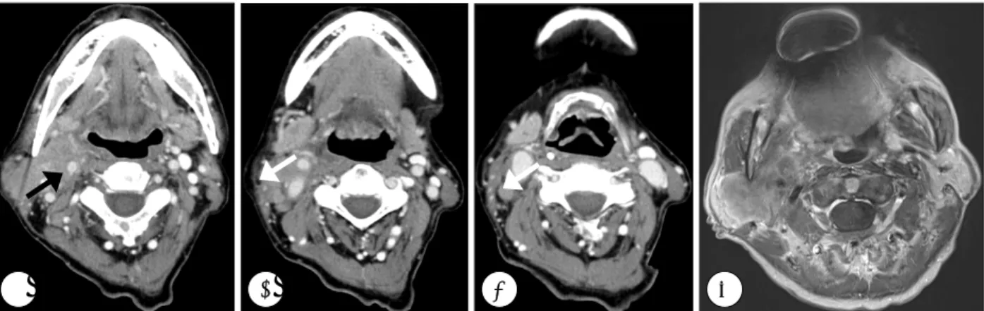
Fig. 1. Preoperative axial CT scans and MRI. CT scans show a relatively homogenous enhanced parotid mass enclosing a external carotid artery(black arrow) and multiple metastatic lymph nodes with extracapsular spread at level II and III(white arrow)(A- C). T1-weighted enhanced MRI demonstrates a slightly and homogenously enhancing mass that extends to right parotid de- ep lobe and parapharyngeal spaces(D).
A B C D
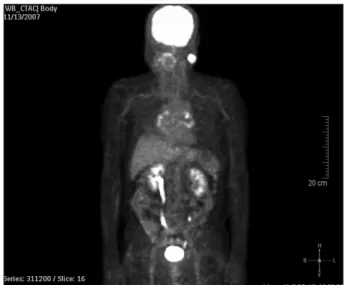
Fig. 2. Preoperative F-18 FDG PET(CT background) of the whole body. It shows intense hypermetabolic lesion in right parot- id gland, mainly in deep lobe, with abnormal FDG uptake suggesting neck metastasis(maxSUV 1.5).
Fig. 3. Intraoperative finding. Parotid tumor was completely re- sected with the whole levels of neck, including marginal mandibular branch of facial nerve(arrow head), glossop- haryngeal nerve, spinal accessory nerve, hypoglossal nerve and external carotid artery(black arrow).
Fig. 4. Histopathologic finding. Sheets of squamoid tumor cells with abdundant cytoplasm, marked pleomorphic nucleoli and distinct cell border, several keratin pearls(black arrow) at the center of tumor cell nest are evidence of squamous cell carcinoma(H & E stain, ×200).
-
158
- 비 소견은 없었다. 종물의 바깥쪽 경계면 피부 및 경부에는 특이 소견이 없었다. 세침흡입세포검사상 악성 의심 소견을 보였고, 컴퓨터 단층 촬영상에서 2.7×2.0cm 크기의 비교적 경계는 좋고 굵은 테증강을 보이며 내부에 괴사를 동반한 종 괴가 관찰 되었으나 주위로 임파선 전이는 없었다(Fig. 5).전신 전이와 이차암의 진단을 위해 시행한 양전자 방출 단층 촬영 검사상 전이가 의심되는 소견은 보이지 않았으며, 위내 시경상 정상 소견 보였다(Fig. 6).
환자는 좌측 이하선 전절제술과 경부 level II, III번에 선택 적 경부 절제술을 시행하였고, 동결 절편 검사상 임파선 전이 가 없음을 확인하였다. 병리 조직학적 검사에서 절제연 음성 인 원발성 편평세포암종으로 진단되었고, 임파선 전이 소견은 없었다(Fig. 7). 술 후 5일에 합병증 없이 환자는 퇴원하였고, 술 후 방사선 치료는 거부하여 시행하지 않은 상태로, 술 후 2년째 외래 추적 관찰상 재발의 증거는 보이지 않았다.
고 찰
이하선에 발생하는 편평세포암종은 그 형성 과정에 따라 병리학적으로 4가지 형태로 분류할 수 있다. 첫째, 악성 편평 세포암종의 요소를 가지고 있는 고등급 점액표피양암종 일부 의 암, 둘째, 피부나 점막의 직접적인 침습을 통해 발생한 이 하선 전이암, 셋째, 원격 장기로부터 전이된 이하선 전이암, 마지막으로 이하선 자체의 원발성 편평세포암종으로 구분할 수 있다.5) 원발성 편평세포암종의 정확한 진단을 위해서는 실 제 병리조직 검사시 정확한 조직학적 평가가 중요하다. 이하 선의 원발성 편평세포암종의 경우 만성 염증을 통한 도관 상피 세포의 이차적인 변화에서 기인한다고 알려져 있으며,1) 이형성 과정을 거쳐 침습성 편평 세포암종으로 변화한다. 따라 서 조직학적으로 도관 상피 세포 주위에서 발견되는 편평세포 암종의 경우, 전이암이 아닌 원발암으로 단정 지을 수 있는 근 거가 된다.6)
Spiro 등은 원발성 편평세포암종의 경부 임파선 전이율을 70%로, 고등급의 점액표피양암종에 비하여 높은 것으로 보 고한바 있으며, Shemen 등도 45%, Gaughan 등에 의하면 임상적으로 N0인 경우에도 30%까지 경부 임파선 전이를 보 인다고 보고한 바 있다.2,4,7) 그러므로 치료 계획을 세울 때 임파선 전이 유무 여부의 확인은 매우 중요하며, 환자의 예 후를 결정하는 인자가 된다. 실제로 이하선에 발생하는 원발 성 편평세포암종의 경우 경부 림프절 전이가 많으므로 N0 병기에서도 선택적 경부 절제술을 시행한다. 수술의 범위는 경부 level II, III번, 상경부군만 절제하는 법, 변형된 광범위 경부 절제술, 상견갑설골 경부 절제술 등의 방법이 있다. 수 술 범위에 대한 결정에 있어 안면 신경의 침범 여부는 과거부 터 논란이 되어 왔다. 과거에는 종양이 안면 신경에 직접적 침윤 소견을 보이지 않더라도 근치적 절제를 통하여 안면신 경을 희생하는 경우가 많았다.8) 하지만 최근에는 안면신경의 희생 여부가 실제로 치료 성적과 무관하다는 보고가 많으며,9)
Fig. 5. Preoperative axial CT scans. Axial CT scans show a 2.7×
2cm sized ill-defined lobulating parotid mass with internal necrosis(white arrow) and no evidence of metastatic lym- ph nodes(A and B).
A B
Fig. 6. Preoperative F-18 FDG PET(CT background) of the whole body. It shows intense hypermetabolic lesion(maxSUV 10.2) in left parotid gland without abnormal FDG uptake suggest- ing metastasis.
Fig. 7. Histopathologic finding. Primary squamous cell carcinoma contains keratin pearl(black arrow)(H & E stain, ×200).
-
159
- 안면 신경 절제 후 생기는 안면 변형 등으로 인한 환자의 삶 의 질의 저하 등 관련하여 현재는 신경 침범이 있을 때 절제 를 하는 것이 추세이다. 따라서 안면 신경에 대한 침범이 없을 경우, 기능적, 미적 측면을 고려하여 보존하는 것이 합당하다.술 후 방사선 치료는 국소 재발률을 낮추고, 정상적인 안면 신 경의 보존을 위하여 추천된다.10-12)
중심 단어:
이하선·편평세포암종.References
1) Batsakis JG, Regezi JA, Luna MA. Histogenesis of salivary gland neoplasms: A postulate with prognostic implications. J Laryngol Otol. 1989;103(10):939-944.
2) Gaughan RK, Olsen KD, Lewis JE. Primary squamous cell car- cinoma of the parotid gland. Arch Otolaryngol Head Neck Surg.
1992;118(8):798-801.
3) Lee SW, Kim GE, Park CS, Choi EC, Yang WI, Lee CG, et al.
Primary squamous cell carcinoma of the parotid gland. Am J Oto- laryngol. 2001;22(6):400-406.
4) Shemen LJ, Huvos AG, Spiro RH. Squamous cell carcinoma of
salivary gland origin. Head Neck Surg. 1987;9(4):235-240.
5) Taxy JB. Squamous carcinoma in a major salivary gland: A re- view of the diagnostic considerations. Arch Pathol Lab Med. 2001; 125(6):740-745.
6) Flynn MB, Maguire S, Martinez S, Tesmer T. Primary squamous cell carcinoma of the parotid gland: The importance of correct histological diagnosis. Ann Surg Oncol. 1999;6(8):768-70.
7) Spiro RH, Huvos AG, Strong EW. Cancer of the parotid gland: A clinicopathologic study of 288 primary cases. Am J Surg. 1975; 130:452-459.
8) Freeman FJ, Beahrs OH, Woolner LB. Surgical treatment of ma- lignant tumors of the parotid gland. Am J Surg. 1965;110:527-533.
9) Ying YL, Johnson JT, Myers EN. Squamous cell carcinoma of the parotid gland. Head & Neck. 2006;28(7):626-632.
10) Sterman BE, Kraus DH, Sebek BA. Primary squamous cell car- cinoma of the parotid gland. Larygoscope. 1990;100:146-148.
11) Marks MW, Ryan RF, Litwin MS, Sonntag BV. Primary squa- mous cell carcinoma of the parotid gland. Plast Reconstrc Surg.
1987;79(4):550-554.
12) Friedman M, Levin B, Grybauskas V. Malignant tumors of the ma- jor salivary glands. Otolarygol Clin North Am. 1986;19(4):625-636.