C A S E R E P O R T Open Access
Orthognathic surgery for patients with fibrous dysplasia involved with dentition
Santhiya Iswarya Vinothini Udayakumar 1 , Jun-Young Paeng 2 , So-Young Choi 1 , Hong-In Shin 3 , Sung-Tak Lee 1 and Tae-Geon Kwon 1*
Abstract
Background: Fibrous dysplasia (FD) is characterized by the replacement of normal bone by abnormal fibro-osseous connective tissue and typically treated with surgical contouring of the dysplastic bone. When dysplastic lesions involve occlusion, not only is surgical debulking needed, orthognathic surgery for correction of dentofacial deformity is mandatory. However, the long-term stability of osteotomized, dysplastic bone segments is a major concern because of insufficient screw-to-bone engagement during surgery and the risk of FD lesion re-growth.
Case presentation: This case report reviewed two patients with non-syndromic FD that presented with maxillary occlusal canting and facial asymmetry. Le Fort I osteotomy with recontouring of the dysplastic zygomaticomaxillary region had been performed. The stability of osseous segments were favorable. However, dysplastic, newly formed bone covered the previous plate fixation site and mild bony expansion was observed, which did not influence the facial profile. Including the current cases, 15 cases of orthognathic surgery for FD with dentition have been reported in the literature.
Conclusion: The results showed that osteotomy did not appear to significantly reduce the long-term stability of the initial fixation insufficiency of the screw to the dysplastic bone. However, based on our results and those of the others, long-term follow-up and monitoring are needed, even in cases where the osteotomized segment shows stable results.
Keywords: Fibrous dysplasia, Orthognathic surgery, Le fort I, Osteotomy, Rigid fixation
Background
Fibrous dysplasia (FD) is a benign, developmental, nonheri- table, and slowly progressing disorder of the bone charac- terized by replacement of the normal bone by gradual abnormal proliferation of immature, irregularly distributed fibro-osseous connective tissue. FD is caused by a gene mutation that affects both bone formation and resorption [1, 2]. The skeletal involvement varies from monostotic (single bone) to polyostotic (multiple bones), leading to progressive functional deficits and reduced esthetics. When FD involves only one bone, and not contiguous multiple bones in the skull, the disease is characterized as monosto- tic rather than polyostotic [3]. Therefore, craniofacial FD without involving other skeleton, such as the femur or rib, would be commonly defined as monostotic FD [4]. The
maxilla and frontal bones are the most commonly involved bones in the craniofacial region. The typical appearance of patients with FD of the maxillofacial bone is facial asym- metry caused by a significant expansion of the bone [2].
When the maxilla is involved, an increase in the promin- ence of cheek is observed. Since the craniofacial FD is not well delineated, conservative surgical debulking is usually performed to restore the facial contour. However, the FD lesion sometimes affects the alveolar bones of the maxilla or mandible, causing discrepancy in the occlusion second- ary to the alveolar bone expansion. In such cases, complete osteotomy and repositioning of the maxilla or mandibular bones must be performed concomitantly with debulking of the lesion [5–10].
The main goals of orthognathic surgery for FD are to contour the excessive bone lesion, correct the dentofa- cial deformities, and restore the occlusion. Because of the frequent obliteration of the maxillary sinus and the anatomical abnormality at the vascular structures, ad- equate osteotomy is challenging [8]. In addition, the
* Correspondence: [email protected]
1
Department of Oral and Maxillofacial Surgery, School of Dentistry, Kyungpook National University, 2177 Dalgubeol-daero, Jung-gu, Daegu 41940, Republic of Korea
Full list of author information is available at the end of the article
© The Author(s). 2018 Open Access This article is distributed under the terms of the Creative Commons Attribution 4.0
International License (http://creativecommons.org/licenses/by/4.0/), which permits unrestricted use, distribution, and
reproduction in any medium, provided you give appropriate credit to the original author(s) and the source, provide a link to
the Creative Commons license, and indicate if changes were made.
dysplastic nature of the FD lesion cannot afford insuffi- cient screw-to-bone engagement [7]. The major con- cerns following orthognathic surgery are suitable bone reunion, quality of the newly formed bone, which can potentially influence the stability of the osteotomized segments, and FD recurrence.
The purpose of this study was to investigate the man- agement of FD in cases with various degrees of dentofa- cial deformity with occlusal discrepancy. Bone healing of osseous segments and the prognosis of orthognathic sur- gery for FD involved with dentition in the current and previous cases is also reviewed.
Case presentation Case 1
A 13-year-old girl visited our department with com- plaints of swelling and facial asymmetry on the left side of the face. The patient was first diagnosed as craniofa- cial monostotic FD involving the left maxilla, subnasal, and zygoma. The patient underwent primary bone con- touring surgery under general anesthesia. At age 18, the patient presented with a swelling on the left side of the mid face region, suggesting lesion re-growth. On clinical extra-oral examination, a significant facial asymmetry was present. Computed tomographic (CT) images showed a typical dysplastic dense mass affecting the maxillary and zygomatic bones. The preoperative ceph- alometric analysis showed canting of 5.5 mm down- wards. After confirming the cessation of the growth, presurgical orthodontic treatment was continued for 14 months. At age 20, final surgical treatment objectives were established. The patient exhibited severe maxillary occlusal canting with gummy smile and compensatory mandibular occlusal canting, but showed a relatively symmetrical outline of the mandible. Le Fort I osteot- omy was performed to correct occlusal canting, and the dysplastic bone obliterating the maxillary sinus was also removed. Concomitantly, a massive amount of friable
dysplastic bone from the maxillary alveolar bone to the zygoma and infraorbital areas was extensively removed.
Mandibular sagittal split ramus osteotomy (BSSRO) was performed to correct compensatory mandibular occlusal canting. To adjust the mandibular chin and body contour, Triaca style mandibular wing osteotomy [11] was also per- formed. Osteotomized maxilla was rigidly fixed with four 1.5-mm-thick microplates whereas BSSRO was fixed with two 2-mm-thick miniplates (Fig. 1a, b). The fixation of the screws was slightly weaker at the FD-involved side than the contralateral normal maxillary bone.
The postoperative course was uneventful, and postop- erative orthodontic treatment was started 4 weeks after the surgery. There was no evidence of skeletal relapse at the postoperative 2-year follow-up (Fig. 1c). The coronal and sagittal views of the CT showed that the bone union at the osteotomized bone was composed of dysplastic bone, and the maxillary sinus was obliterated again. The plates were covered by the newly formed dysplastic bone (Fig. 2). However, slightly expanded external cortex of the left zygoma and maxilla on the left side did not in- fluence the facial symmetry. The patient did not want further surgery and was satisfied with the final outcome.
Case 2
A 29-year-old woman complained of swelling on the left side of the face. CT images showed a radiodense net- work of the expanded dysplastic bone lesion involving maxilla, zygoma, ethmoid bones, orbit, cranial base, and hemimandible. Therefore, the patient was diagnosed as polyostotic craniofacial FD. The patient did not exhibit visual disorders. The preoperative cephalometric analysis showed canting of 5 mm downwards. An orthognathic surgical procedure was planned. Le Fort I osteotomy, sagittal split ramus osteotomy, and genioplasty were performed under general anesthesia (Fig. 3). To ensure maxillary impaction to correct occlusal canting, the FD lesion was extensively removed during the Le Fort I
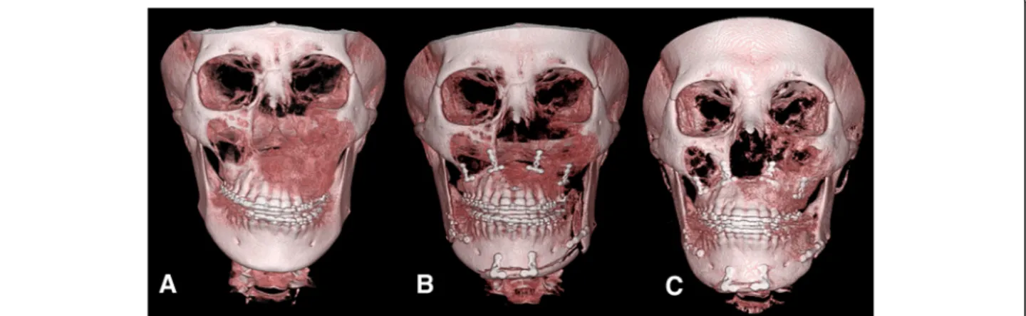
Fig. 1 3D CT image of patient no. 1 preoperative (a) immediately postoperative (b) and 1 year postoperative status (c) showing improvement in
occlusal canting following Le Fort I and wing osteotomy on the mandibular body and chin with rigid fixation
osteotomy. After removal of the dysplastic bone on the maxilla and debulking at the zygomaticomaxillary region, rigid internal fixation was performed with mini- plates (2 mm thick). BSSRO was performed to improve mandibular occlusal canting and remove the dysplastic bone. Additional body shaving and genioplasty were per- formed to improve esthetics. The postoperative recovery was uneventful. The occlusion was stable after 18 months
postoperatively, and there was no evidence of recurrence or relapse. At the time of plate removal, 2 years after the initial surgery, the site of maxillary Le Fort I osteotomy was examined and a significant osseous union was noted between the osteotomized segments (Fig. 4). However, slight expansion of the external cortex of the left maxilla was noted, which did not influence facial symmetry.
Four years after the initial surgery, the patient did not
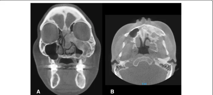
Fig. 2 Coronal (upper panel) and sagittal (lower panel) images of patient no. 1 preoperative (a), immediately postoperative (b), and 2 years postoperative (c). Bone union at the osteotomized bone was composed of dysplastic bone, and the maxillary sinus was obliterated again. The plates were covered by the newly formed dysplastic bone
Fig. 3 Preoperative (a) and 2 years postoperative radiographic image (b) of patient no. 2
show further expansion or re-growth of the dysplastic le- sion (Fig. 5).
Literature review
Including our two cases, 15 documented cases of orthognathic surgery for craniofacial FD involved with dentition have been reported. Most patients were young adults (average age 26.6 ± 5.6 years, range 16 –35), with female predominance (4 males and 11 females). Among these patients, five were monostotic, five were polyosto- tic, and another five were not clearly defined. In most cases, the maxillary or mandibular segment had been rigidly fixed with plates and screws, and orthognathic
surgery for FD showed stable results after Le Fort I or BSSRO (Table 1).
Discussion
One important concern in orthognathic surgery for patients with FD is the long-term stability of the osteo- tomized segments as FD-involved bones are affected by a dysplastic process and are typically soft and friable [12]. Therefore, it is difficult to tightly fix the screws and miniplates to the fibrodysplastic bone. However, long- term stability of the osteotomized segments and occlu- sion was achieved in both our cases and the previously presented reports.
Fig. 4 Intraoperative (a) time of plate removal at 2 years after the initial surgery (b). The previous site of the maxillary Le Fort I osteotomy was revisited, and significant osseous union was noted between the osteotomized segments. Slight expansion of the external cortex of the left maxilla was also noted, which did not affect facial symmetry
Fig. 5 3D CT image of coronal (a) and axial (b) view 4 years postoperative. No further expansion or re-growth of the dysplastic lesion was noted
Table 1 Reported orthognathic surgery for fibrous dysplasia involved with dentition Case Re ference Age /sex Lesion type/s ite Surgical proc edure /metho d of fixat ion Bone heal ing/recurre nce/stability of occlu sion Follow-u p 1 Sachs et al. [ 5 ] 18 /F Polyos totic/R t. max illa, zyg oma, front al, te mporal, sphen oid, rib Le Fort I an d bot h subcondylar ost eoto mies/inteross eous wiring and max illomandib ular fixation (8 weeks) Favorable/n o/stab le 30 mont hs 2 Samm an et al. [ 6 ] 28 /M Polyos totic/R t. max illa, zyg oma Le Fort I/RF (mini plates) Favorable/n o/stab le 30 mont hs 3 Samm an et al. [ 6 ] 24 /M Monos totic/R t. mand ible (body and ramus) Mandibular step ost eotomy /RF with min iplates Favorable/n o/stab le 2 years 4 Cheu ng et al. [ 7 ] 32 /M Unsp ecified/ Rt. max illa, zyg oma, mand ible Maxillary se gment al and mandibular subapical osteoto my/RF with minipl ates Favorable/n o/stab le 26 mont hs 5 22 /F Unsp ecified/ Rt. max illa, zyg oma, Le Fort I, BSSRO, geniopl asty /RF with min iplates Favorable/n o/stab le 9 mont hs 6 30 /F Monos totic/Lt. mand ible (body an d ram us) Maxilla and mandibular segm ental osteoto my, ge nioplasty Favorable/n o/stab le 13 mont hs 7 25 /F Monos totic/R t. mand ible (body and ramus) Mandibular body step ost eotomy Favorable/n o/stab le 32 mont hs 8 Ye ow an d Chen (1999 )[ 8 ] 27 /F Unsp ecified/ Lt. max illa, zyg oma, skul l base Le Fort I (maxi llary ridg e resect ion)/R F with min iplates Favorable/n o/stab le 1~ 9 years 9 35 /F Unsp ecified/ Lt. front o-orbito-zygom atico-max illa 3 piec e Le Fort I (left maxillary intr usion )/RF wit h mi niplates Favorable/n o/stab le 10 31 /F Unsp ecified/ Lt. front o-orbito-zygom atico-max illa Le Fort I (left maxillary intr usion)/RF with mi niplates Favorable/n o/stab le 11 31 /F Monos totic/R t. mand ible (body and ramus) BSSRO, geniopl asty, body shavin g Favorable/n o/stab le 12 Matsuo et al. [ 9 ] 29 /F Polyos totic/R t. max illa, zyg oma, mand ible Le Fort I (nav igati on surge ry) and BSSRO/ RF with min iplates Favorable/n o/stab le 2 years 13 Mag raw et al. [ 10 ] 16 /M Polyos totic/R t. zyg oma, max illa, mand ible BSSRO, geniopl asty/RF Favorable/n o/stab le 6 mont hs 14 Cur rent report 20 /F Monos totic/Lt. maxilla, zygoma Le Fort I cant ing corr ection, surgical cont ouri ng, BSSRO , Mandibular wing ost eotomy /RF with mi niplates Favorable/mi ld bone expan sion/stabl e 2 years 15 31 /F Polyos totic/Lt. naso-e thmoid , max illa, mand ible, zygoma Le Fort I cant ing corr ection, surgical cont ouri ng, BSSRO , geniopl asty /RF with min iplates Favorable/mi ld bone expan sion/stabl e 4 years BSSRO bilateral sagittal split ramus osteotomy, RF rigid fixation
Notably, in cases of lower extremity fracture involved with FD, screw fixation is strongly discouraged. When screws are inserted into the FD-involved bone, the proced- ure is recommended to be used carefully, and only in patients with adequate strength of cortical bone [2]. How- ever, our experience and those of others have noted that the dense residual bone does not usually remain at the FD-involved bones, such as zygomaticomaxillary but- tresses, where conventional rigid fixation cannot be achieved [6–8]. Even under these unfavorable conditions, the successful long-term stability after plate/screw fixation of FD-involved osseous segments can be explained. Yeow and Chen [8] suggested that FD of the craniofacial region tends to be more osseous in nature than FD of long bones.
Another histological study showed that the dysplastic bone typically healed favorably around the biocompatible titan- ium screws. Osteointegration was observed between the screws and dysplastic bone [7]. CT imaging of patient no.
1 (Fig. 2) also showed that the interface of the osteoto- mized bone was healed with dysplastic bone, and the max- illary sinus was obliterated again with a fibrodysplastic lesion. Since the miniplates were covered by the newly formed dysplastic bone, these findings suggest that the dys- plastic bone can contribute to the stability of the osseous segments. Because of poor bone quality, it can be challen- ging, but it is not impossible to achieve adequate fixation on the dysplastic bone intraoperatively, and long-term stability can be expected after the orthognathic surgery. In addition, since FD is not usually bilateral, the normal contralateral side can provide adequate stability if the previous lesion can be healed with softer bone.
Another major concern after FD treatment is recurrence.
Depending on the site and extent of involvement, the rate of growth, clinical behavior of the lesion, esthetic disturb- ance, functional disruption, general health of the patient, and type of surgical intervention can be considered [8, 13].
The prognosis of the monostotic form is reported to be good, whereas prognosis of the polyostotic form is consid- ered to be proportional to the extent of the disease [1].
There is a report that showed that the surgical manipula- tion can accelerate the re-growth of the remaining FD lesion [14].
In the previously reported 13 cases of orthognathic sur- gery for FD, no recurrence was reported (Table 1) regard- less of monostotic or polyostotic FD. Boyce et al. [15]
reported that in patients with craniofacial FD, re-growth and reoperation are more frequent, particularly after debulking procedures, than aggressive reconstructive mea- sures. It has also been suggested that growth hormone ex- cess should be treated prior to surgery to reduce the rate of recurrence after surgery [2, 15]. In some reports, evaluation of biochemical markers, such as serum osteocalcin, and total and bone-specific alkaline phosphatases, has been ad- vised to follow the disease progression [16, 17]. Therefore,
the favorable results of our report and previous findings may be explained by the fact that most patients were not syndromic and did not present with endocrine disorders.
Another factor is that FD resection and recontouring were performed at the same time during the orthognathic sur- gery, which is a more aggressive approach than debulking.
Since the definition of suitable predictors of the recurrence of FD remains controversial, close follow-up in the long term is emphasized [17, 18].
It is has also been suggested that (1) surgical treatment after confirmation of skeletal maturity and (2) absence of further growth of the dysplastic bone are the most important factors in the successful management of FD affecting the occlusion [3, 4].
While the previously reported cases showed no recur- rence of FD, it is unclear whether all cases were examined with CT or the operated site was directly inspected.
Although the amount of newly formed bone did not influ- ence facial appearance, we found that dysplastic bone can grow over the miniplates during the healing process and show mild expansion of the lesion.
Conclusion
Patients with FD of the craniomaxillofacial region often benefit from orthognathic surgery, which may be necessary in cases with higher rates of facial asymmetry and malocclusion. The major concerns in orthognathic surgery for patients with FD are bone reunion, stability, quality of the newly formed bone, and recurrence and relapse follow- ing the osteotomy. In the presented cases, we were able to improve facial deformities and functional disturbances after orthognathic surgery. Bone healing was also favorable, similar to other reports. However, we experienced a case of mild growth of dysplastic bone over the osteotomized seg- ments and miniplates, even though it did not significantly affect facial appearance. Therefore, long-term follow-up and monitoring are needed, even in cases where the osteo- tomized segment shows stable results.
Abbreviations
FD: Fibrous dysplasia; CT: Computed tomography
Acknowledgements Not applicable.
Funding
This research was supported by Basic Science Research Program through the National Research Foundation of Korea (NRF) funded by the Ministry of Education, Science and Technology (NRF-2014R1A1A1008141).
Availability of data and materials
Not applicable. (Data sharing not applicable to this article as no datasets were generated or analyzed during the current study.)
Authors ’ contributions
SIVD and STL drafted the manuscript. TGK, JYP, and STL participated in the
patient treatment. SYC, HIS, and TGK critically revised the manuscript. All
authors read and approved the final manuscript.
Ethics approval and consent to participate Not applicable.
Consent for publication
Written informed consent was obtained from the patient for the publication of this report and any accompanying images.
Competing interests
The authors declare that they have no competing interests.
Publisher ’s Note
Springer Nature remains neutral with regard to jurisdictional claims in published maps and institutional affiliations.
Author details
1