Received: May 28, 2018 Revised: July 7, 2018 Accepted: July 13, 2018
for patients with skeletal class III malocclusion
Seung-Hun Lee‧Jeong-Jae Kim
Department of Dental hygiene, Cheongam College
Corresponding Author: Seung-Hun Lee, Department of Dental Hygiene, Cheongam College 1641 Nokseak-ro, Suncheon, Jeonnam 57997, Korea, Tel: +82-61-740-7382, Fax: +82-61-740-7418, E-mail:
Abstract
Objectives: This retrospective study evaluated the changes in the airway width after the orthognathic surgery associated with the skeletal Class III malocclusion. Methods: The lateral cephalograms of 30 adult patients were taken before and immediately after the operation, and after the orthodontic treatment. The angles and distances of them were measured and compared. Results: Before the surgery, the mean value of mandibular (S-B) setback was 9.66 mm, and moved by 1.56 mm anteriorly after the orthodontic treatment. The ANB increased by 5.42 degrees, since then it decreased by 0.68 degree. The hyoid bone (S-APH) moved by 5.05 mm posteriorly, but then moved by 2.26 mm anteriorly. The soft tissue width of laryngeal pharynx (apw2-ppw2) was narrowed by 1.04 mm, and decreased by additional 0.83 mm after the orthodontic treatment. Conclusions: As the mandible was moved back, the location of hyoid bone and laryngeal pharynx were moved backward.
Key Words: Airway, Malocclusion, Orthodontic treatment, Orthognathic surgery
Introduction
Anairwayisanorganwhichconnectsanasalcavity
,
amaxillatysinus,
apharynx,
andalarynx.
Itis mainlycomposedofamusclesandamucousmembranes,
andhastheimportantfunctionssuchas breathing,
swallowing,
andpronouncing.
Inthedentistry,
anairwayisalsointerestedinanadenoids face,
anobstructivesleepapnea,
andanoralmaxillofacialsurgery.
Besides,
itisassociatedwithanoral andanadjacenttissues,
andiscloselyrelatedtothemaxillofacialgrowthandamalocclusion[
1-
3].
Amalocclusioncanbedividedintothreetypes
,
theClassIIImalocclusionistheanteriorpositionof mandiblethanthenormalocclusion.
Itcausesadysfunctionandanaestheticdefect,
andthesepatients canbeimprovedbyanorthodontictreatmentandsurgery.
However,
themandibularandmaxillasetback osteotomycancausenotonlytheskeletalchangessuchasanocclusion,
amastication,
butalsothesofttissuechanges
[
4-
9].
Therefore
,
whenplanningthesurgery,
asurgeonwillconsiderhowthemovementofmandibleandmaxilla affecttheairway.
YoonandHan[
4]
measuredthechangesofairwayspaceaccordingtotheorthognathic surgerymethod,
Choietal.[
5]
measuredthemafterbilateralsagittalsplitramusosteotomy(
B-
SSRO),
Kimet al.[
6]
measuedthemafterB-
SSROusingcomputedtomography(
CT)
images.
Changetal.[
7]
measuredthe changesintheairwayspaceandcranial,
cervicalangulationaftermandibularsetbackoperation,
Kawakami etal.[
8]
measuredthechangesinthehyoidbone,
tongue,
andairwayfollowingmandibularsetback,
Leeet al.[
9]
measuredthemafterB-
SSROusingthree-
dimensional(
3D)
CT.
Inthepreviousstudyoftheairwaychangesafterorthognathicsurgery
[
4-
9],
therewerethestudiesthat showedthemthetemporarydecreaseand thenreturnagain[
7-
9],
whiletherewereotherstudiesthatthe reducedairwaydidnotreturn[
4-
6].
Thepurposeofthisstudywastomeasurethechangeofairwayafterorthognathicsurgeryinpatientswith theskeletalclassIIImalocclusion
.
Alsoitwastoevaluatethemoveralongperiodoftime.
Methods
1. Study subjects
Thestudy subjectswerethe skeletal classIIImalocclusion patientswhounderwentthe orthognathic surgerywithApoint
-
Nasion-
Bpoint(
ANB)
lessthan1angleamongthepatientswhovisitedKuniversity hospital dentistry.
The patients were consulted and agreed on the surgery,
the judgment sampled 90 radiographswereusedinthefinalanalysis.
ThoseweretakenfromOctober2015toJanuary2016.
Theminimumnumberofstudysampleswerecalculatedas20usingGPower3
.
1[
10-
12]
programby repeatedmeasureanalysisofvariance(
RMANOVA)
withingroup,
0.
05significancelevel,
95%
powerof test,
and0.
73effectsize.
Assumingtheeliminationby20%,
30subjectsweresuitablefortheminimum samplesizefortheanalysis.
Thesizeeffectwasreferredtoanteroposteriordimensionofpharyngealairway-
3(
Pap-
3)
asresultsofLeeetal.[
9].
TheresearcherreceivedtheexemptionletterfromCheongamcollegeInstitutionalReviewBoard
(
CA-
IRB,
CA17-
171023-
HR-
001-
01).
2. Study method
Thesubjectsweretakenacephalometricradiographat85kVp
,
12mA,
and10seconds(
Orthopantomograph OC-
100D,
Instrumentarium Imagin,
Tuusula,
Finland).
The photos were storedasDigitalImaging and CommunicationsinMedicine(
DICOM)
usingPictureArchivingandCommunicationSystem(
PACS).
The fileswereconvertedtoimagefileformat(
JointPhotographiccodingExpertsGroup;
JPEG).
Thephotofiles werereconstructedandmeasuredatcomputer-
aideddesignprogram(
Autodesk,
Inc.,
SanRafael,
CA).
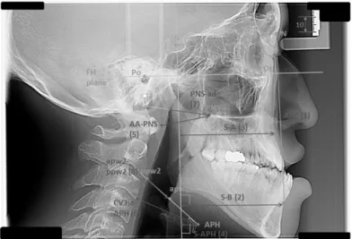
ThecephalometricpointswereSellaturcica
(
S;
thecentralpointofpituitaryglandofsphenoidbone),
Nasion(
N;
thenodalpointbetweenthesuturaofnasalboneandfrontalboneandthesuturaofnasalbone
),
Orbitale(
Or;
themostinferiorpointoforbit),
Posteriornasalspine(
PNS;
themostposteriorpointofhardpalate
),
Apoint(
A;
themostposteriorpointofcurvaturelippingbetweenthefundusofmaxillaandthe alveolarbone),
Bpoint(
B;
themostposteriorpointofmediansuturaofmandibularoutline),
Basion(
Ba;
the mostanteriorinferiorpointofgreateroccipitalforamen),
Porion(
Po;
themostsuperiorpointofexternal acousticmeatus),
themostanteriorpointofhyoidbone(
APH),
themostanteriorpointofanteriorarchof1st atlas(
AA),
themostinferiorpointof3rdatlas(
CV3ia),
thenodalpointbetweentheposteriorpharyngealwall andtheconnectinglinefromPNStoBa(
ad),
thenodalpointbetweentheanteriorpharyngealwallandthe connectinglinefromthemostanteriorpointofhyoidbonetothemostanteriorpointof2ndatlas(
apw2),
and thenodalpointbetweentheposteriorpharyngealwallandtheconnectinglinefromthemostanteriorpointof hyoidbonetothemostanteriorposteriorpointof2ndatlas(
ppw2)<
Fig.
1>.
ThecephalometricplanewasFrankforthorizontal
(
FH)
plane,
andthecephalometricanglewasANB.
The horizontalmeasurementdistancesofskullwerethedistancebetweentheverticallinefromFHplanetoSand theB(
S-
B),
thedistancebetweentheverticallinefromFHplanetoSandtheA(
S-
A),
andthedistance betweentheverticallinefromFHplanetoSandtheAPH(
S-
APH).
Thehardtissueairwaydistanceswere thedistancebetweenAAandPNS(
AA-
PNS)
andthedistancebetweenCV3iaandAPH(
CV3ia-
APH).
The softtissueairwaydistanceswerethedistancebetweenPNSandad(
PNS-
ad),
thedistancebetweenapw2and ppw2(
apw2-
ppw2)<
Fig.
1>.
3. Data analysis
Thecraniometricpoint
,
plane,
angle,
anddistanceofthesubjectswerecalculatedthemeanandthestandard deviation.
Inaddition,
thosewereanalyzedbyRMANOVAwithingroupdependingonthemeasurement period(
beforethesurgery,
afterthesurgery,
andaftertheorthodontic).
AlsoitwasanalyzedbyRMANOVA betweengroupaccordingtothegenderandthesurgicalmethod.
ThedatawereanalyzedusingStatisticalPackagefortheSocialSciences
(
SPSSver.
18.
0,
Chicago,
Illinois,
USA).
Fig. 1. The craniometric point, plane, angle, and distance.
Results
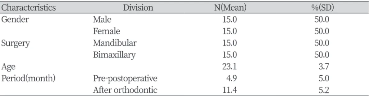
1. The general characteristics of the study subjects
Thestudysubjectswere30patientswiththeorthognathicsurgery
.
Theywere15males,
and15females.
15 patientsunderwentthemandibularsurgery,
and15patientsunderwentthebimaxillarysurgery.
Theaverage ageofthepatientwas23.
1years,
andunderwentthesurgeryafteranaverageof4.
9months.
After11.
4 monthsontheaverage,
theorthodontictreatmentwascompleted<
Table1>.
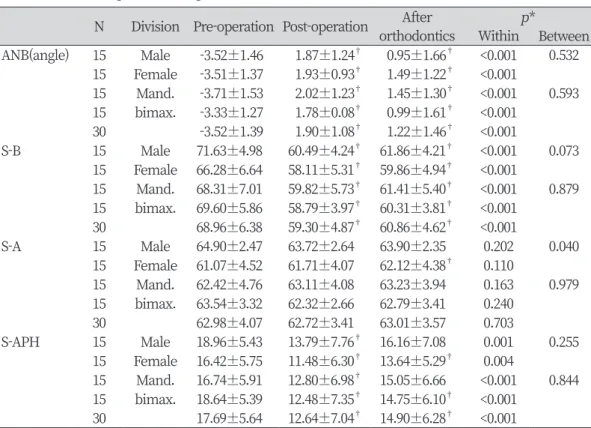
2. The changes in the angle and distance measurements of skull after the surgery
Itwasshownin
<
Table2>
thatwasthechangesintheangleanddistancemeasurementsofskull.
ANBwas–
3.
52anglepreoperatively,
1.
90anglepostoperatively,
and1.
22angleaftertheorthodontic treatment.
ANBincreased5.
42anglepostoperativelycomparedwiththepreoperative,
butdecreased0.
68 angleaftertheorthodontictreatment(
p<
0.
01).
Therewasnosignificantdifferenceaccordingtothegender andthesurgicalmethod.
S
-
Bwas68.
96mm,
59.
30mmand60.
86mm,
respectively.
S-
Bdecreased9.
65mmafterthesurgery,
but increased1.
56mmaftertheorthodontics.
Therewasnosignificantdifferenceaccordingtothegenderandthe surgicalmethod.
TherewasnostatisticallysignificantchangeinS
-
A,
buttherewasthesignificantdifferenceaccordingto thegender(
p<
0.
05).
Thepre-
operativeS-
Awasgreaterinthemalesthanthefemales,
themalewasdecreased andthenincreasedwhilethefemalewasincreasedcontinually(
p<
0.
05).
S
-
APHwas17.
69mmpreoperatively,
12.
64mmpostoperatively,
and14.
90mmaftertheorthodontic treatment.
S-
APHwasstatisticallysignificantlydecreased5.
05mmafterthesurgery,
butincreased2.
26mm aftertheorthodontictreatment(
p<
0.
01).
Therewasnosignificantdifferenceaccordingtothegenderandthe surgicalmethod<
Table2>.
3. The changes in the oropharynx after the surgery
AA
-
PNSwas28.
45mmpreoperatively,
29.
10mmpostoperatively,
and28.
81mmaftertheorthodontic treatment.
AA-
PNSwasstatisticallyincreased0.
65mmafterthesurgery(
p<
0.
05).
Therewasnosignificant differenceaccordingtothegenderandtheoperativemethod.
Table 1. The characteristics of the study subjects
Characteristics Division N(Mean) %(SD)
Gender Male 15.0 50.0
Female 15.0 50.0
Surgery Mandibular 15.0 50.0
Bimaxillary 15.0 50.0
Age 23.1 3.7
Period(month) Pre-postoperative 4.9 5.0
After orthodontic 11.4 5.2
TherewasnostatisticallysignificantchangesinPNS
-
ad<
Table3>.
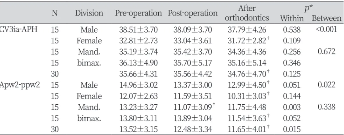
4. The changes in the laryngopharynx after the surgery
TherewasnostatisticallysignificantdifferenceinCV3ia
-
APH,
buttherewasthesignificantdifference accordingtothegender.
Thepre-
operativeCV3ia-
APHwasgreaterinthemalesthanthefemales,
themale wasdeceasedcontinuallywhilethefemalewasincreasedandthendecreased(
p<
0.
01).
Inaddition,
thefemaleTable 2. The changes in the angle and distance measurements of skull (Unit: mm)
N Division Pre-operation Post-operation After p*
orthodontics Within Between ANB(angle) 15 Male -3.52±1.46 1.87±1.24† 0.95±1.66† <0.001 0.532
15 Female -3.51±1.37 1.93±0.93† 1.49±1.22† <0.001 15 Mand. -3.71±1.53 2.02±1.23† 1.45±1.30† <0.001 0.593 15 bimax. -3.33±1.27 1.78±0.08† 0.99±1.61† <0.001 30 -3.52±1.39 1.90±1.08† 1.22±1.46† <0.001 S-B 15 Male 71.63±4.98 60.49±4.24† 61.86±4.21† <0.001 0.073
15 Female 66.28±6.64 58.11±5.31† 59.86±4.94† <0.001 15 Mand. 68.31±7.01 59.82±5.73† 61.41±5.40† <0.001 0.879 15 bimax. 69.60±5.86 58.79±3.97† 60.31±3.81† <0.001 30 68.96±6.38 59.30±4.87† 60.86±4.62† <0.001
S-A 15 Male 64.90±2.47 63.72±2.64 63.90±2.35 0.202 0.040
15 Female 61.07±4.52 61.71±4.07 62.12±4.38† 0.110 15 Mand. 62.42±4.76 63.11±4.08 63.23±3.94 0.163 0.979 15 bimax. 63.54±3.32 62.32±2.66 62.79±3.41 0.240
30 62.98±4.07 62.72±3.41 63.01±3.57 0.703
S-APH 15 Male 18.96±5.43 13.79±7.76† 16.16±7.08 0.001 0.255 15 Female 16.42±5.75 11.48±6.30† 13.64±5.29† 0.004 15 Mand. 16.74±5.91 12.80±6.98† 15.05±6.66 <0.001 0.844 15 bimax. 18.64±5.39 12.48±7.35† 14.75±6.10† <0.001 30 17.69±5.64 12.64±7.04† 14.90±6.28† <0.001
* by RM ANOVA
†post-hoc analysis at α=0.05: Bonferroni correction with the value of pre-operative
Table 3. The changes in the oropharynx (Unit: mm)
N Division Pre-operation Post-operation Afterorthodontics p*
Within Between AA-PNS 15 Male 28.06±4.03 28.85±3.74 28.60±3.86 0.075 0.644
15 Female 28.85±2.98 29.36±2.78 29.01±2.83 0.190 15 Mand. 29.60±3.08 30.04±2.87 29.74±3.11 0.157 0.095 15 bimax. 27.31±3.62 28.17±3.43 27.87±3.38 0.083 30 28.45±3.50 29.10±3.25† 28.81±3.33 0.015 PNS-ad 15 Male 21.95±3.47 21.09±3.54 21.34±3.48 0.216 0.487
15 Female 22.38±3.24 21.94±3.23 22.52±2.99 0.250 15 Mand. 22.72±3.02 21.97±3.22 22.67±3.02 0.192 0.607 15 bimax. 21.61±3.59 21.06±3.54 21.19±3.39 0.406
30 22.17±3.31 21.51±3.36 21.93±3.24 0.110
* by RM ANOVA
†post-hoc analysis at α=0.05: Bonferroni correction with the value of pre-operative
wasmoredifferencethanthemalebetweenbeforetheoperationandaftertheorthodontictreatment
.
Apw2-
ppw2was13.
52mmpreoperatively,
12.
48mmpostoperatively,
and11.
65mmaftertheorthodontic treatment.
Apw2-
ppw2wasstatisticallysignificantlydecreased1.
87mmaftertheorthodontic treatment comparedwiththeprioroperation(
p<
0.
05).
Therewasalsoasignificantdifferenceaccordingtothegender(
p<
0.
05).
The pre-
operativeApw2-
ppw2wasgreaterinthemalesthanthe females,
themale wasmore differencethanthefemalebetweenbeforetheoperationandaftertheorthodontictreatment<
Table4>.
5. The correlation between the variables
Thecorrelationbetweenthevariables wasshownin
<
Table 5>.
The ANBdecreased(
p<
0.
01)
asthe mandibularanteriorposition(
S-
B),
thehyoidbone(
S-
APH)
waslocatedanteriorly(
p<
0.
01),
andthewidthof hardoropharynx(
AA-
PNS)
wasincreased(
p<
0.
05).
Thewidthofsoftlaryngopharynx(
apw2-
ppw2)
also increased(
p<
0.
01).
S
-
BhadasomewhathighernegativecorrelationwithANB(
r=-
0.
634)
andasomewhathigherpositive correlationwithS-
APH(
r=
0.
662),
butalowerpositivecorrelationwithAA-
PNS(
r=
0.
225)
andappw2-
ppw2(
r=
0.
369).
Discussion
For the cephalometry
,
Broadbent[
13]
used the lateral cephalometric radiographs,
King[
14]
used the roentgenographwhichstudiedthegrowthofpharynx.
Recently,
thelateralcephalogramshavebeenusedforTable 4. The change in the laryngopharynx (Unit: mm)
N Division Pre-operation Post-operation Afterorthodontics p*
Within Between CV3ia-APH 15 Male 38.51±3.70 38.09±3.70 37.79±4.26 0.538 <0.001
15 Female 32.81±2.73 33.04±3.61 31.72±2.82† 0.109 15 Mand. 35.19±3.74 35.42±3.70 34.36±4.36 0.256 0.672 15 bimax. 36.13±4.90 35.70±5.17 35.16±5.14 0.346 30 35.66±4.31 35.56±4.42 34.76±4.70† 0.125 Apw2-ppw2 15 Male 14.96±3.02 13.37±3.00 12.99±4.50† 0.051 0.022
15 Female 12.07±2.63 11.59±3.51 10.31±3.03† 0.144 15 Mand. 13.23±3.27 11.07±3.09† 11.75±4.48 0.003 0.338 15 bimax. 13.80±3.11 13.89±3.04 11.54±3.63† 0.052 30 13.52±3.15 12.48±3.34 11.65±4.01† 0.015
* by RM ANOVA
†post-hoc analysis at α=0.05: Bonferroni correction with the value of pre-operative
Table 5. The correlation coefficients between the variables
S-B ANB S-APH AA-PNS apw2-ppw2
S-B 1
ANB -0.634** 1
S-APH 0.662** -0.301** 1
AA-PNS 0.225* 0.129 0.013 1
Apw2-ppw2 0.369** -0.223* 0.176 0.236* 1
* p<0.05, **p<0.01 by pearson correlation analysis
themeasurements
[
4-
5,
7-
8].
Thisstudyalsousedthattomeasure.
Becausetheairwaymaychangewithgrowing
,
thesubjectsweretheadults.
ANBwas
–
3.
52anglebeforethesurgery,
thenitwasimprovedto1.
90angleafterthesurgery,
butwas slightlyreturnedto1.
22angleaftertheorthodontictreatment.
ThechangeinANBisthoughttobedueto changeinS-
B.
BecausetherewasthenegativecorrelationwithS-
B(
p<
0.
01,
r=-
0.
634).
Accordingtothe previousstudy[
15],
ANBdidnotthecorrelationwiththepositionofmaxilla,
butthepositivecorrelationwith themandible.
Themandibularposition
(
S-
B)
transferredposteriorlyafterthesurgery,
butslightlymovedanteriorlyafter theorthodontictreatment.
LeeandHan[
4]
reportedthatthemandibularmovedtotheposteriorposition,
and thenregressedtotheanteriorposition.
InthestudyofChangetal.[
7],
themandiblemovedtoposterosuperior position,
itregressedslightly.
ButChoietal.[
5]
reportedthatthemandiblemovedposteriorlyandfurtherto theposteriorposition.
Thisstudyresultwassimilartosomepreviousstudies[
4,
7].
S
-
APHdecreased5.
05mmafterthesurgerybutincreased2.
26mmaftertheorthodontictreatment.
The changeinS-
APHisthoughttobeduetochangeinS-
B.
BecausetherewasthenegativecorrelationwithS-
B(
p<
0.
01,
r=-
0.
662).
Thehyoidbonewasregressed,
butitdidnotreturntoitspreoperativeposition.
Inthestudy ofLeeandHan[
4],
thehyoidbonewasrecoveredafteroneyear,
butthehyoidbonewaslocatedposteriorly beforethesurgery.
Choietal.[
5]
reportedthatthehyoidbonemovedtotheposteriorbutwasrestoredto preoperativeposition.
ThestudyresultwassimilartothatofLeeandHan[
4].
However,
somestudieshave reportedthatthehyoidbonemovedinferiorlyafterthesurgery[
7,
9].
Therefore,
thereareneedthevarious follow-
upstudiesonthepositionofhyoidbone.
TherewasastatisticallysignificantdifferenceinAA
-
PNS,
butthechangebetweenthebeforeandafterthe surgerywasminimal
(
0.
65mm).
TherewasnostatisticallysignificantdifferenceinPNS-
ad.
Itwasprobably duetothefactthatthepositionofmaxilla(
S-
A)
didnotchangesignificantly.
LeeandHan[
4]
reportedthatthe oropharyngealwidthwasreduced2.
3mmafterthesurgeryanditwaslengthened0.
9mmafter6months,
but itwasreduced1.
1mmcomparedtobeforethesurgery.
Choietal.[
5]
showeditwasdecreasedafterthe surgeryandwasfurtherdecreasedatthefollow-
up.
InthestudyofZhangetal.[
7],
theoro-
pharygealairway wasdecreasedafterthesurgery,
butmostofthatwasreturnedafteroneyear.
Lee[
9]
wasalsoreportedto recoverafter6months.
Thisstudyresultwassimilartosomepreviousstudies[
7,
9].
TherewasnostatisticallysignificantdifferenceinCV3ia
-
APH,
butapw2-
ppw2wassignificantlydecreased(
p<
0.
05).
Thereasonwasthatthemandible(
S-
B)
andhyoidbone(
S-
APH)
movedbackward.
Intheprevious study[
4,
7,
9],
thelaryngopharyngealwidthdecreasedandthenregressed.
However,
Choietal.[
5]
reportedthat itwasdecreasedafterthesurgery,
andfurtherdecreasedinthefollow-
up.
Thisstudyresultwassimilartothat ofChoietal.[
5].
Inthisstudy
,
thechangesofairwayweremeasuredaftersurgery.
Beside,
itwasthefollow-
upinvestigation aftertheorthodontictreatment.
Inaddition,
thosewereanalyzedthedifferenceinchangesaccordingtothe surgicalmethodandthegender.
Itwasconsideredtobeaseriesofstepstocompensateforthemandible movedbackwardthatwasthenarrowingofairwaywidthaftertheorthognathicsurgery.
However,
thisstudy hasthelimitationswhichwasdifficulttogeneralizebecauseoffewstudysubjectsintheir20s,
hadnotbeenabletousemoreaccuratemeasurementtoolssuchas3DCT
,
andcouldnotdirectlyconductaquestionnaire orinterviewwiththepatienttocheckavariousproblemsafterthesurgery.
afollow
-
upstudyisneededtostudythechangesofairwayaccordingtovarioussubjectsandsurgical methodssuchasgenioplasty,
forwardmovementsurgeryofmaxillaryormandible,
andthepositionofthe tongue.
Whenasurgeonplansanorthognathicsurgeryforapatientswithriskfactorswhichanairwayisnarrowor theposteriormovementofmandibleislarge
,
thesechangesshouldbereferredto.
Inaddition,
asaspecialist whocooperateswithadentalcareandeducatesaoralhealth,
adentalhygienistsneedtoexplainandeducate apatientswithreferencetothesechanges.
Conclusions
Thepurposeofthisstudywastomeasurethechangesofairwayaftertheorthognathicsurgeryinthe patientswith skeletalClass IIImalocclusion
.
Inaddition,
itwasalsofollowedup after theorthodontic treatment.
Itwasusedforfinalanalysisthatwasthetotalof90lateralcephalometricradiographsof30judgement sampledsubjects
.
Themeasurementangle and distanceswere calculatedasthe meanandthe standard deviation,
andthechangesofvariablesaccordingtothemeasurementperiodwereanalyzedbyrepeated measuresANOVA.
Alsothevariableswereanalyzedbycorrelation.
1
.
Thesubjectswere23.
1yearsoldonaverage,
underwentthesurgeryafteranaverageof4.
9months,
and completedtheorthodontictreatmentafteranaverageof11.
4months.
2
.
ANBwasincreasedafterthesurgery,
butwasdecreasedaftertheorthodontictreatment.
3
.
The mandible(
S-
B)
moved posteriorly after the surgery,
but partially moved anteriorlyafter the orthodontictreatment.
4
.
Thehyoidbone(
S-
APH)
alsowentbackpostoperatively,
sincethensomewentahead.
5.
Thelocationoftheoropharynxdidnotchangewidelyafterthesurgery.
6
.
Thesofttissuewidth(
apw2-
ppw2)
oflaryngealpharynxwasnarrowedafterthesurgery,
andfurther decreasedaftertheorthodontictreatment.
Asthemandiblewastransferredaftertheorthognathicsurgery