• 검색 결과가 없습니다.

Double primary lung adenocarcinoma diagnosed by epidermal growth factor receptor mutation status

N/A
N/A
Protected

Academic year: 2021

Share "Double primary lung adenocarcinoma diagnosed by epidermal growth factor receptor mutation status"

Copied!
5
0
0

로드 중.... (전체 텍스트 보기)

전체 글

(1)

Yeungnam Univ J Med 2017;34(2):270-274

Double primary lung adenocarcinoma diagnosed by epidermal growth factor receptor mutation status

Oh Jung Kwon

1

, Min Hyeok Lee

1

, Sung Ju Kang

1

, Seul Gi Kim

1

, In Beom Jeong

1

, Ji Yun Jeong

2

, Eun Jung Cha

3

, Do Yeun Cho

1

, Young Jin Kim

4

, Ji Woong Son

1

1

Department of Internal Medicine, Konyang University Hospital, Daejeon;

2

Department of Pathology, Kyungpook National University Medical Center, Kyungpook National University School of Medicine, Daegu;

Departments of

3

Pathology and

4

Thoracic Surgery, Konyang University Hospital, Daejeon, Korea

A nodular density was detected on a chest radiograph taken from a 57-year-old Korean woman who was visiting a hospital for a routine check. Chest computed tomography revealed a 4.8 cm lobulated mass in the right lung and another focal nodular lesion in the left lung; biopsies of both lungs revealed adenocarci- noma. We conducted DNA sequencing and peptide nucleic acid clamping to investigate the potential double primary lung cancer. The results verified that the mass in the right lung had a mutation in the epidermal growth factor receptor, whereas the nodule in the left lung had a wild-type sequence, showing that these two were genetically different cancers from one another. Thus, we demonstrate that genetic testing is useful in determining double primary lung cancer, and we herein report on this case.

Keywords: Adenocarcinoma; Differential diagnosis; Lung cancer; DNA sequence analysis; Mutations

Copyright ©2017 Yeungnam University College of Medicine

This is an Open Access article distributed under the terms of the Creative Commons Attribution Non-Commercial License (http://creative- commons.org/licenses/by-nc/4.0/) which permits unrestricted non-commercial use, distribution, and reproduction in any medium, provided the original work is properly cited.

Received: July 11, 2016, Revised: September 13, 2016 Accepted: September 26, 2016

Corresponding Author: Ji Woong Son, Deprartment of Internal Medicine, Konyang University Hospital, Gwanjeo dong-ro 158, Seo-gu, Daejeon 35365, Korea

Tel: +82-42-600-8817, Fax: +82-42-600-9090 E-mail: sk1609@hanmail.net

INTRODUCTION

The number of patients with multiple primary lung cancer (MPLC) is increasing [1]. The concept of MPLC was intro- duced in 1924 [2], and second primary tumors are either me- tachronous — being detected after the primary lesion, or syn- chronous — being resected or detected simultaneously. Syn- chronous MPLC was presumed to be uncommon; however, in reality its true incidence, which ranges from 0.2% to 20%

[3], is increasing owing to the wide usage of positron emission tomography (PET) scanning and improvements in computed tomography (CT) scanning and other diagnostic modalities.

When multiple synchronous lung tumors have been identi- fied, the differentiation of multi-centric cancers from single cancers with pulmonary metastases or intrapulmonary meta- stases from primary cancers in different organs can often be difficult, especially if the histologic characteristics of synchro- nous tumors are the same.

We present a case of synchronous double primary lung cancers; an adenocarcinoma with an epidermal growth factor receptor (EGFR) mutation in the right lung and an adenocar- cinoma without mutation in the left lung.

CASE

A 57-year-old Korean woman visited a hospital for a rou-

tine check-up. A nodular density was detected on a chest

radiography. She was healthy with no symptoms and had

an unremarkable family or medical history. In addition, she

had no history of smoking. Her vital signs revealed a blood

pressure of 110/71 mmHg, a heart rate of 81 beats per mi-

(2)

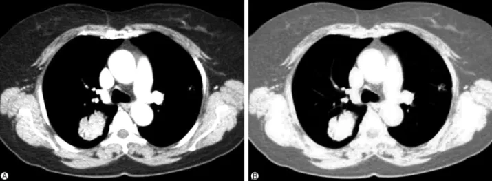
Fig. 1. Computed tomography scan showing a 4.8 cm lobulated mass with air bronchogram in the right posterior segment (A). Another focal nodular lesion is evident in the left anterior segment with a lucent internal bubble (B).

Fig. 2. The left lung adenocarcinoma shows an acinar and lepidic pattern (A), while adenocarcinoma in the right lung consists of tumor cell nests with poorly-formed and small glandular spaces (B). (A, B, H&E stain, ×200).

nute, and a respiration rate of 17 breaths per minute; her laboratory findings were all normal. Her general condition was good, and a physical examination showed no abnorma- lities. Chest X-ray showed an abnormal shadow in the right middle lung field. A chest CT scan revealed a 4.8 cm lobulated mass in the right upper lobe (RS3) and another focal nodular lesion in the left upper lobe (LS3) (Fig. 1). We observed no mediastinal lymph node enlargement and noted no metastatic lesions on brain magnetic resonance imaging san, PET-CT scan, or whole-body bone scan. The levels of serum tumor markers, such as carcinoembryonic antigen, cytokeratin frag- ment, and neuron-specific enolase, were within normal range.

We performed two percutaneous needle aspiration biopsies (NABs) for each of the nodules and collected biopsy speci- mens from both lesions. There were no complications during NAB, and the patient had an uneventful recovery. Histolo- gical examinations of both specimens revealed adenocarcino- mas in both lungs (Fig. 2).

To determine whether the lesions arose independently or

as a result of metastasis, we conducted direct sequencing of

the deoxyribonucleic acid (DNA) from both tumors. Exon se-

quencing of the genomic DNA revealed deletion and substi-

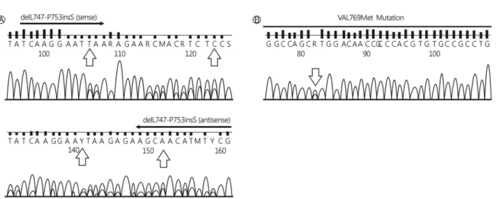
tution mutations of EGFR in the right lung nodule and a

wild-type sequence in the left lung nodule (Fig. 3). Given the

(3)

Fig. 3. Direct sequencing DNA demonstrating an exon 19 deletion (A) and an exon 20 substitution (B) in the tumor in the right lung.

Fig. 4. PNA-mediated real-time PCR clamping. A PNA/DNA hy- brid with a single base pair mismatch did not suppress annealing of the PCR primer or amplification of mutant alleles (exon 19 deletion). PNA, peptide nucleic acid; PCR, polymerase chain reaction; RFU, relative fluorescence unit.

low sensitivity of direct sequencing, especially for a small mass, a peptide nucleic acid (PNA)-mediated real-time poly- merase chain reaction (PCR) clamping was performed, which revealed a positive signal in exon 19 (deletion) from the sam- ple of the right lung (Fig. 4) and no mutation in the left lung (data not shown). Although the NAB test was not able to represent the overall characteristics of the lesion due to the small size of the sample, additional highly-specific DNA anal- ysis and highly-sensitive PNA test provided reliability to the results.

We performed a right upper lobectomy in March 2014 and a left upper lobe anterior and superior lingular segmentec-

tomy in April, 2014. There were no intraoperative complica- tions, and the patient fully recovered. Pathologic staging of the nodule in the right lung was pT2aN2 (stage IIIA), whereas that in the left lung was pT2aN0 (stage IB). Both nodules were positive for thyroid transcription factor-1. Adjuvant con- current chemoradiation therapy was administered after the surgery.

During the follow-up period, there was recurrence only in the right-side (lower right lung, RS6). After the treatment with an EGFR tyrosine kinase inhibitor, a partial response state was maintained.

DISCUSSION

Lung cancer can be divided into four main types: squamous cell carcinoma, small cell carcinoma, large cell carcinoma, and adenocarcinoma. Especially in adenocarcinoma, a more pre- cise subtyping is made available due to the molecular analytic techniques. According to the presence or absence of a driving mutation, the clinical characteristics and optimal treatments are different. Therefore, genetic analysis of the driving muta- tion is an essential tool in lung cancer diagnosis and treatment.

The diagnosis of a second primary lung cancer is complex

for a number of reasons. It can be difficult to differentiate

a metastasis from the primary lesion, if the latter occurs with-

in 2 years of the initial tumor. Differentiation is also hindered

when the initial tumor and second primary lesion have a simi-

lar histological subtype and when the second lesion is located

in an area previously exposed to radiotherapy. This is the

(4)

case because changes in tissue morphology can complicate the differential diagnosis.

Direct sequencing and PNA clamping are approved by the Korean Food and Drug Administration. A PNA is an artifi- cially synthesized polymer that can bind to a complementary sequence in DNA; the binding capacity of PNA is stronger than that of DNA. PNA clamping can detect 29 target muta- tions of clinical significance among approximately 250 known EGFR mutations [4], but it cannot detect unknown EGFR mu- tation sites. The weakness of PNA clamping is that this meth- od can only be used to detect mutations for which the primers have been individually designed; conversely, direct sequencing can uncover novel mutations. In this case, PNA clamping did not detect substitution mutations in exon 20.

The diagnosis is complicated by difficulties in clearly mak- ing a distinction between MPLC and metastatic lung cancer.

Clinicopathologic diagnosis, assessment, and management have all evolved, but still remain severely limited by a lack of dependable and robust molecular markers for the differ- ential diagnosis of MPLC and metastasis. The Martini and Melamed clinicopathological assessment criteria are based on tumor characteristics that include, but are not limited to, the location, morphology, absence or presence of carcinoma in situ, metastasis, and vascular invasion [5]. However, these cri- teria lack the power to differentiate between a second primary lung cancer and metastasis [6,7]. Genomic analysis of patients is useful to distinguish between a second primary lung cancer and metastasis. Girard et al. found that genomic profiling contradicted the clinicopathological diagnosis in 18% of tu- mors [8]. That same group reported that EGFR and K-ras mu- tation status could be used to determine the existence of clo- nal relationships between MPLCs. EGFR/K-ras mutational profiling was useful in improving the discrimination between intrapulmonary metastases and MPLC. In a search of the lit- erature, we found four cases with simultaneous EGFR muta- tions in multiple lesions. Two cases had the same mutation, while the other two cases had different mutations. In the case of simultaneous EGFR mutations, examination of addi- tional somatic tumor gene alterations by next generation se- quencing is useful in differentiating between metastasis and primary lung cancer [9]. However, next generation sequenc- ing may be limited as it is very expensive and is rarely used in clinical settings. Developing rapid, inexpensive, and reli- able molecular tools may allow the refinement of the clini-

copathologic criteria that are currently used in MPLCs [10].

Precise staging and an aggressive surgical approach that in- cludes tissue-sparing procedures for early-stage cancers should be adapted as the standard clinical practice to increase long- term survival and maintain respiratory function in patients with MPLC. The present case used direct sequencing and PCR clamping to diagnose primary adenocarcinomas in both lungs based on the EGFR mutation status. Genetic analysis presents an extremely attractive method to diagnose double primary cancer.

CONFLICT OF INTEREST

No potential conflict of interest relevant to this article was reported.

REFERENCES

1. Xue X, Liu Y, Pan L, Wang Y, Wang K, Zhang M, et al. Dia- gnosis of multiple primary lung cancer: a systematic review.

J Int Med Res 2013;41:1779-87.

2. Beyreuther H. Multiplicität von Carcinomen bei einem Fall von sog. “Schneeberger” Lungenkrebs mit Tuberkulose. Vir- chows Arch Path Anat 1924;250:230-43.

3. Rea F, Zuin A, Callegaro D, Bortolotti L, Guanella G, Sartori F. Surgical results for multiple primary lung cancers. Eur J Cardiothorac Surg 2001;20:489-95.

4. Lee HJ, Xu X, Kim H, Jin Y, Sun P, Kim JE, et al. Com- parison of direct sequencing, PNA clamping-real time poly- merase chain reaction, and pyrosequencing methods for the detection of EGFR mutations in non-small cell lung carcino- ma and the correlation with clinical responses to EGFR tyro- sine kinase inhibitor treatment. Korean J Pathol 2013;47:

52-60.

5. Martini N, Melamed MR. Multiple primary lung cancers. J Thorac Cardiovasc Surg 1975;70:606-12.

6. Chang YL, Wu CT, Lee YC. Surgical treatment of synchro- nous multiple primary lung cancers: experience of 92 pati- ents. J Thorac Cardiovasc Surg 2007;134:630-7.

7. Ostrovnaya I, Olshen AB, Seshan VE, Orlow I, Albertson DG, Begg CB. A metastasis or a second independent cancer? Evalu- ating the clonal origin of tumors using array copy number data. Stat Med 2010;29:1608-21.

8. Girard N, Ostrovnaya I, Lau C, Park B, Ladanyi M, Finley D, et al. Genomic and mutational profiling to assess clonal relationships between multiple non-small cell lung cancers.

Clin Cancer Res 2009;15:5184-90.

9. Chuang JC, Shrager JB, Wakelee HA, Neal JW. Concordant and discordant EGFR mutations in patients with multifocal adenocarcinomas: implications for EGFR-targeted therapy.

Clin Ther 2016;38:1567-76.

(5)

10. Girard N, Deshpande C, Azzoli CG, Rusch VW, Travis WD, Ladanyi M, et al. Use of epidermal growth factor receptor/

Kirsten rat sarcoma 2 viral oncogene homolog mutation test-

ing to define clonal relationships among multiple lung adeno- carcinomas: comparison with clinical guidelines. Chest 2010;

137:46-52.

수치

Fig. 1. Computed tomography scan showing a 4.8 cm lobulated mass with air bronchogram in the right posterior segment (A)
Fig. 3. Direct sequencing DNA demonstrating an exon 19 deletion (A) and an exon 20 substitution (B) in the tumor in the right lung.

참조

관련 문서

Vertical ridge augmentation by means of deproteinized bovine bone block and recombinant human platelet-derived growth factor-BB: a histologic study in a

Results: 44 of 1048 patients with gastric cancer(4.1%) had synchronous and metachronous cancers. The average time interval between gastric cancer and secondary primary cancer

Since it is found that the self-regulation counseling program has an positive effect on the reduction of addiction in psychological factor, behavior․social

Objective :::: The neovascularization is an essential a factor for the growth of solid organ cancer, and especially vascular endothelial growth factor (VEGF)

It might also act in conjunction with a growth factor, such as bone morphogenic protein (BMP), insulin-like growth factor, or platelet-derived growth factor.

First, as for the relationship between interest factor and self-esteem formation, human relation factor as an interest factor had a significant effect at

For example, the public’s sentiments of risk regarding nuclear power plants exceeds the risk felt by an expert in the area, and therefore the public sentiment is a negative

In this study, platelet-derived growth factors-BB (PDGF-BB), vascular endothelial growth factor (VEGF), and insulin-like growth factor-1 (IGF-1) were quantified in the PRF