• 검색 결과가 없습니다.

Large Thrombus Formation from Right Atrial Incision Site after Closure of Atrial Septal Defect

N/A
N/A
Protected

Academic year: 2022

Share "Large Thrombus Formation from Right Atrial Incision Site after Closure of Atrial Septal Defect"

Copied!
3
0
0

로드 중.... (전체 텍스트 보기)

전체 글

(1)

842 Copyright © 2013 The Korean Society of Cardiology Korean Circulation Journal

Introduction

Atrial septal defect (ASD) is a tissue defect which allows blood passing between both atria, and accounts for approximately 5-10%

of all congenital heart defects.

1)

Under clinical situations such as high proportions of left-to-right shunts, right ventricular volume overloads and paradoxical embolisms, the closure of ASD may be required. For the treatment of ASD, depending on the type and an- atomical characteristics, percutaneous closure or surgical repair techniques are applied. The surgical approach is based on closing the defects with a direct suture or patch.

2)

Embolic complications can occur after surgical interventions,

3)

but cases reported after pri- mary sutures are uncommon.
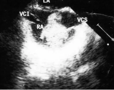
4)5)

Herein, we report two cases of large right atrial thrombus developed in the late stage after repairs of ASDs which were treated surgically.

Case Report

http://dx.doi.org/10.4070/kcj.2013.43.12.842 Print ISSN 1738-5520 • On-line ISSN 1738-5555

Large Thrombus Formation from Right Atrial Incision Site after Closure of Atrial Septal Defect

Olcay Murat Disli, MD 1 , Nevzat Erdil, MD 1 , Barıs Akca, MD 1 , Yılmaz Omur Otlu, MD 2 , and Bektas Battaloglu, MD 1

1

Departments of Cardiovascular Surgery and

2

Cardiology, Inonu University Faculty of Medicine, Malatya, Turkey

Atrial septal defect (ASD) is the common congenital anomaly which requires surgical interventions. Right atrial thrombus formations after primary suture repairs of the ASD and evidences of thromboembolic complications are extremely rare. Specifically, the cases of thrombo- embolic complications have high mortality and morbidity risks. Two cases of giant intra-atrial thrombus formation detected in the late stage after primary repairs of ASDs are being discussed. (Korean Circ J 2013;43:842-844)

KEY WORDS: Heart septal defects; Thrombosis; Heart surgery; Heart atrium.

Received: February 3, 2013 Revision Received: May 16, 2013 Accepted: July 26, 2013

Correspondence: Olcay Murat Disli, MD, Department of Cardiovascular Surgery, Inonu University Faculty of Medicine, Elazig Road, Malatya 44280, Turkey

Tel: 90 422 3410660, Fax: 90 422 3410036 E-mail: [email protected]

• The authors have no financial conflicts of interest.

This is an Open Access article distributed under the terms of the Creative Commons Attribution Non-Commercial License (http://creativecommons.

org/licenses/by-nc/3.0) which permits unrestricted non-commercial use, distribution, and reproduction in any medium, provided the original work is properly cited.

Cases

Case 1

A 24-year-old female patient with a history of primary repair for

ASD performed 8 years ago admitted to our clinic for her annual ch-

eckups. In her previous primary repair surgery, the defect was clos-

ed directly with an atriotomy incision followed by 5/0 polypropyl-

ene and 4/0 polypropylene sutures. The patient had attended her en-

suing controls every three months for a period of one year, and no

pathological findings were identified during this period. There was

no history of any drugs being used recently. Physical examination

was normal, and routine hematological and biochemical laboratory

analysis were within normal levels. Electrocardiogram revealed nor-

mal sinus rhythm and right bundle branch block. Transthoracic echo-

cardiography (TTE) revealed normal left ventricular systolic func-

tions, mildly dilated right chambers, minimal mitral regurgitation,

mild tricuspid regurgitation, systolic pulmonary artery pressure of

30 mm Hg, and an irregular-shaped mass in the right atrium. On her

transesophageal echocardiography (TEE) examination, a 3.7×3.5

cm sized giant pedunculated mobile mass was observed being at-

tached to the septum in the right atrium (Fig. 1). After the proce-

dure, the patient was hospitalized. Chest computed tomography (CT)

showed no evidence of pulmonary embolism, and ventilation/perfu-

sion scans indicated no problems. Protein C, protein S, and antith-

rombin III levels were within the normal range. Venous bilateral Dop-

pler of lower extremity and abdominal ultrasonography results were

normal. The patient was scheduled for excision of the mass. Surgery

was performed via a median sternotomy by utilizing the cardiopul-

monary bypass. Venous drainage was via the superior vena cava

(2)

843 Olcay Murat Disli, et al.

http://dx.doi.org/10.4070/kcj.2013.43.12.842 www.e-kcj.org

and the right femoral vein. The right atrium was being opened, and a large mass filling the entire atrium and obstructing the tricuspid valve was observed. The mass was intimately attached to the free atrial wall, it was irregularly-shaped, and 2.2×4.1 cm in dimension.

It had a tanned, gelatinous appearance, and showed multifocal ar- eas of calcification. The entire free wall of the right atrium mass was resected. The patient was weaned off with cardiopulmonary bypass without any difficulty. The mass consistent with thrombus forma- tion originating from the suture line was excised (Fig. 2). Histopa- thological evaluation was consistent with the organized thrombus.

Case 2

A 42-year-old female, without cardiac complaints, was admitted to our clinic presented with chest discomforts, one month history of exertional dyspnea and persistent dry coughs. She had a history

of primary ASD repair 3 years ago, and quitted routine follow-ups.

In her previous primary repair surgery, the defect was closed directly with an atriotomy incision followed by 5/0 polypropylene and 4/0 polypropylene sutures. The patient had attended her ensuing con- trols every two months for a period of one year, and no pathological findings were identified during this period. Biochemical and hema- tological values were within normal limits. Cardiovascular examina- tion was normal and no pathologic sounds or murmurs were de- tected. The patient was in normal sinus rhythm and not on any me- dications. Chest examination elicited few scattered crepitations bi- laterally. An image from contrast-enhanced CT pulmonary angio- gram demonstrated multiple bilateral pulmonary emboli. TTE re- vealed normal left ventricular systolic functions, heart chambers within normal size, and a mass in the right atrium. On TEE examin- ation, a 3.2×2.4 cm mobile and irregularly-shaped mass was ob- served in the right atrium. The patient’s bilateral Doppler of lower extremity, abdominal ultrasonography, and hypercoagulability sc- reening panel was negative. Excision of the mass with redo sterno- tomy was decided for treatment. During the surgery, exploration of the right atrium revealed a 3.2×2.3 cm globular mass with a tanned, gelatinous appearance and multifocal areas of calcification. The mass was attached to the free wall of the right atrium by a 1-cm stalk. This mass originating from the free wall of the right atrium was then excised and its histological examination later revealed it to be an organized thrombus. Both patients had uneventful post- operative courses, and were discharged with warfarin (interna- tional normalized ratio: 2-3) combined with acetylsalicylic acid therapy on postoperative seventh and sixth day, respectively. The TTE and TEE showed no thrombus after 6 months of follow-up for both patients.

Discussion

The right atrium contains crista terminalis, eustachian valve and chiari network, and these anatomical structures often cause incor- rect interpretations for the evaluation of masses.

6)

Thrombi, myxo- mas, and vegetations should be kept in mind for the differential di- agnosis of right atrial masses. Fifteen percent of the myxomas derived from the right atrium and are usually linked to the interatrial septum with a broad base and a narrow stalk.

7)

Myxomas are usually slow- growing, but sometimes may show rapid progressions and may oc- cur after repairs of ASD.

8)

We offered possible diagnoses among our cases, the right atrial myxoma and thrombus were definitely diag- nosed by histopathological examination after surgery.

After the ASD closure, possible complications include pericardial effusion, arrhythmias and thrombus formation. Embolization com- plications have been reported after ASD closure with transcatheter Fig. 1. The right atrial thrombus in the image of transesophageal echocar-

diography. LA: left artrium, VCI: vena cava inferior, VCS: vena cava superior, RA: right atrium.

Fig. 2. Intraoperative view of the thrombus as shown in Fig. 1.

(3)

844 Thrombus Formation in Right Atrium after Atrial Septal Defect Operation

http://dx.doi.org/10.4070/kcj.2013.43.12.842 www.e-kcj.org

devices,

9)

prosthetic patches

10)

and pericardial patches.

11)

However, cases of embolization or intracardiac thrombus developed after pri- mary closure of ASD are less common. Right atrial thrombi appear- ing in the postoperative period is a rare finding and possibly origi- nated from the site of interatrial trauma by suction and devices or suture line. Previously published cases of atrial thrombus detected in the free wall of the right atrium were also detected in the post- operative third month and third year.

4)5)

In addition, in the report of thrombus on the primary sutured patent foramen ovale site and complicated with systemic embolization by Rodriguez et al.,

12)

the atrial thrombus was detected six months later after the operation.

In the cases which we presented, thrombus were detected at lon- ger periods such as 8 years and 3 years after surgery, and they were treated with surgical interventions.

Right atrial thrombus, which are almost always accompanied by pulmonary embolism, carry a higher mortality rate in the preopera- tive and perioperative period. Treatment of right chamber thrombi is controversial; medical, surgical procedures and percutaneous inter- ventional techniques can be applied.

13)

When using thrombolytic agents for the treatment of immobile intra-cardiac thrombus, there is a risk that the dissolution of the peduncule might lead to the mi- gration of the thrombus, which in return may result in emboliza- tion.

14)15)

In addition, one of our patients were complicated with pul- monary embolism. Thrombolytic therapy might have partially dis- integrated the thrombus, thus, leaving smaller thrombi. These could have easily migrated into both pulmonary arteries that were nearly completely occluded, therefore, we preferred surgical treatments.

There is no consensus on the duration of anticoagulant therapy after repair of ASD. Anticoagulation treatment for 3 months was previously proposed and implemented.

4)16)

As seen in our cases, for the detection of thrombus formations over the years after opera- tion, further studies on long-term antiplatelet and anticoagulant therapy are necessary. Right atrial incisions are used in many other intracardiac procedures, such as congenital repair. This complication can thus occur in all surgeries requiring right atrial incision. There- fore, it is important to bear in mind and apply the above mentioned precautions following the completion of these procedures.

The long-term detection of atrial thrombus after operation shows the importance of routine echocardiography. Echocardiographic follow-ups after the procedure are recommended, usually at 24 hours, 1 month, 6 months, 1 year and then, at regular intervals.

17)

As a result, thrombosis may occur after closure of ASD in the right atrium. Caution should be exercised in the long-term follow-up period, especially in terms of intra-atrial thrombus.

References

1. Ferencz C, Rubin JD, Loffredo CA, Magee CM. The epidemiology of

congenital heart disease: the Baltimore-Washington Infant Study 1981-1989. Perspectives in Pediatric Cardiology. Vol 4. Mount Kisco, NY: Futura Publishing Co Inc;1993. p.33-73.

2. Alpert JS, Rippe JM. Congenital heart disease. In: Manual of cardio- vascular diagnosis and therapy. Boston: Little Brown;1988. p.235-54.

3. Hawe A, Rastelli GC, Brandenburg RO, McGoon DC. Embolic compli- cations following repair of atrial septal defects. Circulation 1969;39(5 Suppl 1):I185-91.

4. Dinckal MH, Davutoglu V, Soydinc S, Akdemir I, Aksoy M. Large throm- bus at the site of primary sutured atrial septal defect associated with pulmonary embolism and treatment by thrombolysis. Echocardiogra- phy 2003;20:535-8.

5. Sheikh AY, Schrepfer S, Stein W, et al. Right atrial mass after primary repair of an atrial septal defect: thrombus masquerading as a myxo- ma. Ann Thorac Surg 2007;84:1742-4.

6. Armstong WF, Ryan T. Feigenbaum’s Echocardiography. 7th ed. Phila- delphia, PA: Lippincott Williams and Wilkins;2010.

7. Cianciulli TF, Saccheri MC, Redruello HJ, et al. Right atrial thrombus mimicking myxoma with pulmonary embolism in a patient with sys- temic lupus erythematosus and secondary antiphospholipid syndrome.

Tex Heart Inst J 2008;35:454-7.

8. Suzuki I, Koide S, Odagiri S, Shohtsu A. Right atrial myxoma develop- ing 4 years following patch closure of an atrial septal defect: report of a case. Surg Today 1994;24:176-8.

9. Chessa M, Carminati M, Butera G, et al. Early and late complications associated with transcatheter occlusion of secundum atrial septal de- fect. J Am Coll Cardiol 2002;39:1061-5.

10. Haro M, Ruiz J, Ribas J, Maestre J, Pérez-Piteira J, Morera J. Massive haemoptysis complicating prosthetic patch pulmonary embolism after atrial septal defect repair. Thorax 1995;50:911-2.

11. Backer CL, Hartz RS, Meyers SN, Davis G. Coronary embolism follow- ing atrial septal defect repair. Ann Thorac Surg 1988;45:561-3.

12. Rodriguez CJ, Di Tullio MR, Sacco RL, Homma S. Intra-atrial thrombus after surgical closure of patent foramen ovale. J Am Soc Echocardiogr 2001;14:63-6.

13. Chartier L, Béra J, Delomez M, et al. Free-floating thrombi in the right heart: diagnosis, management, and prognostic indexes in 38 consec- utive patients. Circulation 1999;99:2779-83.

14. Thompson CA, Skelton TN. Thromboembolism in the right side of the heart. South Med J 1999;92:826-30.

15. Hwang YJ, Ahn YC, Lee CH, et al. Surgical removal of large thrombus at the suture site of the right atriotomy after atrial septal defect closure associated with pulmonary embolism: 1 case. Korean J Thorac Cardio- vasc Surg 2004;37:448-51.

16. Kirklin JW, Barratt-Boyes BG. Cardiac Surgery. New York: Wiley;1986.

p.488.

17. Warnes CA, Williams RG, Bashore TM, et al. ACC/AHA 2008 Guide-

lines for the Management of Adults with Congenital Heart Disease: a

report of the American College of Cardiology/American Heart Associ-

ation Task Force on Practice Guidelines (writing committee to devel-

op guidelines on the management of adults with congenital heart

disease). Circulation 2008;118:e714-833.

수치

Fig. 2. Intraoperative view of the thrombus as shown in Fig. 1.

참조

관련 문서

Fig. After fine grinding of hardness test specimens 29 Fig. Result of tensile strength according to number of welding pass 34 Fig. Result of elongation according

The purpose of this study is to analyze the situations of violating the right to learn and violence in student-athletes from an ethnographic view and

A stress component is positive when it acts in the positive direction of the coordinate axes, and on a plane whose outer normal points in one of the positive coordinate

OECD모델협약은 로열티를 “a payment of any kind received as a consideration for the use of or the right to use, any copyright of any literary, artistic

Fig.11 Temperature histories of the hot side and cold side heat exchangers according to time with speaker power(10W).. Fig.12 Temperature histories of the hot

Bone transplants, autogenous bones, allogenic bones, xenogenic bones, and synthetic bone substitutes have all been used as bone graft materials.. However,

Rate of callus formation from Ginkgo leaf disks grown on MS medium supplemented with 2,4-D and Kinetin after 4 weeks of culture under the light... Rate of callus formation

After the vehicle has become a large-formation and high-speed, loud noise from an express highway is coming to be higher and the number of large-scale