• 검색 결과가 없습니다.

Intermediate and Long-Term Results of Transcatheter Closure of Patent Foramen Ovale Using the Amplatzer Patent Foramen Ovale Occluder: One Case of Pulmonary Embolism Irrespective of Patent Foramen Ovale Closure

N/A
N/A
Protected

Academic year: 2022

Share "Intermediate and Long-Term Results of Transcatheter Closure of Patent Foramen Ovale Using the Amplatzer Patent Foramen Ovale Occluder: One Case of Pulmonary Embolism Irrespective of Patent Foramen Ovale Closure"

Copied!
7
0
0

로드 중.... (전체 텍스트 보기)

전체 글

(1)

356

Open Access

Intermediate and Long-Term Results of Transcatheter Closure of Patent Foramen Ovale Using the Amplatzer Patent Foramen Ovale Occluder: One Case of Pulmonary Embolism Irrespective of Patent Foramen Ovale Closure

Ju Hee Yoon, MD, Joon Sik Kim, MD, Dae Hyung Lee, MD, Eun Jung Shim, MD, So Yeon Lee, MD, Ki Sik Min, MD, Do Jun Cho, MD, and Hye Ran Lee, MD Department of Pediatrics, College of Medicine, Hallym University, Anyang, Korea ABSTRACT

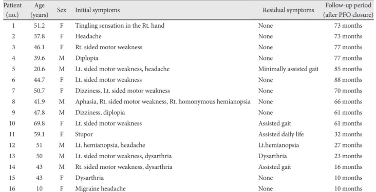
Background and Objectives: Patent foramen ovale (PFO) has been implicated in the pathogenesis of cryptogenic stroke or transient ischemic attack (TIA) due to paradoxical embolism, and in the pathogenesis of migraine. This paper reports the intermediate and long-term results of transcatheter closure of PFO associated with cerebrovascular accidents (CVAs), TIAs and migraine, using the Amplatzer PFO occluder. This paper also reports a case of pulmonary embolism which developed in one patient after PFO closure. Subjects and Methods: From January 2003 to May 2010, 16 patients with PFO (seven males and nine females) with a history of at least one episode of cryptogenic stroke/TIA, CVA, or migraine and who underwent per- cutaneous transcatheter closure of PFO using the Amplatzer occluder. All the procedures were performed under general anesthesia and were assisted by transesophageal echocardiography. Results: The device was implanted without any signifi- cant complications in all the patients, and the PFOs were effectively closed. At an average follow-up period of 54 months, the 15 patients with TIA/CVA had no recurrence of any thromboembolic event. The symptoms in one patient with migraine sub- sided after occlusion of the PFO. In this study, pulmonary embolism occurred five months after PFO closure in one patient, but the cause of pulmonary embolism was not identified. However, it is believed that the pulmonary embolism occurred with- out stroke recurrence because occlusion of the PFO was performed when the patient had a stroke event. Conclusion: It can be concluded that according to the intermediate and long-term follow-up results, transcatheter PFO closure is an effective and safe therapeutic modality in the prevention of thromboembolic events, especially in the patients with cryptogenic st- roke/TIA, and PFO closure is helpful in the treatment of migraine. However, this study involved a small number of patients and also the follow-up period was not long enough. Hence, randomized, controlled trials are necessary to determine if this ap- proach is preferable to medical therapy for the prevention of recurrent stroke or as primary treatment for patients with mi- graine headache. (Korean Circ J 2011;41:356-362)

KEY WORDS: Patent foramen ovale; Stroke; Transient ischemic attack; Migraine; Pulmonary embolism.

Received: July 16, 2010 / Revision Received: October 5, 2010 / Accepted: October 27, 2010

Correspondence: Do Jun Cho, MD, Department of Pediatrics, College of Medicine, Hallym University, 896 Pyeongchon-dong, Dongan-gu, Anyang 431- 070, Korea

Tel: 82-31-380-3730, Fax: 82-31-380-4118, E-mail: [email protected]

• The authors have no financial conflicts of interest.

cc

This is an Open Access article distributed under the terms of the Creative Commons Attribution Non-Commercial License (http://creativecommons.org/licens- es/by-nc/3.0) which permits unrestricted non-commercial use, distribution, and reproduction in any medium, provided the original work is properly cited.

Introduction

The etiology of ischemic stroke remains unknown in upto 40% of patients, despite an extensive diagnostic evaluation, and is referred to as cryptogenic.

1)

Patients with cryptogenic

stroke have a higher prevalence of patent foramen ovale (PFO)

than patients with stroke of determined cause.

2)

The relation-

ship between paradoxical (right-to-left) embolism through a

PFO and stroke remains controversial because of variability

in the reported stroke risk in patients with PFOs.

3)

A high risk

(2)

assessment study with transesophageal echocardiography (TEE) found a PFO in 25.6% of healthy subjects.

4)

Excess at- rial septal tissue in the region of the fossa ovalis causes incr- eased movement of the septum during respiration. This find- ing is classified as atrial septal aneurysm (ASA) when it had an excursion greater than 10 mm into the right or left atrium.

PFO with and without ASA was recognized as a potential risk factor for ischemic stroke only in the past two decades. Also, patients who have suffered a cryptogenic stroke related to PFO may have a stroke recurrence,

5)6)

a risk that is particularly pronounced in patients with PFO and associated ASA.

7)8)

PFO is also associated with migraine with aura. Recent retrospec- tive studies indicate a reduction in migraine frequency after PFO closure. Therapeutic options for PFO range from antipla- telet therapy to surgical or endovascular closure of the atrial shunt. Transcatheter closure of PFO was recently introduced in clinical practice. The goals of PFO closure are to prevent neurological events and to avoid the need for long-term an- ticoagulant therapy.

9)

This study reports the intermediate and long-term results of transcatheter closure of PFO associated with paradoxical embolism, which led to cryptogenic stroke or transient ischemic attack (TIA), and migraine, using the Amplatzer PFO occluder at the authors’ hospital. This paper also reports a case of pulmonary embolism that developed in one patient irrespective of PFO closure.

Subjects and Methods

Patient population

From January 2003 to June 2010, of a total of 16 patients, 15 patients with PFO (seven males, and eight females) were re- ferred for percutaneous closure of PFO. The primary reasons for referral were stroke {12/16 (75%)}, and TIA {3/16 (18%)}.

The symptoms included motor deficit in eight patients (50%), aphasia in one patient (6%), visual disturbances in four pa- tients (25%), headache in four patients (25%), stupor in one patient (6%), and dysarthria in three patients (18%) (Table 3).

The patients’ conditions were initially diagnosed by neurolo- gists. One patient who had migraine was referred for percu- taneous PFO closure from pediatric outpatient department.

Multiple thromboembolic events occurred in six patients prior to transcatheter device closure of PFO. The vascular risk factors were infrequent (Table 1). Cryptogenic stroke/TIA was present in eight patients (53%). ASA was present in sev- en patients (43%). The electrocardiographic findings before the procedure were all non-specific. Cryptogenic stroke was defined by the presence of a transient or permanent neuro- logical deficit with associated magnetic resonance imaging (MRI) evidence of an embolic lesion, in the absence of a clear cause.

10)

Ischemic stroke was defined as a cerebrovascular event with neurological symptoms lasting more than 24 hours without any sign of a primary cerebral hemorrhage. TIA was

defined as a new neurological deficit with complete cessa- tion of symptoms for 24 hours in the absence of lesions in the MRI.

11)

Migraine was defined as a severe recurring headache lasting from 4 to 24 hours, characterized by sharp pain, and is often accompanied by nausea, vomiting, and visual distur- bances.

Echocardiography

TEE was performed to detect PFO. An agitated saline bub- ble study was conducted to evaluate the degree of the right-to- left shunt at rest and after the Valsalva maneuver. The air bub- ble test was performed in all the patients, and all of them sh- owed positive results except for one patient, wherein the result was not positive because of poor co-operation.

Device

The Amplatzer PFO occluder device was used in this study.

It was constructed from the nitinol wires and consisted of two flat disks with a 3-mm long thin connecting waist. The right atrial disk was larger than the left atrial disk, which made it suitable for the occlusion of PFO. In this study, three sizes of the device were used, 18 mm, 25 mm, and 35 mm (Fig. 1).

Implantation procedure

The full procedure was performed under general anesthe- sia and was assisted by TEE through the femoral artery and vein. The NIH catheter (Cordis Europa N.V., Roden, The Ne- therlands) was advanced through an 8-Fr sheath, and pul- monary venous angiography was performed in LAO 45° view via the femoral vein. During TEE monitoring, the size of PFO with or without ASA was estimated using a balloon catheter (Boston Medical Corporation, Boston, MA, USA), and the size of the device was selected accordingly. The Amplatzer PFO occluder was implanted in the interatrial septum with great care under the water seal to prevent air embolism.

Before and after the procedure, all the patients received in- travenous heparin to prevent thrombus formation, and the transseptal sheaths were removed after the activated coagu- lation time stabilized (activated clotting time <140 seconds)

Fig. 1. Amplatzer PFO occluder (AGA Medical Corporation) is a self expandable, double disc device made from Nitinol wire mesh.

The two discs are linked together by a short connecting waist al-

lowing free motion of each disc.

(3)

with manual compression for achieving hemostasis, and an antidote was not used.

TEE was performed before the patient was discharged, mostly at two days after the procedure. After the occlusion of PFO, the air bubble test was performed again and all the pa- tients showed negative results. After the procedure, 100 mg/

day of aspirin for six months was prescribed for all the pa- tients.

Follow-up

All the patients returned for regular check-up visits at one, two, six and twelve months after the procedure, and yearly th- ereafter. Each patient underwent at least one TEE examina- tion during the first six months after the procedure to confirm the adequacy of placement of the device and to document the absence of a residual shunt or thrombus formation on the de- vice. Frequent clinical follow-ups at six- to 12-month inter- Table 1. Clinical characteristics of patients

Patient (no.) Age (years) Sex Cardiac abnormality Vascular risk factor Qualifying event

1 51 F PFO (30 mm) with ASA Hypercholesterolemia TIA

2 37 F PFO (25 mm) None CVA, TIA

3 46 F PFO (12 mm) None CVA

4 39 M PFO (21 mm) with ASA Alcohol, smoking CVA

5 20 M PFO (18.7 mm) with ASA Smoking CVA

6 44 F PFO (12.5 mm) None TIA

7 50 F PFO (18.7 mm) with ASA None CVA

8 41 M PFO (18 mm) Alcohol, smoking CVA

9 47 M PFO (20 mm) Alcohol, smoking, hypertension TIA

10 69 F PFO (17 mm) with ASA None CVA

11 59 F PFO (13.5 mm) with ASA None CVA

12 51 M PFO (12.5 mm) Hypertension CVA

13 50 M PFO (13 mm) None CVA

14 43 M PFO (12.5 mm) Alcohol, smoking CVA

15 43 F PFO (11.5 mm) with ASA None CVA

16 10 F PFO (9 mm) None Migraine

PFO: patent foramen ovale, ASA: atrial septal aneurysm, TIA: transient ischemic attack, CVA: cerebrovascular accident Table 2. Results of the transcatheter closure of PFO with the amplatzer PFO occluder

Patient (no.)

Fluoroscopy time (minutes)

Procedural complication

Follow-up period (after PFO closure)

Recurrence of TIA/Stroke

Residual shunt

Amplatzer device size

1 35.6 Transient ST elevation 73 months None None 35 mm

2 20.5 None 73 months None None 35 mm

3 28.0 None 77 months None None 25 mm

4 33.1 None 77 months None None 35 mm

5 24.6 None 85 months None None 35 mm

6 29.5 None 88 months None None 25 mm

7 16.0 None 70 months None None 25 mm

8 12.0 None 66 months None None 25 mm

9 10.3 None 61 months None None 25 mm

10 10.0 None 61 months None None 25 mm

11 20.0 None 32 months None None 18 mm

12 15.0 None 27 months None None 18 mm

13 15.0 None 23 months None None 18 mm

14 15.0 None 16 months None None 25 mm

15 10.0 None 10 months None None 25 mm

16 15.0 None 10 months None None 18 mm

PFO: patent foramen ovale, TIA: transient ischemic attack, Lt: left, Rt: right

(4)

vals were performed with a questionnaire to evaluate the sy- mptoms, and evidence of recurrent thromboembolic events.

A detailed history of new neurological incidents was taken, and physical as well as neurological examinations were per- formed during each follow-up visit.

Results

Patient characteristics

The clinical variables of the group that underwent PFO clo- sure are listed in Table 1. There were seven male, and nine fe- male patients. Their median age was 43.8 years (range, 10.2- 69.8 years).

Procedure results

The procedure results are shown in Table 2. The device was successfully implanted in all the patients under general anes- thesia. The sizes of the Amplatzer devices used were 18 mm (n=4), 25 mm (n=8), and 35 mm (n=4). The mean duration of the fluoroscopy time was 19.3 minutes (range, 10.0-35.6 minutes). In all the patients, the PFO was closed completely (Fig. 2A). TEE and chest PA, which were performed before the patients were discharged from the hospital, confirmed the correct placement of the device in the interatrial septum in all the patients. No procedural complications were observed.

Follow-up

All the patients were followed up for a mean period of 54 months (range: 10-88 months). There were no recurrent epi- sodes of cerebrovascular accidents or TIA. Of all the 16 pa-

tients, the initial symptoms almost subsided in 10 patients (62.5%), due to which the anticoagulation therapy was dis- continued. Three patients were in a minimally assisted gait state, and three patients were in a assisted daily life state, or had lt. hemianopsia, and dysarthria, respectively (Table 3).

Three patients had taken anticoagulation therapy to prevent the recurrence of cerebrovascular events. A follow-up TEE was performed during the first six months after the proce- dure, and no residual shunt was observed. None of the pa- tients documented atrial fibrillation before or after the im- plantation of the device. No dislocation of the device or fracture of the occluder construction was detected (Fig. 2B).

The symptoms in the patient who had migraine subsided af- ter occlusion of the PFO. However, the patient who develop- ed pulmonary embolism was an exceptional case. A 55-year- old woman who had an acute infarction in the left middle ce-

Fig. 2. PFO occlusion using the Amplatzer PFO occluder. A: the selective superior vena cava angiography after the implantation of Amplatzer PFO occluder (arrow). B: the simple chest radiogra- phy, lateral view 5 months after PFO closure (arrow). PFO: patent foramen ovale.

A B

Table 3. Presenting symptoms of patients with TIA or CVA Patient

(no.) Age

(years) Sex Initial symptoms Residual symptoms Follow-up period

(after PFO closure)

1 51.2 F Tingling sensation in the Rt. hand None 73 months

2 37.8 F Headache None 73 months

3 46.1 F Rt. sided motor weakness None 77 months

4 39.6 M Diplopia None 77 months

5 20.6 M Lt. sided motor weakness, headache Minimally assisted gait 85 months

6 44.7 F Lt. sided motor weakness None 88 months

7 50.7 F Dizziness, Lt. sided motor weakness None 70 months

8 41.9 M Aphasia, Rt. sided motor weakness, Rt. homonymous hemianopsia None 66 months

9 47.8 M Dizziness, diplopia None 61 months

10 69.8 F Lt. sided motor weakness Assisted gait 61 months

11 59.1 F Stupor Assisted daily life 32 months

12 51 M Lt. hemianopsia, headache Lt.hemianopsia 27 months

13 50 M Lt. sided motor weakness, dysarthria Dysarthria 23 months

14 43 M Rt. sided motor weakness, dysarthria Assisted gait 16 months

15 43 F Dysarthria None 10 months

16 10 F Migraine headache None 10 months

TIA: transient ischemic attack, CVA: cerebrovascular accident, PFO: patent foramen ovale, Lt: left, Rt: right

(5)

rebral artery was admitted to the authors’ hospital due to the development of dysarthria and right-sided weakness, and recurrence of stroke was detected on an MRI scan (Fig. 3).

The patient was referred to the Pediatric Department and she underwent PFO closure with an 18 mm Amplatzer PFO oc- cluder. During a follow-up check-up at five months after the PFO closure, the patient had dyspnea and was readmitted to the hospital via the ER. The patient’s oxygen saturation was found to be 94-95%, and chest X-ray showed a prominent right hilum (Fig. 4). The 2D echocardiogram revealed severe pulmonary hypertension, right ventricular dysfunction, and tricuspid regurgitation {TR G (Ш)/VI; TR peak pressure:

82.46 mmHg} (Fig. 5). Neither thromboembolism nor a sh- unt was observed in this patient treated using the PFO Am- platzer occluder (Fig. 6). However, acute pulmonary embo- lism was found on the chest CT angiogram (Fig. 7). Hence, 25000 UI bolus of heparin was injected, following which 12 UI/kg/hr of heparin was infused. This protocol was discon- tinued after the fourth day of heparin administration be- cause the aPTT was 60-90 seconds and the international nor- malized ratio was 2-3. The patient’s symptoms improved after two days of hospital admission. A lower extremity doppler sonography was performed to identify the cause of the pul- monary embolism, but no deep vein thrombosis was detect- ed. No sources for embolism were found either on the abdo-

minal pelvic CT or. Ten days after the pulmonary embolism, the previous RV dysfunction was no longer observed on the 2-D echo, and the patient had slight sequelae.

Discussion

PFO has been implicated in the pathogenesis of cryptoge- nic stroke and TIA due to paradoxical embolism, and in the pa- thogenesis of migraine.

12)

The increased risk of stroke in pa- tients with migraine could be explained by the patients’ in- creased propensity to paradoxical embolism.

2)

Meta-analyses of observational studies have indicated that the prevalence of PFO is approximately three times higher in patients with cry- ptogenic stroke and migraine than in the controls. Converse- ly, the observational evidence indicates a two to three fold in- creased prevalence of migraine and cerebrovascular events in PFO carriers.

12)

The analysis of the 16 patients who under- went percutaneous closure of PFO with Amplatzer devices demonstrated the relative safety of percutaneous transcathe-

Fig. 3. Recurrence stroke was detected on an MRI scan. A: 1st acute infarction in the left middle cerebral artery-left fronto-tempo- ral lobe, insular cortex, and basal ganglia (arrow). B: 2nd acute in- farction (2 months later)-left frontal lobe (arrow).

A B

Fig. 4. Chest X-ray (sitting position) shows prominent right hilum and decreased pulmonary vascularity when the patient was admit- ted again via emergency room with dyspnea, five months after the patent foramen ovale closure.

A B

Fig. 5. Patientsʼ 2D echocardiogram revealed severe pulmonary hypertension, right ventricular dysfunction, and tricuspid regurgitation. A:

the 2D echocardiogram (2 chamber view) revealed right ventricular dysfunction (right ventricle is larger than the left ventricle). B: pulmonary hypertension and tricuspid regurgitation (arrow). 2D: 2-dimensional, RV: right ventricle, LV: left ventricle.

RV

LV

(6)

ter PFO closure in patients with cryptogenic stroke or TIA.

Over the course of 54 months, 16 patients were referred for PFO closure. The primary reasons for referral was the history of embolic events in 15 patients, which was determined by the clinical diagnosis of TIA or stroke, as confirmed via MRI, and migraine in one patient. All the patients had PFO, and seven patients (43%) had ASA. Two recent TEE studies con- firmed the increased prevalence of PFO and ASA in young patients who had experienced stroke due to unexplained cau- ses.

13)14)

In a meta-analysis of case control studies, Overell et al.

15)

concluded that the relative risk of stroke compared with non-stroke controls increased by a factor of 1.83 {confidence interval (CI): 1.25-2.66} if a PFO was present. In the patients with both PFO and ASA, the relative risk of stroke was 4.96 (CI: 2.36-10.39). Previous trials of transcatheter PFO closure (on a total of 538 patients) have documented a recurrence rate of 0-3.2% of stroke or embolism after a one-year follow- up, depending on the device used.

16-19)

Studies that involved newer devices, including the Amplatzer PFO occluder, have shown a recurrence rate of <1% of stroke and embolism and an occlusion rate of 96-100%.

17-19)

The authors’ experience re- vealed a 0% recurrence of embolic events in these 16 patients who underwent PFO closure, a 100% procedural success rate, and a combined complication rate of 0%, with a mean follow- up period of 54 months (range: 10-88 months). However, em- bolization remains one of the possible complications. Other serious complications that occur in less than 1% of cases are infection, erosion into the pericardium or aorta at the rim of

the device, a new ASD caused by the tearing of the thin sep- tum primum by the lower rim, atrial fibrillation, and palpit- ations that are not common and usually subside spontane- ously.

20)

In this study, one patient developed pulmonary embolism without a known cause, despite an extensive evaluation. If this patient had not been treated with the transcatheter closure device; the Amplatzer PFO occluder when she had a stroke recurrence (second attack), she would have had a third attack of stroke, rather than developing only pulmonary embolism.

Therefore, it can be concluded that transcatheter closure of PFO with the Amplatzer PFO occluder appears to be a promis- ing therapy for prevention of stroke in patients with a history of cryptogenic stroke or TIA. Also, this procedure had a high success rate, a low rate of complications, and a low frequency of recurrent systemic thromboembolic events, and it avoids the need for lifelong anticoagulation therapy. It especially seemed to have more benefits in those cases in which the pa- tient was younger than 55 years, and had no identifiable cause of his/her thromboembolic event, except for a PFO with/with- out ASA. In this study, the symptoms in one patient who had migraine subsided after the occlusion of PFO. Earlier studies have also indicated that migraine frequency might decrease after PFO closure.

21)22)

Migraine improves spontaneously with age,

23)

however, and any kind of intervention has a high placebo response in migraine, which can reduce the migraine frequency by upto 70%.

24)25)

Thus, there is a debate on perform- ing PFO closure for the prevention of migraine.

However, this study had several limitations. It involved a small number of patients who had cryptogenic stroke (n=

12) or TIA (n=3) {eight patients (53%)} and migraine (n=1), and also the follow-up period was not long enough. Hence, randomized, controlled trials are necessary to determine if the proposed approach is preferable to medical therapy for the prevention of recurrent stroke or as a primary treatment for patients with migraine headache.

REFERENCES

1) Di Tullio M, Sacco RL, Gopal A, Mohr JP, Homma S. Patent foramen ovale as a risk factor for cryptogenic stroke. Ann Intern Med 1992;

117:461-5.

2) Diener HC, Weimar C, Katsarava Z. Patent foramen ovale: paradoxi- cal connection to migrane and stroke. Curr Opin Neurol 2005;18:

299-304.

3) Oh BH, Park SW, Choi YJ, et al. Prevalence of the patent foramen ovale in young patients with ischemic cerebrovascular disease: tr- ansesophageal contrast echocardiographic study. Korean Circ J 1993;

23:217-22.

4) Meissner I, Whisnant JP, Khandheria BK, et al. Prevalence of potential risk factors for stroke assessed by transesophageal echocardiography and carotid ultrasonography: the SPARC study. Stroke Prevention:

Assessment of Risk in a Community. Mayo Clin Proc 1999;74:862-9.

5) Homma S, Sacco RL, Di Tullio MR, Sciacca RR, Mohr JP. Effect of medical treatment in stroke patients with patent foramen ovale: patent foramen ovale in Cryptogenic Stroke Study. Circulation 2002;105:

Fig. 7. Chest CT angiography shows acute pulmonary embolism (arrow).

Fig. 6. When the patient was admitted again via emergency room

with dyspnea, five months after PFO closure, neither thromboem-

bolism nor shunt was observed in this case treated using the Am-

platzer PFO occluder (arrow). PFO: patent foramen ovale.

(7)

2625-31.

6) Bogousslavsky J, Garazi S, Jeanrenaud X, Aebischer N, Van Melle G.

Stroke recurrence in patients with patent foramen ovale: the Laus- anne Study. Lausanne Stroke with Paradoxal Embolism Study Group.

Neurology 1996;46:1301-5.

7) Mas JL, Zuber M. Recurrent cerebrovascular events in patients with patent foramen ovale, atrial septal aneurysm, or both and cryptoge- nic stroke or transient ischemic attack. French Study Group on Patent Foramen Ovale and Atrial Septal Aneurysm. Am Heart J 1995;130:

1083-8.

8) Agmon Y, Khandheira BK, Meissner I, et al. Frequency of atrial sep- tal aneurysms in patients with cerebral ischemic events. Circulation 1999;99:1942-4.

9) Chun KJ. Patent foramen ovale and cryptogenic stroke. Korean Circ J 2008;38:631-7.

10) De Castro S, Cartoni D, Fiorelli M, et al. Morphological and func- tional characteristics of patent foramen ovale and their embolic im- plications. Stroke 2000;31:2407-13.

11) Kerut EK, Norfleet WT, Plotnick GD, Giles TD. Patent foramen ovale: a review of associated conditions and the impact of physiolog- ical size. J Am Coll Cardiol 2001;38:613-23.

12) Gaspardone A, Iani C, Papa M. Percutaneous closure of patent fora- men ovale: awise approach. G Ital Cardiol 2008;9:593-602.

13) Mattioli A, Bonetti L, Aquilina M, Oldani A, Longhini C, Mattioli G.

Association between atrial septal aneurysm and patent foramen ovale in young patients with recent stroke and normal carotid arteries. Ce- rebrovasc Dis 2003;15:4-10.

14) Lamy C, Giannesini C, Zuber M, et al. Clinical and imaging findings in cryptogenic stroke patients with and without patent foramen ovale:

the PFO-ASA Study. Atrial Septal Aneurysm. Stroke 2002;33:706-11.

15) Overell JR, Bone I, Less KR. Interatrial septal abnormalities and stroke: a meta-analysis of case-control studies. Neurology 2000;55:

1172-9.

16) Onorato E, Melzi G, Casilli F, et al. Patent foramen ovale with para- doxical embolism: mid-term results of transcatheter closure in 256 pa- tients. J Interv Cardiol 2003;16:43-50.

17) Windecker S, Wahl A, Chatterjee T, et al. Percutaneous closure of patent foramen ovale in patients with paradoxical embolism: long- term risk of recurrent thromboembolic events. Circulation 2000;101:

893-8.

18) Du ZD, Cao QL, Joseph A, et al. Transcatheter closure of patent fo- ramen ovale in patients with paradoxical embolism: intermediate- term risk of recurrent neurological events. Catheter Cardiovasc In- terv 2002;55:189-94.

19) Bruch L, Parsi A, Grad MO, et al. Transcatheter closure of interatri- al communications for secondary prevention of paradoxical embo- lism: single center experience. Circulation 2002;105:2845-8.

20) Meier B. Closure of patent foramen ovale: technique, pitfalls, com- plications, and follow up. Heart 2005;91;444-8.

21) Morandi E, Anzola GP, Angeli S, Melzi G, Onorato E. Transcatheter closure of patent foramen ovale: a new migraine treatment. J Interv Cardiol 2003;16:39-42.

22) Wilmshurst PT, Nightingale S, Walsh KP, Morrison WL. Effect on mi- graine of closure of cardiac right-to-left shunts to prevent recurrence of decompression illness or stroke or for haemodynamic reasons.

Lancet 2000;356:1648-51.

23) Stewart WF, Shetcher A, Rasmussen BK. Migraine prevalence: a re- view of population-based studies. Neurology 1994;44(6 Suppl 4):S17- 24) Migraine-Nimodipine European Study Group M. European multi- 23.

center trial of nimodipine in the prophylaxis of common migraine (mi- graine without aura). Headache 1989;29:633-8.

25) Migraine-Nimodipine European Study Group M. European multi- center trial of nimodipine in the prophylaxis of classic migraine (mi- graine with aura). Headache 1989;29:639-42.

수치

Fig. 1. Amplatzer PFO occluder (AGA Medical Corporation) is a  self expandable, double disc device made from Nitinol wire mesh
Table 3. Presenting symptoms of patients with TIA or CVA  Patient
Fig. 5. Patientsʼ 2D echocardiogram revealed severe pulmonary hypertension, right ventricular dysfunction, and tricuspid regurgitation
Fig. 7. Chest CT angiography shows acute pulmonary embolism  (arrow).

참조

관련 문서

It considers the energy use of the different components that are involved in the distribution and viewing of video content: data centres and content delivery networks

After first field tests, we expect electric passenger drones or eVTOL aircraft (short for electric vertical take-off and landing) to start providing commercial mobility

1 John Owen, Justification by Faith Alone, in The Works of John Owen, ed. John Bolt, trans. Scott Clark, &#34;Do This and Live: Christ's Active Obedience as the

Fig. Identification of the round-shaped accessory foramen.. 기계적 기구조작을 통한 완전한 잔사제거는 불가능하고, 근관의 근단부에 괴사된 조직 잔사들과

The new activation programs of industry academic cooperation are delivered, that is, The bonus payments system of technology development patent and free

Therefore, the aim of this study was to investigate the morphologic characteristics of the interalveolar foramen and to analyze according to sex and age using cone

In teeth which have lateral foramen, major foramen deviated from the main axis of root, radiographic appearance of working length determined by electronic apex locator

 Euler buckling strength of long column cannot be improved by higher- strength material since it is due to the instability of the column as a whole.  For a column