INTRODUCTION
Increased carotid intima-media thickness (CIMT) in asymp-
tomatic patients is known for its association with elevated car- diovascular (CV) and cerebrovascular disease risk.1-6 Recently, the cost-effectiveness of routine screening of general asymp- tomatic individuals has been questioned due to its low health benefits and inconclusive results from meta-analyses.7-12 The reason behind the low-cost-effectiveness of routine CIMT screening is the high cost of CIMT measurements using stan- dard console ultrasound devices and the large number of as- ymptomatic patients required for screening one patient with coronary artery disease (CAD). As a strategy to resolve such problems, we undertook a study using an economical device to lower costs and evaluated CIMT in a symptomatic popula- tion of patients visiting the outpatient department to identify the number of screened patients needed to detect CAD. A wire-
Feasibility and Applicability of Wireless Handheld Ultrasound Measurement of Carotid Intima-Media Thickness in Patients with Cardiac Symptoms
Albert Youngwoo Jang
1,2, Jeongwon Ryu
2,3,4, Pyung Chun Oh
1,2, Jeonggeun Moon
1,2, and Wook-Jin Chung
1,21Department of Cardiovascular Medicine, Gachon University Gil Medical Center, Incheon;
2Gachon Cardiovascular Research Institute, Gachon University, Incheon;
3Department of Medical Engineering, School of Medicine, Gachon University, Incheon;
4Healcerion Co., Ltd., Seoul, Korea.
Purpose: Routine screening for carotid intima-media thickness (CIMT) and cardiovascular (CV) disease in asymptomatic patients has been criticized for the high costs and large number of patients required for detecting one patient with coronary artery disease (CAD). In order to overcome the low cost-effectiveness thereof, we investigated the feasibility of an economic wireless handheld ultrasound (WHUS) device for CIMT measurement in symptomatic patients.
Materials and Methods: A total of 100 consecutive patients with cardiac symptoms were enrolled. CIMT was measured in all pa- tients. Coronary angiography was performed in 75 patients indicated for the exam.
Results: The mean of maximal CIMT measured from left/right common carotid artery and bulb (max-CIMT) by the WHUS device showed excellent agreement [intraclass correlation coefficient (ICC)=0.960] with a standard ultrasound device and great interob- server repeatability (ICC>0.9 between all observers). Receiver operating characteristic curve analysis showed that the predictive power for CAD was improved when max-CIMT and plaque information (plaque≥2) was added [area under the curve (AUC):
0.838] to the traditional clinical CV risk factors (AUC: 0.769). The cutoff values for CAD prediction with the standard device and the WHUS device were 1.05 mm (AUC: 0.807, sensitivity: 0.78, specificity: 0.53) and 1.10 mm (AUC: 0.725, sensitivity: 0.98, specificity:
0.27), respectively.
Conclusion: max-CIMT measured by a WHUS device showed excellent agreement and repeatability, compared with standard ul- trasound. Combined max-CIMT and plaque information added predictive power to the traditional clinical CV risk factors in de- tecting high-risk CAD patients.
Key Words: Carotid intima-media thickness, coronary artery disease, wireless technology, ultrasonography
pISSN: 0513-5796 · eISSN: 1976-2437
Received: July 10, 2019 Revised: December 28, 2019 Accepted: December 31, 2019
Corresponding author: Wook-Jin Chung, MD, PhD, FACC, Department of Cardio- vascular Medicine, Gachon University Gil Medical Center, 21 Namdong-daero 774beon-gil, Namdong-gu, Incheon 21565, Korea.
Tel: 82-32-460-3663, Fax: 82-32-469-1906, E-mail: [email protected]
•The authors have no potential conflicts of interest to disclose.
© Copyright: Yonsei University College of Medicine 2020
This is an Open Access article distributed under the terms of the Creative Com- mons Attribution Non-Commercial License (https://creativecommons.org/licenses/
by-nc/4.0) which permits unrestricted non-commercial use, distribution, and repro- duction in any medium, provided the original work is properly cited.
Yonsei Med J 2020 Feb;61(2):129-136 https://doi.org/10.3349/ymj.2020.61.2.129
less handheld ultrasound (WHUS) device was used in the study to reduce the cumbersomeness of either sending the patient to the ultrasound department or bringing the bulky standard con- sole ultrasound device into the outpatient office and to further extend the use of the device not only in symptomatic patients visiting the clinic but also outside the clinic, such as the emer- gency department. The WHUS used in this study is currently approved by the US, Korea, and China Food and Drug Admin- istration, is officially supplied for humanitarian aid activities by the United Nations Office for Project Services, and has also been validated for its utility in routine clinical practice.13-16 Here- in, we investigated the feasibility of WHUS in CIMT measure- ment, compared with standard console ultrasound, and wheth- er such data could be translated into outpatient clinic settings for predicting the presence of CAD in symptomatic patients.
MATERIALS AND METHODS
Study sample
A total of 100 consecutive Korean patients with cardiac symp- toms were enrolled through the outpatient Department of Car- diology at Gachon University Gil Medical Center between Jan- uary 1, 2017 and December 31, 2017. This study conformed to the ethical guidelines of the 1975 Declaration of Helsinki as re- flected in its priori approval by the Institutional Review Board of Gachon University Gil Hospital (IRB No. GDIRB2017-030).
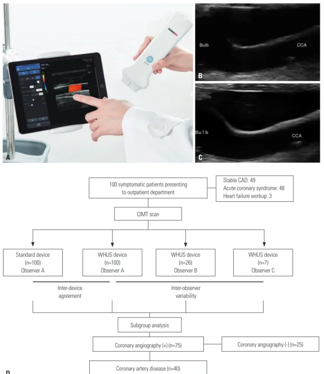
Informed consent was obtained from all enrolled patients. In- clusion criteria were as follows: 1) age ≥18 years and 2) pres- ence of anginal symptoms or dyspnea regardless of the history of underlying CAD. Exclusion criteria were as follows: 1) de- bilitating underlying comorbidities, such as cancer, end stage heart failure, and aortic dissection, and 2) structural deformi- ties in the neck that hampered ultrasonographic access to the carotid arteries. All enrolled patients underwent carotid ultra- sound scans by both the WHUS (Fig. 1A and B) and a standard device (Fig. 1C). Detailed information regarding enrollment is shown in Fig. 1D.
Ultrasonographic assessment of CIMT
All 100 patients underwent carotid scans by a standard ultra- sonographic device (Alpha 7, Aloka, Tokyo, Japan) with a 12- mHz linear probe (UST-5412, Aloka) and the WHUS (SONON 300L, Healcerion, Seoul, Korea) device equipped with a built- in 10-mHz linear probe. The left and right longitudinal projec- tion of the common carotid artery (CCA), the carotid bulb, and the internal carotid artery were observed where the site of the greatest thickness (excluding the plaque lesion) was measured bilaterally along both the near and far walls. The mean value of maximal CIMT measured from four sites (left/right CCA and bulb) was defined as max-CIMT17 and was investigated for its association with CAD. Carotid plaque was defined as a focal region either with more than 50% or 0.5-mm thickening of the
CIMT, compared with the surrounding tissue. The CIMT of the CCA was measured 1 cm proximal to the bifurcation, whereas the CIMT of the bulb was examined 1 cm distal to the bifurca- tion. One highly trained and experienced sonographer per- formed all of the exams for both the standard and the WHUS device. The acquired images were then measured once manu- ally through a standard computer monitor (SyncMaster 2333, Samsung, Seoul, Korea). A total of three observers were allocat- ed for interdevice and interobserver variability. As shown in Fig. 1D, one observer was designated for interdevice variabili- ty from all 100 patients (observer A), while the two other inde- pendent observers (observers B and C) measured max-CIMT for the assigned 26 patient images and 7 patient images, respec- tively, that were attained from the WHUS device to assess in- terobserver variability. All observers were experienced sonog- raphers and were unaware of each patient’s clinical information or status.
Coronary angiography
For subgroup analysis, 75 patients with stable angina with a positive stress test, acute coronary syndrome (ACS), or indica- tion for coronary evaluation during heart failure workup under- went coronary angiography (CAG). CAG was performed using standard Judkins or Amplatz diagnostic catheters. Angiograph- ic CAD was defined as moderate (50–70%) stenosis or severe (≥70%) CAD or a vessel with a previous stent. Minimal athero- sclerosis was defined as <50% stenosis of the coronary arteries.
Statistical analysis
Data analysis was performed using IBM SPSS Statistics for Win- dows (Version 23.0, IBM Corp., Armonk, NY, USA). Continu- ous, normally distributed data are expressed as a mean±SD. A paired sample t-test was used to compare differences in max- CIMT values between the standard device and the WHUS de- vice. Categorical values are expressed as percentages. The dif- ference between the WHUS and the standard device was defined as max-CIMT of WHUS subtracted by the max-CIMT of the standard device. The Bland-Altman plot was used to assess in- terdevice agreement and interobserver repeatability. Linear logistic regression was utilized to determine proportional bias between the two devices. Interdevice repeatability and interob- server variability were examined by Pearson’s correlation and intraclass correlation coefficient (ICC). The correlation coeffi- cient of Pearson’s correlation was denoted as R. Student’s one- way ANOVA test was applied to compare intergroup differenc- es in CIMT values in patients with different numbers of pathologic coronary arteries. Predictive power for clinical factors with or without max-CIMT or plaque information was tested by the re- ceiver operating characteristic (ROC) curves. The optimal cut- off value for max-CIMT in predicting CAD was also examined using the ROC curves.
RESULTS
Baseline characteristics
A total of 100 patients with cardiac symptoms, such as chest pain, dyspnea, palpitation, and syncope, were enrolled. As shown in Table 1, the mean age of the total patient population was 65±15 years. Men comprised 66% of the study population.
The prevalences of hypertension, diabetes, and dyslipidemia were 59, 30, and 31%, respectively. Of all patients, 75 were in-
dicated for CAG (Table 1).
Agreement between the standard device and WHUS device
The comparison between the standard device and the WHUS device is shown in Table 2. The max-CIMT of the standard de- vice ranged from 0.70 mm to 2.80 mm, with a mean value of 1.447±0.443 mm. The max-CIMT of the WHUS ranged from 0.72 mm to 2.50 mm, and the mean value was 1.289±0.393 mm.
Fig. 1. Images of the WHUS device, carotid scans from both devices, and details on patient enrollment. (A) The WHUS device can be connected to cell- phones or tablets through its built-in Wi-Fi feature from virtually anywhere. The carotid bulb and CCA of the same patient acquired by the (B) WHUS and (C) standard device. (D) The carotid intima-media thickness of a total of 100 patients with cardiac symptoms was evaluated both by the standard and WHUS devices. WHUS, wireless handheld ultrasound; CIMT, carotid intima-media thickness; CAD, coronary artery disease; CCA, common carotid artery.
A
B
C
Standard device (n=100) Observer A
Inter-device agreement
Subgroup analysis
Coronary artery disease (n=40)
Coronary angiography (+) (n=75) Coronary angiography (-) (n=25) Inter-observer
variability 100 symptomatic patients presenting
to outpatient department
Stable CAD: 49
Acute coronary syndrome: 48 Heart failure workup: 3
WHUS device (n=100) Observer A
CIMT scan
WHUS device (n=26) Observer B
WHUS device (n=7) Observer C
D
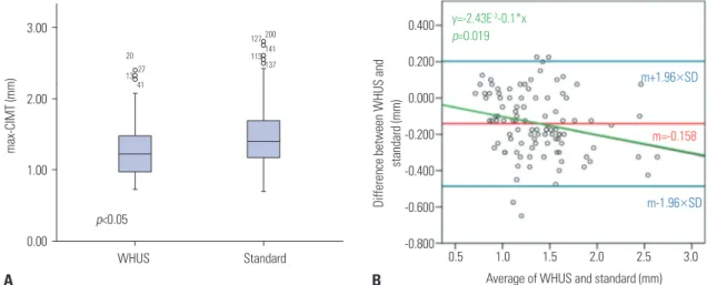
The absolute difference between two devices was -0.158±0.228 mm (p<0.05) by the paired sample t-test, although Pearson’s correlation coefficient and ICC were both above 0.9, suggestive of excellent interdevice correlation and agreement (Table 3, Fig. 2A).
Repeatability of the WHUS device
Data regarding interobserver variability of the WHUS is shown in Table 3. The max-CIMT between three observers were comparable, and the ICC for the interobserver variability was over 0.9, suggesting excellent agreement between observers (Table 3). In the Bland-Altman plot, the red middle line repre- sents the mean value (m=-0.158 mm) of the difference be- tween two devices, whereas the two blue lines indicates the 1.96×SD above and below the mean value (Fig. 2B). A linear equation (y=-2.43E-3-0.1*x) that was derived from linear logis- tic regression, shown as a green line, suggested that the differ- ences between two devices were negatively proportional (p=
0.019) to the average max-CIMT values between two devices:
the discrepancy between two devices was bigger with decreas- ing max-CIMT (Fig. 2B).
CIMT and number of diseased coronary arteries The max-CIMT of patients with different numbers of diseased coronary arteries is shown in Supplementary Table 1 (only on-
line) and Fig. 3. A significantly increasing trend of max-CIMT with increasing number of diseased coronary arteries was ob- served (p=0.004).
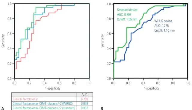
Predictive power for CAD using ROC curves
As a subgroup analysis, patients who underwent CAG were evaluated to analyze the predictive power of WHUS-measured max-CIMT. The baseline characteristics between CAD (+) and CAD (-) groups are presented in Table 1. Except for age, base- line characteristics between the CAD (+) and CAD (-) groups were comparable.
ROC curves constructed using conventional clinical risk fac- tors (age, hypertension, diabetes, smoking, and dyslipidemia) with or without max-CIMT and carotid plaque information are shown in Fig. 4A. The area under the curve (AUC) for the clini- cal risk factors only was 0.769. When CIMT information (max- CIMT and plaque ≥2) was added to clinical factors, the AUC was increased to 0.838 and 0.867 for WHUS and the standard device, respectively.
The AUC and cutoff value of max-CIMT for predicting CAD are presented in Fig. 4B. The AUC values of the standard device and the WHUS device were 0.807 and 0.725, respectively, while the cutoff values were 1.05 and 1.10 mm, respectively. The WHUS device had a higher sensitivity (0.976 to 0.781) and lower speci- ficity (0.265 to 0.781) than the standard device (Table 4). The es- Table 1. Baseline Characteristics of Patients with or without CAD
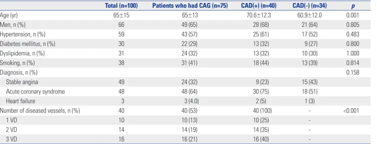
Total (n=100) Patients who had CAG (n=75) CAD(+) (n=40) CAD(-) (n=34) p
Age (yr) 65±15 65±13 70.6±12.3 60.9±12.0 0.001
Men, n (%) 66 49 (65) 28 (68) 21 (64) 0.805
Hypertension, n (%) 59 43 (57) 25 (61) 17 (52) 0.483
Diabetes mellitus, n (%) 30 22 (29) 13 (32) 9 (27) 0.800
Dyslipidemia, n (%) 31 24 (32) 13 (32) 10 (30) 1.000
Smoking, n (%) 38 31 (41) 18 (44) 13 (39) 0.814
Diagnosis, n (%) 0.158
Stable angina 49 24 (32) 9 (23) 15 (43)
Acute coronary syndrome 48 48 (64) 30 (75) 18 (51)
Heart failure 3 3 (4.0) 2 (5) 1 (3)
Number of diseased vessels, n (%) 40 40 (53) 40 (100) - <0.001
1 VD 10 10 (13) 10 (25) -
2 VD 14 14 (19) 14 (35) -
3 VD 16 16 (21) 16 (40) -
CAG, coronary angiography; CAD, coronary artery disease; VD, vessel disease.
Table 2. Measured CIMT from Three Observers and the Standard Device
Sample size (n) max-CIMT (mm) CCA CIMT (mm) Bulb CIMT (mm) Number of plaques
WHUS observer A 100 1.289±0.393 1.036±0.344 1.542±0.527 0.9±1.3
WHUS observer B 26 1.378±0.502 - - -
WHUS observer C 7 1.940±0.603 - - -
Standard (reference) 100 1.447±0.443 1.289±0.393 1.289±0.393 1.1±1.4
CIMT, carotid intima-medial thickness; max-CIMT, mean of the maximal values of carotid intima-medial thickness of a patient; WHUS, wireless handheld ultra- sound; CCA, common carotid artery.
timated numbers of patients with CAD using the cutoff values were 63 and 60 for the WHUS and standard device, respective- ly (Table 4). Both devices were able to detect 39 (97.5%) out of the 40 patients who had angiographic CAD.
DISCUSSION
In this study, we demonstrated that WHUS-measured CIMT values have great repeatability and agreement with a standard ultrasound device. The WHUS-measured CIMT information
also improved predictive power when added to the traditional CV risk factors in detecting CAD, compared with clinical CV risk factors alone. To our knowledge, this is the first study to investigate the feasibility and clinical applicability of WHUS- measured CIMT for point-of-care in symptomatic patients pre- senting to the clinic.
Our data showed that WHUS-measured CIMT results are re- producible. The ICCs between three observers, which is a ba- rometer of combined agreement and correlation, were all over 0.9, suggestive of excellent interobserver variability. This indi- cates that WHUS-generated data may be consistent between different users and therefore may be a legitimate diagnostic tool. The gap observed between the conventional and the WHUS device (Table 3, Fig. 2A) may be explained by the rela- tively blurry image quality of the WHUS in smaller CIMT val- ues (Fig. 1B), compared with the standard device (Fig. 1C). The negative slope (-2.43) of the linear regression for proportional bias suggested that the gap between two devices is wider with smaller CIMT (Fig. 2B).
Previous studies regarding the relationship between CIMT or carotid artery stenosis and CV outcomes have generally in- vestigated asymptomatic patients with or without traditional CV risk factors.5-10,18 Moreover, CIMT evaluation for CV screen- ing in such large asymptomatic populations requires many pa- tients for the purpose of preventing a single CV event. The high prices of the conventional ultrasound devices (approximate- ly 150000 USD)19 may also lead to high CIMT test costs (330 Table 3. Absolute Difference and ICC between the WHUS and the Standard Device
Sample size (n) Absolute difference (mm) pabs R pR ICC
WHUS vs. Standard (reference) 100 -0.158±0.228 <0.050 0.858 <0.001 0.960
Observer A vs. observer B 26 0.024±0.078 0.109 0.991 <0.001 0.988
Observer A vs. observer C 7 0.028±0.116 0.406 0.777 0.005 0.998
Observer B vs. observer C 7 -0.009±0.035 0.445 0.760 0.007 0.978
WHUS, wireless handheld ultrasound; ICC, intraclass correlation coefficient; pabs, p value of absolute difference; pR, p value of correlation coefficient R.
Fig. 2. Interdevice agreement and proportional bias. (A) A plot showing the CIMT of the WHUS and the standard device. (B) A Bland-Altman plot of the rela- tionship between the average of WHUS and standard device and differences between the two devices. m, mean of differences between the WHUS and standard device; max-CIMT, mean of the maximal values of carotid intima-medial thickness of a patient; WHUS, wireless handheld ultrasound.
3.00
2.00
1.00
0.00
max-CIMT (mm)
p<0.05 WHUS
20 27
200 141 127 113137 1341
Standard A
0.400 0.200 0.000 -0.200 -0.400 -0.600 -0.800 Difference between WHUS and standard (mm)
0.5 1.0 1.5 2.0 2.5 3.0 Average of WHUS and standard (mm)
m-1.96×SD m+1.96×SD y=-2.43E-3-0.1*x
p=0.019
m=-0.158
B
2.50 2.00 1.50 1.00 0.50 0.00
max-CIMT (mm)
0 1 2 3 Number of diseased arteries
p=0.004
Fig. 3. Relationship between maximal carotid intima-media thickness (max- CIMT) and the number of each patient’s diseased coronary vessels.
USD).11 Most governments or insurance companies world- wide, including the US and Korea, do not reimburse for CIMT:
the state of Texas was the only state in the US that covered CIMT for CV screening in 2009.20 Major clinical guidelines also no longer recommend routine CIMT screening for similar rea- sons.8,12 Accordingly, using a readily accessible economical WHUS device (8000 USD), which costs approximately 5% of the standard device, may dramatically lower the expenses for each CIMT exam. Evaluating symptomatic populations in the outpatient clinic may also reduce the number of patients re- quired to detect a single CV event. Reducing the device cost and focusing on symptomatic populations may collectively improve cost-effectiveness.
The WHUS device may also improve timely decision-mak- ing process adding to clinical information gathered by patient examination or history taking. For physicians, determining whether a patient is at high-risk for CAD can be challenging in an outpatient clinical setting. Our data show that WHUS-mea- sured max-CIMT and plaque information (number of plaques
≥2) may improve the precision of detecting CAD patients. As
both devices detected 39 patients (97.5%) out of 40 patients with CAD, there was no underestimation by the WHUS device, although the underestimation may have been masked by the small sample size. The high sensitivity and negative predictive value of the WHUS-generated max-CIMT may help physicians to effectively rule out patients at low risk of CAD. With its addi- tive predictability and high sensitivity, physicians may effec- tively focus their diagnostic process on higher risk patients.
These promising features, however, are only useful when the ultrasound can be readily accessible. Conventional ultrasound devices may not meet such necessities in the clinic, as they are usually heavy and bulky: it may be cumbersome to bring a con- ventional ultrasound console in and out of the clinic every time a patient with cardiac symptoms present or to send the patient to the ultrasound department. Additionally, the current WHUS device can be connected to cellphones or tablets through its built-in Wi-Fi feature, which enables physicians to evaluate CIMT of patients from virtually anywhere. Such accessibility may improve point-of-care quality for cardiologists, general practitioners, or emergency physicians not only in clinics but Table 4. Diagnostic Properties Regarding the WHUS and Standard Devices
AUC Cutoff
value (mm) Sensitivity Specificity PPV NPV Estimated number of CAD patients using cutoff
Number of true CAD
max-CIMT of standard device 0.807 1.05 0.78 0.53 0.98 0.29 60 39 (97.5%)
max-CIMT of WHUS 0.725 1.10 0.98 0.27 0.78 0.53 63 39 (97.5%)
max-CIMT, mean of maximal value of carotid intima-medial thickness of a patient; AUC, area under the curve; PPV, positive predictive value; NPV, negative pre- dictive value; WHUS, wireless handheld ultrasound; CAD, coronary artery disease.
Fig. 4. ROC curves for predicting high-risk CAD patients. (A) A plot showing the predictive power of traditional clinical risk factors alone (red), mean maximal CIMT measured from left/right CCA and bulb (max-CIMT), and carotid plaques ≥2, in addition to clinical risk factors, using the WHUS and the standard de- vice, shown in blue and green, respectively. (B) The cutoff max-CIMT values and in predicting high-risk CAD patients using the standard (1.05 mm) and WHUS (1.10 mm) device. ROC, receiver operating characteristic; AUC, area under the curve; CAD, coronary artery disease; WHUS, wireless hand-held ultrasound.
1.0
0.8
0.6
0.4
0.2
0.0
Sensitivity
0.0 0.2 0.4 0.6 0.8 1.0 1-specificity
AUC
Clinical factors only 0.769
Clinical factors+max-CIMT+plaques≥2 (WHUS) 0.838 Clinical factors+max-CIMT+plaques≥2 (standard) 0.867 A
1.0
0.8
0.6
0.4
0.2
0.0
Sensitivity
0.0 0.2 0.4 0.6 0.8 1.0 1-specificity
WHUS device AUC: 0.725 Cutoff: 1.10 mm Standard device
AUC: 0.807 Cutoff: 1.05 mm
B
in triaging systems in underprivileged regions throughout the world.
The significant proportional bias between the two devices, especially in the lower values, may indicate that the WHUS de- vice may not be able to accurately depict lower CIMT values due to lower resolution. Future efforts for fine-tuning image quality may improve the proportional bias and eventually close the gap between conventional ultrasound devices. Addition- ally, the small sample size (n=75) for CAD prediction may be a limitation of our study. The non-association between the presence of carotid plaque and CAD risk is inconsistent with previous findings, which may have been caused by the small sample size. Further investigation using a larger sample size in a more homogeneous patient population, such as patients with ACS, for identifying the association between WHUS-measured CIMT and coronary artery plaque burden using intravascular ultrasound is warranted to add precision to the device.
The current study showed that the novel WHUS device has excellent agreement with the standard ultrasound device and offers repeatability among observers. Additionally, CIMT in- formation combined with clinical CV risk factors improved predictability for detecting high-risk patients for CAD, com- pared with the CV clinical risk factors alone.
ACKNOWLEDGEMENTS
This research was partly supported by the Gachon Universi- ty Gil Medical Center (Grant number: FDR2015-02) and the Next-Generation Medical Device Development Program for Newly-Created Market of the NRF funded by the Korean gov- ernment, MSIT (No. 2015M3D5A1066043). This research was also supported by grants [2018-ER6304-00 (2018) and 2018- ER6304-01 (2019)] for research from the Korea Centers for Disease Control, Prevention and partially supported by KSC 200403-17 from the Korean Society of Cardiology, and Seoul R&D Program (BT190153) through the Research and Develop- ment for Regional Industry. Wook-Jin Chung works as a non- profit consultant for Healcerion Co., Ltd. Wook-Jin Chung and the CEO of Healcerion, Jeongwon Ryu, have jointly applied and received a research grant (MSIT No. 2015M3D5A1066043) from NRF of Korea. No fundingbodies or Jeongwon Ryu were involved in either the measurement/analysis of CIMT data of the current research project or writing/revising the manu- script. No other author reported potential conflicts of interest relevant to this article.
We thank Hanul Choi, Kyoung Min You, and Je Hyeong Park for performing ultrasonography for the study.
AUTHOR CONTRIBUTIONS
Conceptualization: Wook-Jin Chung and Jeongwon Ryu. Data cura- tion: Wook-Jin Chung, Pyung Chun Oh, and Jeunggeun Moon. Formal analysis: Albert Youngwoo Jang and Wook-Jin Chung. Funding acqui- sition: Wook-Jin Chung and Jeongwon Ryu. Investigation: Wook-Jin
Chung. Methodology: Wook-Jin Chung. Project administration:
Wook-Jin Chung. Resources: Wook-Jin Chung. Software: Wook-Jin Chung. Supervision: Wook-Jin Chung. Validation: Albert Youngwoo Jang and Wook-Jin Chung. Visualization: Albert Youngwoo Jang and Wook-Jin Chung. Writing—original draft: Albert Youngwoo Jang and Wook-Jin Chung. Writing—review & editing: Albert Youngwoo Jang and Wook-Jin Chung. Approval of final manuscript: All authors.
ORCID iDs
Albert Youngwoo Jang https://orcid.org/0000-0002-8802-268X Jeongwon Ryu https://orcid.org/0000-0002-0741-2142 Pyung Chun Oh https://orcid.org/0000-0002-9955-223X Jeonggeun Moon https://orcid.org/0000-0001-6431-2802 Wook-Jin Chung https://orcid.org/0000-0002-9767-7098
REFERENCES
1. Nambi V, Chambless L, Folsom AR, He M, Hu Y, Mosley T, et al.
Carotid intima-media thickness and presence or absence of plaque improves prediction of coronary heart disease risk: the ARIC (Ath- erosclerosis Risk In Communities) study. J Am Coll Cardiol 2010;
55:1600-7.
2. Cao JJ, Arnold AM, Manolio TA, Polak JF, Psaty BM, Hirsch CH, et al. Association of carotid artery intima-media thickness, plaques, and C-reactive protein with future cardiovascular disease and all- cause mortality: the Cardiovascular Health Study. Circulation 2007;
116:32-8.
3. Øygarden H. Carotid intima-media thickness and prediction of cardiovascular disease. J Am Heart Assoc 2017;6:e005313.
4. Zanchetti A, Hennig M, Hollweck R, Bond G, Tang R, Cuspidi C, et al. Baseline values but not treatment-induced changes in carotid intima-media thickness predict incident cardiovascular events in treated hypertensive patients: findings in the European Lacidipine Study on Atherosclerosis (ELSA). Circulation 2009;120:1084-90.
5. Kwon HJ, Park JH, Lee JH, Jeong HS, Song HJ, Kim J, et al. Low com- mon carotid artery systolic occlusion pressure and symptomatic carotid artery stenosis are associated with development of neuro- logic intolerance during proximal protected carotid artery stent- ing. Korean Circ J 2018;48:217-26.
6. Dwivedi A, Al’Aref SJ, Lin FY, Min JK. Evaluation of atherosclerotic plaque in non-invasive coronary imaging. Korean Circ J 2018;48:
124-33.
7. Bots ML, Groenewegen KA, Anderson TJ, Britton AR, Dekker JM, Engström G, et al. Common carotid intima-media thickness mea- surements do not improve cardiovascular risk prediction in indi- viduals with elevated blood pressure: the USE-IMT collaboration.
Hypertension 2014;63:1173-81.
8. Williams B, Mancia G, Spiering W, Agabiti Rosei E, Azizi M, Burni- er M, et al. 2018 ESC/ESH Guidelines for the management of arte- rial hypertension. Eur Heart J 2018;39:3021-104.
9. Costanzo P, Perrone-Filardi P, Vassallo E, Paolillo S, Cesarano P, Brevetti G, et al. Does carotid intima-media thickness regression predict reduction of cardiovascular events? A meta-analysis of 41 randomized trials. J Am Coll Cardiol 2010;56:2006-20.
10. Lorenz MW, Polak JF, Kavousi M, Mathiesen EB, Völzke H, Tuo- mainen TP, et al. Carotid intima-media thickness progression to predict cardiovascular events in the general population (the PROG- IMT collaborative project): a meta-analysis of individual partici- pant data. Lancet 2012;379:2053-62.
11. den Ruijter HM, Vaartjes I, Sutton-Tyrrell K, Bots ML, Koffijberg H.
Long-term health benefits and costs of measurement of carotid
intima-media thickness in prevention of coronary heart disease. J Hypertens 2013;31:782-90.
12. Goff DC Jr, Lloyd-Jones DM, Bennett G, Coady S, D’Agostino RB Sr, Gibbons R, et al. 2013 ACC/AHA guideline on the assessment of cardiovascular risk: a report of the American College of Cardiolo- gy/American Heart Association Task Force on Practice Guidelines.
J Am Coll Cardiol 2014;63:2935-59.
13. Kim EY, Park KH, Choi SJ, Chung WJ. Educational value of pocket- sized ultrasound devices to improve understanding of ultrasound examination principles and sonographic anatomy for medical stu- dent. PLoS One 2017;12:e0185031.
14. Lee SH, Kim YJ. Managing a seroma with wireless mobile ultra- sound device. J Plast Reconstr Aesthet Surg 2017;70:e7-9.
15. Kim J, Kim S, Jeon S, Jung S. A longitudinal study investigating cer- vical changes during labor using a wireless ultrasound device. J Matern Fetal Neonatal Med 2018;31:1787-91.
16. Oh NR, Woo JH, Kim DY, Baek MK. Wireless mobile ultrasonogra-
phy-assisted parotid duct stone removal. Ear Nose Throat J 2018;
97:E36-8.
17. Dogan S, Kastelein JJ, Grobbee DE, Bots ML. Mean common or mean maximum carotid intima-media thickness as primary out- come in lipid-modifying intervention studies. J Atheroscler Thromb 2011;18:946-57.
18. Seo J, Kim GS, Lee HY, Byun YS, Jung IH, Rhee KJ, et al. Prevalence and clinical outcomes of asymptomatic carotid artery stenosis in patients undergoing concurrent coronary and carotid angiogra- phy. Yonsei Med J 2019;60:542-6.
19. Tse KH, Luk WH, Lam MC. Pocket-sized versus standard ultra- sound machines in abdominal imaging. Singapore Med J 2014;55:
325-33.
20. Polak JF, Pencina MJ, Pencina KM, O’Donnell CJ, Wolf PA, D’Agostino RB Sr. Carotid-wall intima-media thickness and cardiovascular events. N Engl J Med 2011;365:213-21.