of Cancer Prevention 2003; 8(4): 261-266
INTRODUCTION
Stem cells are key players in the development and maintenance of specific tissues in animals, and the discovery of them in the central and peripheral nervous systems (CNS and PNS) is a relatively recent
event.1) Neural progenitor cells are maintained in the central nervous system throughout life. These pre- cursor cells migrate through the rostral migratory stream to the olfactory bulb, differentiating into granule and periglomerular neurons.2) Neural progeni- tor cells can be isolated from embryonic or adult brain, and maintained and manipulated in vitro as
Establishment of Immortal Neuronal Stem Cell Line from Embryonic Day 17 Rat Brain
Se-Ran Yang1, Sung-Dae Cho1, Nam-Shik Ahn1, Ji-Won Jung1, Joon-Suk Park1, Nguyen Ba Tiep1, Ki-Su Park1, In-Sun Hong1, Eun-Hye Jo1, Min-Su Seo1,
Byong-Su Yoon2, Yong-Soon Lee1 and Kyung-Sun Kang1
1Department of Veterinary Public Health, College of Veterinary Medicine, Seoul National University, Seoul 151-742, Korea;
2Department of Biology, College of Natural Science, Kyonggi University, Suwon, 442-760, Korea
We have recently characterized Neurosphere-derived cells from embryonic day (ED) 17 rat fetus brain. Neural stem cells (NSCs) have been shown to reside in both fetal and adult brain leading to formation of cell clusters termed “neurospheres” and the neurosphere-derived cells can differentiate into neurons, oligodendrocytes and astrocytes.
In this study, we reported the establishment of neuronal stem cell line using SV40 large T antigen. The results show that the cells were acquired extended life span and constant increase at early (∼30 cumulative population doubling level (cpdl)), middle (∼60 cpdl), and late (∼100 cpdl) passages. To characterize the established immortal cell, immuno- fluorescent staining was performed using GFAP antibody for astrocyte, NSE for neuron, and Nestin for neuronal stem cells. These results showed that the established immortal cell line expressed nestin. These results suggest that the established cell line might be a neuronal stem cells, and stem cells might be also major target cells for the carcinogenesis.
ꠏꠏꠏꠏꠏꠏꠏꠏꠏꠏꠏꠏꠏꠏꠏꠏꠏꠏꠏꠏꠏꠏꠏꠏꠏꠏꠏꠏꠏꠏꠏꠏꠏꠏꠏꠏꠏꠏꠏꠏꠏꠏꠏꠏꠏꠏꠏꠏꠏꠏꠏꠏꠏꠏꠏꠏꠏꠏꠏꠏꠏꠏꠏꠏꠏꠏꠏꠏꠏꠏꠏꠏꠏꠏꠏꠏꠏꠏꠏꠏꠏꠏꠏꠏꠏꠏ
Key Words: Immortalization, Stem cells, SV40 Large T-antigen, Nestin, Neurosphere- derived cells
Corresponding author: Kyung-Sun Kang, or Yong-Soon Lee, Department of Veterinary Public Health, College of Veterinary Medicine, Seoul National University, Silim 9-dong, Gwanak-gu, Seoul 151-742, Korea, Tel: +82-2-880-1246, Fax: +82-2- 876-7610, E-mail: kangpub@snu.ac.kr or leeys@snu.ac.kr,
Received : July 2, 2003, Accepted : September 1, 2003
either primary cultures or immortalized cell lines.3,4) In primary culture, nestin-positive cells are grown in the presence of EGF and/or FGF, generating clones of cells.5) The discovery of neural precursors with the ability both to self-renew and to generate progenitors for neurons, astrocytes, and oligodendrocytes in vitro,6) and the progenitors formed the clusters termed
“neurospheres.” A latent precursor of the neurosphere- derived cells is believed to exist in the brain (i.e.
embryonic CNS, including basal forebrain, cerebral cortex, hippocampus.7) Nestin is a 220 kDa class VI intermediate filament protein originally found to be expressed early in central nervous system (CNS) development. Initially nestin expression appears dur- ing the time of CNS stem and progenitor cell prolif- eration and neuronal migration, and subsequently decreases as the brain develops.8) Although nestin is expressed in undifferentiated CNS precursors, little information is available about the temporal expression of nestin. Nestin has also been detected in neuroect- odermal and glial cell-derived tumors.9) The maintena- nce and culture of stem cell on neurosphere is very difficult in vitro. Therefore, it is important the estab- lish stem cell line for facilitate neuronal stem cell research.
In this study, we are able to establish an immortal cell line using SV40 Large T Antigen and also show that this established cell line express a stem cell marker.
MATERIALS AND METHODS 1) Primary rat brain culture
The Pregnant Sprague-Dawly rat (Bio Genemics, Inc., Seoul, Korea) were sacrificed at embryonic day (ED) 17,10) embryos placed in a petri dish containing Hanks balanced salt solutions (HBSS, Gibco, USA).
For dissociation and plating cells, the cortex was dissected from the rest of the brain and isolated cortex transferred to a 0.5% trypsin solution (Gibco, USA).
To obtain small clumps of cells the solution was gently pipetted up and down about 20 times in 5ml pipette until it attained a milky, homogeneous appear-
ance. The suspension was incubated for 30 min at 37oC. There after 1ml of PBS containing 0.04% deox- yribonuclease (DNase, type I, 650 KU/mg, Sigma, USA) was added to the tissue. The soultion was pipetted up and down several times. Cells were plated in 100 mm dished (Nunc, Denmark) and maintained in a 1:1 mixture of Dulbeco's Modified Eagle Medium and F12 medium (DMEM/F12, Gibco, USA) supple- mented with 5% fetal bovine serum (Gibco, USA), basic fibroblast growth factor (bFGF, Boehringer mannheim, USA), epidermal growth factor (EGF).
Dishes were incubated at 37oC in humidified 5% CO2:
95% air.
2) Immortalization
The neurosphere-derived cells were transfected with a plasmid carrying an origin-defective SV40 genome expressing a wild-type large T-antigen (PRNS-1; a gift from James E Trosko, Michigan State Univ., East Lansing, MI), by DNA superfect (Qiagen, USA). The actively proliferating colonies were selected by their resistance to G418 (Gibco, USA) 0.4 mg/ml for 7 days. The proliferation potential of transformed clones was determined by their total cumulative population doubling level (cpdl) using formula cpdl=ln (Nf/
Ni)ln2, where Ni and Nf are initial and final cell numbers, respectively, and ln is the natural log.11) The initial cell number was 2×10⁵ for each propagation.
During the course of determining the potential cpdl for each SV40-transformed cell line, the populations of cells at different cpdls were preserved in liquid nitrogen. To measure a constant increase, the cells at early (22∼30 cpdl), middle (50∼60 cpdl), and late (100∼110 cpdl) passages were grown and harvested to prepare cell lysates.
3) Immunocytochemistry
The procedures were slightly modified from the method described previously. Cells were grown on eight well Lab-Tek slide (Nunc, USA). Cells on the slide were fixed in 4% paraformaldehyde (in PBS, pH 7.2) for 30 min at room temperature followed by 3
washed in PBS. Cells were the permeabilized for 30 sec in 3% Triton-X and washed in PBS, and blocked for 1 hr in PBS containing 10% normal goat serum (NGS) (Zymed, USA). After blocking, slides were incubated in anti-nestin mouse monoclonal (Phar- mingen, USA), anti-glial fibrillary acidic protein (GFAP) rabbit polyclonal, and rabbit anti-neuron specific enolase (NSE) diluted in PBS containing 1%
NGS for 2 hr at room temperature. Slides were then washed in PBS 3 times and incubated in TRITC goat anti-rabbit or mouse (Zymed, CA, USA) secondary antibodies for 1 hr at room temperature. Slides were washed 3 times in PBS and mounted in Gelvatol (Lab vision, USA).
Fig. 2. Proliferation potential of SV40-transformed cell line using the cumulative population doubling level (cpdl) formula.
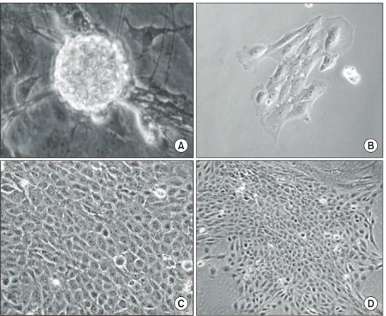
Fig. 1. Morphology of neurosphere-derived cells (A) and formation of colony selected by G418 after transfected with SV40 large T antigen (pRNS-1) (B∼D).
A D C
A B
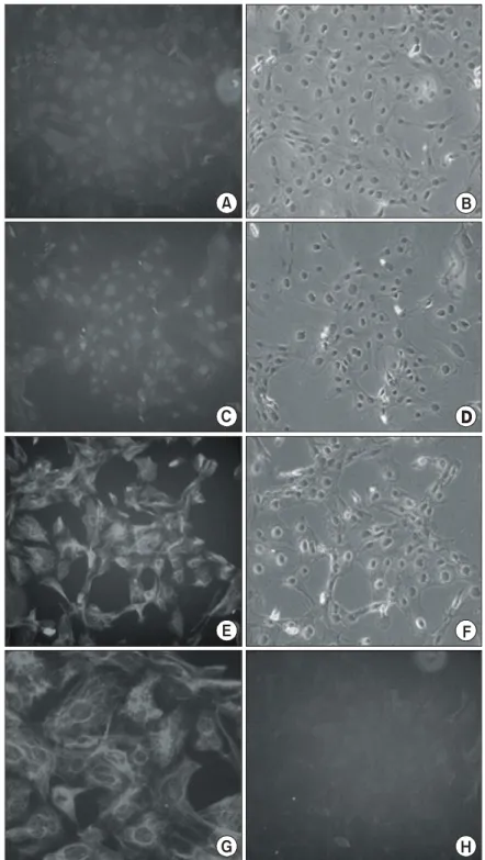
Fig. 3. Characterization of immortalized cells. Immortalized cells exhibited immunoreactivity for Nestin (E); magnification of Nestin-positive cells (G), not NSE (A) and GFAP (C). Phase-contrast for each antibodies (B, D, F) and control (H).
A B
G H
C
E F
RESULTS 1) Differentiation of neurospheres
Cells derived from the dissociation of embryonic day (ED) 17 rat brain were in presence of bFGF, EGF, and 5% fetal bovine serum (FBS, Gibco, USA) leading to the formation of neurospheres. Until 72 h, neurospheres were shown the largest sphere formation and the highest number, whereas over 72 h, the number of neurospheres was decreased and at the same time, spheres started to exhibiting outgrowth of process and differentiated (Fig. 1A).
2) Development of SV40 Large T-antigen- transformed neurosphere-derived cells
We next determined neurosphere-derived cells were transfected with an origin-defective SV40 genome expressing the wild-type large T-antigen (PRNS-1) and selected by G418. Three independent clones were isolated from neurosphere-derived cells, however, 2 clones of them were unable to grow more than 20 cpdl. A selected clone proliferate larger colony (Fig.
1B∼D), and showed a constant increase for all each early (cpdl∼30), middle (cpdl∼60), and late stages (cpdl >∼100) (Fig. 2). Therefore, this clone was able to reach to immortal stage.
3) Characterization of immortalized cells
To characterize the immortalized cells transfected with SV40 large T antigen, the cells were plate on chamber slides and processed for immunocytochemis- try. Cells were immunoreactive for nestin (Fig. 3E and 3G), whereas didn't express the immunoreactivity for neuron specific enolase (NSE) (Fig. 3A) and glial fibrillary acidic protein (GFAP) (Fig. 3C).
DISCUSSION
EGF and FGF has been shown to induce the proliferation of multipotent precursor cells from either embryonic or adult mouse striatum, leading to forma-
tion of cell clusters termed neurospheres.3)
A number of growth factors support the prolifer- ation of neural precursor cells and the differentiation of their progenitors, they contain, in addition to neurospheres expressed Nestin, in differentiate state of differentiation. Studies that sought the influence of extrinsic factors on the differentiation of precursors, clearly show that the origin of these cells and the conditions in which they have been cultured, modulate the differentiation process and the effect of growth factors,12,13) and these neurospheres differentiated into neurons, oligodendrocytes, and astrocytes. Here, we have used the SV40 large T-antigen to immortalize primary rat neurosphere-derived cells. These cells could reach to grow at ∼100 cpdl. This results suggest that the established cells were in immortal stage. Normal cells can not grow until ∼100 cpdl.
And these cells were positive for the expression of neuronal stem cell marker, Nestin. Several studies have reported adenoviral infection of neural progenitor cells transplanted into embryos differentiated into neurons, oligodenderocytes, and astrocytes.14) Hughes and co-workers demonstrated neurosphere-derived cells up to 800 um from the injection site in the rostral-caudal direction at all time points tested.15) Nestin-positive cell line originated form neurospheres could have the potential to replace degenerating cells, and may allow a more complete therapy for neuro- degenerative disorders study. The availability of an immortalized line representing the Nestin-positive cells of neurosphere-derived cells should be of great value to help define biological function of these cells and their tissue distribution.
ACKNOWLEDGEMENTS
This work was supported by a grant (R01-2000- 000-00213-0) of the Korea Science & Engineering Foundation and by a grant (01-PJ10-PG8-01EC01- 0007) of the Korea Health 21 R&D Project, Mistry of Health & Welfare, Republic of Korea. This work was also partly supported by Institute for Veterinary
Science from Seoul National University of Korea.
REFERENCES
1) Mahendra S. Rao. Stem cells and CNS development, Humana Press, Nw Jersey 2001; pp 1-19.
2) Altman J. Autoradiographic and histological studies of postnatal neurogenesis. J Comp Neurol 1969; 137:
433-458.
3) Reynolds BA, Weiss S. Generation of neurons and astrocytes from isolated cells of the adult mammalian central nervous system. Science 1992; 255: 1707-1710.
4) Bae MH, Song HS, Bae SK, Lee SK, Lee SW, Kim CW, Kim KW. Regulation of transcriptional activity of insulin-like growth factor II (IGF-II). J Cancer Prevention 2002; 7(1): 27-33.
5) Gage FH, Coates PW, Palmer TD, Kuhn HG, Fisher LJ, Suhonen JO, Peterson DA, Suhr ST, Ray J.
Survival and differentiation of adult neuronal progen- itor cells transplanted to the adult brain. Proc Natl Acad Sci USA 1995; 92: 11879-11883.
6) Cattaneo, E, MaKay R. Proliferation and differen- tiation of neuronal stem cells regulated by nerve growth factor. Nature 1990; 347: 762-765.
7) Johe KK, Hazel TG, Muller T, Dugich-Djordjevic MM, and McKay RD. Single factors direct the differentiation of stem cells from the fetal and adult central nervous system. Genes Dev 1996; 10: 3129-3140.
8) Dahlstrand J, Lardelli M, and Lendahl U. Nestin mRNA expression correlates with the central nervous
system progenitor cell state in many, but not all, regions of developing central nervous system. Brain Res Dev Brain Res 1998; 84: 109-129.
9) Tohyama T, Lee VM, Rorke LB, Marvin M, McKay RD, Trojanowski JQ. Nestin expression in embryonic human neuroepithelium and in human neuroepithelial tumor cells. Lab Invest 1992: 66; 303-313.
10) Kang KS, Park JE, Ryu DY, and Lee YS. Effects and neuro-toxic mechanism of 2,2',4,4',5,5'-hexachlorob- iphenyl and endosulfan in neuronal stem cells. J Vet Med Sci 2001; 63: 1183-1190.
11) Sun W, Kang KS, Morita I, Trosko, JE, Chang CC.
High susceptibility of a human breast epithelial cell type with stem cell characteristics to telomerase acti- vation and immortalization. Cancer Res 1999; 59:
6118-6123.
12) Alain T. Angiogenesis and P-glycoprotein: Their roles in cancer. J Cancer Prevention 2001; 6(4): 248-255.
13) Castillo L, Martinez L, Grygar E, Hutter-Paier B.
Characterization of proliferation and differentiation of EGF-responsive striatal and septal precursor cells. Int J Dev Neurosci 2003; 21: 41-47.
14) Brustle O, Choudhary K, Karram K, Huttner A, Murray K, Dubois-Dalcq M, McKay RD. Chimeric brains generated by intraventricular transplantation of fetal human brain cells into embryonic rats. Nat Bio- technol 1998; 16: 1040-1044.
15) Hughes SM, Moussavi-Harami F, Sauter SL, Davidson BL. Viral-mediated gene transfer to mouse primary neural progenitor cells. Mol Ther 2002; 5: 16-24.