대한안과학회지 제 49 권 제 3 호 2008 J Korean Ophthalmol Soc 49(3):514-518, 2008 DOI : 10.3341/jkos.2008.49.3.514
급성림프구성백혈병환자에서 동종골수이식 후 발생한 골수이식망막병증
조영준․왕선진․김정열 충남대학교 의과대학 안과학교실
목적 : 급성림프구성백혈병으로 동종 골수이식을 시행받고 18개월이 경과한 후 골수이식망막병증이 발생한 예가 있어
보고하고자 한다.
증례요약 : 20세의 남자환자가 급성림프구성백혈병으로 골수이식을 받고 18개월 후 좌안의 시력저하를 주소로 내원하
였다. 교정시력은 우안 1.0, 좌안 0.6이었다. 안저검사에서 양안에 다수의 면화반 및 점상출혈이 있었고, 좌안에 황반 부종이 관찰되었다. 형광안저혈관조영에서 좌안 상비측에 모세혈관비관류가 관찰되었다. 4개월 후 좌안 교정시력이 0.3으로 감소되었고 안저검사상 신생혈관이 관찰되었으며, 형광안저혈관조영에서 증가된 모세혈관비관류, 신생혈관, 그리고 황반부 허혈을 확인할 수 있었다. 신생혈관 및 모세혈관비관류가 있는 부위에 2차례에 걸쳐 레이저광응고술을 시행하였다. 1년 후 좌안 교정시력 0.6으로 다소 회복되었으나 형광안저혈관조영에서 황반부 허혈의 부위에는 변함이 없었고, 새로운 부위에 신생혈관 및 모세혈관비관류가 관찰되어 2회의 추가 레이저광응고술을 시행하였다.
<한안지 49(3):514-518, 2008>
<접수일 : 2007년 5월 15일, 심사통과일 : 2007년 9월 21일>
통신저자 : 김 정 열
대전시 중구 대사동 640 충남대학교병원 안과
Tel: 042-280-8433, Fax: 042-255-3745 E-mail: kimjy@cnu.ac.kr
* 본 논문의 요지는 2007년 대한안과학회 제97회 춘계학술대회 에서 포스터로 발표되었음.
백혈병은 백혈병세포가 직접 안조직에 침착되어 문 제를 일으키기도 하지만, 빈혈, 혈액점도증가, 혈소판 감소증과 같은 혈액학적인 이상도 눈에 영향을 미친다.
또한 백혈병의 치료로 사용되는 항암화학요법, 방사선 요법, 골수이식도 눈에 악영향을 끼칠 수 있다.1
골수이식의 전신적 부작용으로는 혈액무형성증으로 인한 출혈과 감염, 면역억제로 인한 부작용 등이 있다.2 골수이식 후 발생할 수 있는 안과적 문제로 전안부합병 증으로는 이식편대숙주반응에 의한 건성각결막염, 백내 장, 각막감염 등이 있으며,3-7 후안부 합병증으로는 흔 하지는 않지만, 유리체 출혈 또는 망막출혈, 미세혈관 장애 망막병증, 시신경 유두부종, 감염 등이 있다.1,8-11 본 증례에서는 항암화학요법과 방사선 요법 후 동종
골수이식을 시행받은 환자에서 18개월 후 황반부종과 모세혈관비관류를 동반하는 골수이식망막병증이 발생 한 환자가 있어 이를 보고하고자 한다.
증례보고
18개월 전에 급성림프구성백혈병으로 동종골수이식 을 받은 20세 남자환자가 좌안 시력저하를 주소로 본원 안과에 내원하였다.
이 환자는 4년 전 급성림프구성백혈병으로 진단받고 Vincristine, Adriamycin, Cytarabine, Etopo- side로 6개월간의 전신항암화학요법 후 완전관해되었 다. 항암요법 15개월 후 중추신경계 재발이 발생하여 Methotrexate, Cytarabine을 뇌척수액내에 2회 주사하고, 두경부 방사선요법을 200 cGy로 12회 조사 받았다. Vincristine, Daunorubicin, Cytarabine 으로 전신항암요법을 재시행하여 2차 관해를 유도한 후 동종골수이식을 받았다. 골수이식을 시행받고 2개월 후 이식편대숙주반응이 피부에 발생하여 지속적으로 Cy- closporine을 복용하였다.
내원시 교정시력은 우안 1.0, 좌안 0.6이었다. 양안 모두 대광반사는 정상 반응을 보였고, 구심동공운동장 애는 없었다. 골드만압평안압계로 측정한 안압은 우안
= 증례보고 =
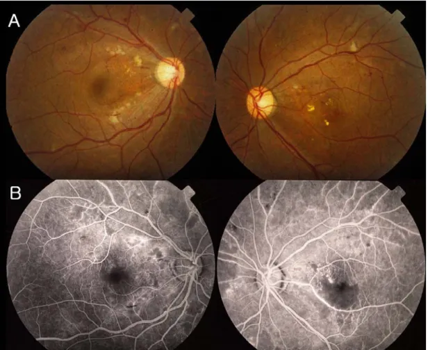
Figure 1. Fundus photographs and fluorescein angiographs at the initial visit. (A) Fundus photographs show multiple cotton- wool spots and dot-like hemorrhages. (B) Fluorescein angiographs show capillary nonperfusion in both eyes and macular ischemia in the left eye.
12 mmHg, 좌안 11 mmHg로 정상범위였다. 전안부 에는 특이소견 없었으며 안저검사에서 양안에 다수의 면화반 및 점상출혈이 있었고, 좌안에 황반부종이 관찰 되었다. 형광안저혈관조영에서 좌안 상비측에 모세혈관 비관류가 관찰되었다(Fig. 1). 4개월 후 좌안 교정시 력이 0.3으로 감소되었고 안저검사에서 신생혈관이 관 찰되었으며, 형광안저혈관조영상 증가된 모세혈관비관 류, 신생혈관, 그리고 황반부 허혈을 확인할 수 있었다.
신생혈관 및 모세혈관비관류가 있는 부위에 2차례에 걸 쳐 레이저광응고술을 시행하였다(Fig. 2). 1년 후 좌 안 교정시력은 0.8로 회복되었으나 형광안저혈관조영 에서 황반부 허혈의 부위에는 변함이 없었고, 새로운 부위에 신생혈관 및 모세혈관 비관류가 관찰되어 2회의 추가 레이저광응고술을 시행하였다(Fig. 3).
고 찰
골수이식망막병증은 골수이식 후 발생하는 합병증의
하나로서 면화반, 망막출혈, 미세혈관류, 지질삼출물, 망막신생혈관, 유리체출혈, 견인망막박리, 시신경위축, 홍채신생혈관, 신생혈관녹내장 등의 소견을 보이는 폐 쇄성 망막미세혈관병증이다.1,9-12 이러한 망막증은 방 사선조사, 고용량의 항암제사용, 이식편대숙주반응 및 이식편대숙주반응의 예방 및 치료를 위해 사용하는 면 역억제제 등이 복합적으로 작용하여 발생하는 것으로 알려져 있다.9
일부에서는 골수이식망막병증이 단순한 방사선 망막 병증의 일종이라고 얘기하기도 하지만, 방사선조사를 받지 않은 눈에서도 골수이식망막병증이 발생하기 때문 에 유일한 원인으로 볼 수는 없다.
Cunningham et al12은 방사선 요법을 시행하지 않은 백혈병 환자에서 Cytarabine과 Daunorubi- cin으로 관해 후, 동종골수이식을 시행받고 Busulfan 과 Etoposide 투여 1년 후 양안에 면화반과 망막내 출혈이 발생한 증례를 보고하였고, Johnson et al13 은 방사선 요법은 받지 않고 Cyclophosphamide,
― 조 영 준 외 : 골 수 이 식 후 골 수 이 식 망 막 병 증 ―
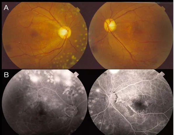
Figure 2. Fundus photographs and fluorescein angiographs at 4 months after the initial visit. (A) Fundus photographs show new vessels. (B) Fluorescein angiographs show increased capillary nonperfusion and leakage from new vessels in both eyes.
Cisplatin을 고용량 투여받은 자가조혈모세포이식을 받은 환자 140명 중 34명에서 면화반, 망막출혈, 시신 경유두부종이 발생한 것을 보고한 바 있다. 이는 방사 선 조사가 없어도 항암화학요법과 골수이식이 망막혈관 에 독성을 나타낼 수 있음을 보여주는 것이라 하겠다.
방사선 조사량과 망막증의 발생에 관한 많은 연구가 보고되어 왔는데 보통 3,000 cGy 이하의 방사선 요 법으로는 방사선 망막병증의 발생은 드문 것으로 알려 져 있고,14 6,000 cGy의 방사선 요법 후에는 50%가, 7,000~8,000 cGy 후에는 85-95%의 환자가 방사선 망막병증으로 진행한다는 보고가 있다.15 본 증례는 방 사선 조사량이 2,400 cGy로 비교적 적은 양의 방사선 이 조사되었으므로 방사선 요법 단독에 의한 것이라기 보다는 다른 요소들과 병합되어 망막증이 발생하였다고 볼 수 있다.
이식편대숙주반응은 골수이식 후 흔하게 발생하는 부 작용으로 만성 안구 이식편대숙주반응은 60~90%까 지 발생하는 것으로 보고되어 있다.3,4 안과적으로 건성 각결막염, 공막염, 토안, 망막미세혈관질환 등을 일으 킬 수 있으며,5-7 안증상의 치료를 위해 인공누액, 스테 로이드 및 Cyclosporine 점안액 등을 보조적으로 사
용하며, 전신적으로 장기간 Cyclosporine을 투여한다.
Cyclosporine은 골수이식 후 이식편대숙주반응의 억제를 위해 가장 널리 쓰이는 약물로서 신경독성, 이 를테면 경련, 구음장애, 뇌피질맹, 뇌압상승, 시신경유 두부종을 일으키는 것으로 알려져 있다.8 Coskuncan et al2은 동종골수이식을 받은 환자 372명을 대상으로 한 연구에서 8명(2%)이 Cyclosporine으로 인한 것 으로 추정되는 양안 시신경유두부종이 발생한 것을 보 고하였다. 또한 전신방사선 요법과 Cyclosporine의 병합시 허혈성 망막병증이 일어난 예가 수차례 보고되 었다.9-11,16,17
이 밖에 골수이식 후 생기는 합병증으로는 면역력의 저하로 인한 안구 감염,2 거대세포바이러스18나 대상포 진 바이러스에 의한 망막염,19 candida나 aspergi- llus에 의한 망막염과 안내염도 보고되고 있다.20 또한 골수이식 후 고용량의 스테로이드, 스트레스, 고혈압, Cyclosporine과 연관되어 드물게 중심성 장액 맥락망 막병증이 발생하였다는 보고도 있다.21-23
본 증례에서 골수이식망막병증은 중추신경계 재발의 치료로 시행한 두경부 방사선 요법, 백혈병 관해를 위 해 사용한 Vincristine, Cytarabine 등의 항암요법,
Figure 3. Fundus photographs and fluoresein angiographs at 1 year after the initial visit. (A) Fundus photographs show new vessels in the other areas. (B) Fluoresein angiographs show leakage from new vessels.
이식편대숙주반응 및 이식편대숙주반응의 치료를 위해 사용한 Cyclosporin이 복합적으로 작용하여 허혈성 망막병증을 일으킨 것으로 생각된다.
결론적으로 골수이식 후 드물지만 진행성의 망막허 혈을 보이는 골수이식망막병증이 발생할 수 있음을 알 수 있었고, 골수이식 후 주기적인 안과적 검사가 필요 할 것으로 생각된다.
참고문헌
1) Jack MK, Hicks JD. Ocular complications in high-dose che- moradiotherapy and marrow transplantation. Ann Ophthalmol 1981;13:709-11.
2) Coskuncan NM, Jabs DA, Dunn JP, et al. The eye in bone marrow transplantation.Ⅵ. Retinal complications. Arch Oph- thalmol 1994;112:372-9.
3) Fisher VL. Long-term follow-up in hematopoietic stem cell transplant patient. Pediatr Transplant 1999;3:122-9.
4) Franklin RM, Kenyon KR, Tutschka PJ, et al. Ocular mani- festations of graft-vs-host disease. Ophthalmology 1983;90:4- 13.
5) Kim RY, Anderlini P, Naderi AA, et al. Scleritis as the initial
clinical manifestation of graft versus host disease after allogenic bone marrow transplantation. Am J Ophthalmol 2002;133:843-5.
6) Arocker-Mettinger E, Skorpik F, Grabner G, et al. Mani- festations of graft-versus-host disease following allogenic bone marrow transplantation. Eur J Ophthalmol 1991;1:28-32.
7) Anderson NG, Regillo C. Ocular manifestations of graft- host-versus disease. Curr Opin Ophthalmol 2004;15:503-7.
8) Avery R, Jabs DA, Wingard JR, et al. Optic disc edema after bone marrow transplantation. Possible role of cyclosporine toxicity. Ophthalmology 1991;98:1294-301.
9) Bernauer W, Gratwohl A, Keller A, Daicker B. Microvascul- opathy in the ocular fundus after bone marrow transplantation.
Ann Intern Med 1991;115: 925-30.
10) Gloor B, Gratwohl A, Hahn H, et al. Multiple cotton wool spots following bone marrow transplantation for treatment of acute lymphatic leukaemia. Br J Ophthalmol 1985;69:320-5.
11) Lopez PF, Sternberg P Jr, Dabbs CK, et al. Bone marrow transplant retinopathy. Am J Ophthalmol 1991;112:635-46.
12) Cunningham ET Jr, Irvine AR, Rugo HS. Bone marrow transplantation retinopathy in the absence of radiation therapy.
Am J Ophthalmol 1996;122:268-70.
13) Johnson DW, Cagnoni PJ, Schossau TM, et al. Optic disc and retinal microvasculopathy after high-dose chemotherapy and
― 조 영 준 외 : 골 수 이 식 후 골 수 이 식 망 막 병 증 ―
autologous hematopoietic progenitor cell support. Bone Mar- row Transplant 1999;24:785-92.
14) Perrers-Taylor M, Brinkley D, Reynolds T. Choroido-retinal damage as a complication of radiotherapy. Acta Radiol Ther Phys Biol 1965;3:431.
15) Nakissa N, Rubin P, Strohl R. Keys H. Ocular and orbital complications following radiation therapy of paranasal sinus malignancies and review of literature. Cancer 1983;51:980-6.
16) Vogler WR, Winton EF, Helffner LT, et al. Ophthalmological and other toxicities related to cytosine arabinoside and total body irradiation as preparative regimen for bone marrow transplantation. Bone Marrow Transplant 1990;6:405-9.
17) Tiscelli A. Late ocular complications after bone marrow trans- plantation. Nouv Rev Fr Hematol 1994;36:S79-82.
18) Wingard JR, Piantadosi S, Burns WH, et al. Cytomegalovirus infections in bone marrow transplant recipients given intensive cytoreductive therapy. Rev Infect Dis 1990;12:793-804.
19) Walton RC, Reed KL. Herpes zoster ophthalmicus following bone marrow transplantation in children. Bone Marrow Trans- plant 1999;23:1317-20.
20) Rao NA, Hidayat A. A comparative clinicopathologic study of endogenous mycotic endophthalmitis: variations in clinical and histopathologic changes in candidiasis compared to aspergil- losis. Trans Am Ophthalmol Soc 2000;98:183?93.
21) Karashima K, Fujioka S, Harino S. Two cases of central serous chorioretinopathy treated with photocoagulation after bone marrow transplantation. Retina 2002;22:651-3.
22) Fawzi AA, Cunningham ET Jr. Central serous chorioretino- pathy after bone marrow transplantation. Am J Ophthalmol 2001;131:804-5.
23) Cheng LL, Kwok AK, Wat NM, et al. Graft-vs-host-disease- associated conjunctival chemosis and central serous choriore- tinopathy after bone marrow transplant. Am J Ophthalmol 2002;134:293-5.
=ABSTRACT=
Bone Marrow Transplantation Retinopathy in a Patient with Acute Lymophocytic Leukemia Following Bone Marrow Transplantation
Young Joon Jo, M.D., Seon Jin Wang, M.D., Jung Yeul Kim, M.D.
Department of Ophthalmology, Chungnam National University College of Medicine, Daejeon, Korea
Purpose: To report a case in of a patient who developed bone marrow transplantation retinopathy at 18 months after receiving allograft bone marrow transplantation for acute lymphocytic leukemia.
Case summery: A 20-year-old male patient complained of a decrease in visual activity in his left eye 18 months after receiving a bone marrow transplantation for acute lymphocytic leukemia. The corrected visual activity was 1.0 for the right eye and 0.6 for the left. On fundus examination, both eyes showed cotton wool patches and dot hemorrhage, and the left eye showed macula edema. On fluorescein angiography, capillary nonperfusion was observed in the superior nasal area of the left eye. Four months after initial examination, the corrected visual activity of the left eye decreased to 0.3 and neovascularization was observed on fundus examination. On fluorescein angiography, capillary nonperfusion, neovascularization, and macular ischemia were observed. Laser photocoagulation was performed twice on the area with neovascularization and capillary nonperfusion. One year later, the corrected visual activity of the left eye recovered to 0.8. However, the area of macular ischemia on fluorescein angiography showed no change, and neovascularization and capillary nonperfusion were observed in new areas, which were treated with two additional laser photocoagulations.
J Korean Ophthalmol Soc 49(3):514-518, 2008
Key Words: Acute lymphocytic leukemia, Bone marrow transplantation retinopathy
Address reprint requests to Jung Yeul Kim, M.D.
Department of Ophthalmology, Chungnam National University Hospital
#640 Daesa-dong, Jung-gu, Daejeon 301-721, Korea
Tel: 82-42-280-8433, Fax: 82-42-255-3745, E-mail: kimjy@cnu.ac.kr