pISSN 2234-3180 / eISSN 2234-2591 http://dx.doi.org/10.12771/emj.2013.36.1.35
Microarray Analysis after Intravenous Immunoglobulin Treatment in Patients with Kawasaki Disease
Hyo Yeon Lee, Jung Hyun Kwon, Hae Soon Kim, Sejung Sohn, Young Mi Hong
Department of Pediatrics, School of Medicine, Ewha Womans University, Seoul, Korea
Objectives: The etiology for Kawasaki disease (KD) remains unknown, but several studies have suggested the involvement of immune dysregulation and genetic factors. The purpose of this study is to compare gene ex- pressions before and after an infusion of intravenous immunoglobulin (IVIG) in KD patients.
Methods: Blood was obtained from both acute and sub-acute phases of 4 patients with KD and febrile control children. Blood was collected in PAXgene blood RNA tubes and RNA was extracted using a PAXgene blood RNA isolation kit. Labeled RNAs were analyzed using Roche NimbleGen human whole genome 12-plex array.
Results: KD patients prior to IVIG injection showed more than a two-fold increase in the expression of 88 genes and more than a two-fold decrease in the expression of 98 genes compared to the control group. They also showed more than two-fold increase in the expression of 226 genes and more than a two-fold decrease in 117 genes in KD patients after IVIG treatment compared to the patients before IVIG injection. Through microarray evalua- tion, the expressions of genes involved in proliferation, translation, inflammatory response, immune response, cell adhesion, cell migration, cell differentiation, apoptosis, cell growth, transport, cell cycle, transcription, signal transduction and metastasis were observed.
Conclusion: Changes in gene expressions in pediatric patients with KD before and after IVIG were observed via microarray evaluation. (Ewha Med J 2013;36(1):35-42)
Key Words: Gene expression; Intravenous immunoglobulin; Kawasaki disease; Microarray analysis
Received: January 21, 2013, Accepted: March 11, 2013
Corresponding author: Young Mi Hong, Department of Pediatrics, School of Medicine, Ewha Womans University, 911-1, Mok-dong, Yangcheon-gu, Seoul 158-710, Korea
Tel: 82-2-2650-2841, Fax: 82-2-2653-3718 E-mail: [email protected]
Introduction
Kawasaki disease (KD) is an acute febrile disorder characterized by systemic vasculitis of infants and chil- dren, manifested as prolonged fever and signs of muco- cutaneous inflammation which are polymorphous skin rashes, injected conjunctiva, erythematous edema in the palms and soles [1,2]. Coronary artery lesions are
the most important complication of KD [3,4]. As the current first-line therapy, IVIG, at a dose of 2 g/kg in combination with aspirin, has been shown to reduce the risk of coronary artery complications [5-7]. Even though treatment with IVIG reduces the development of aneurysm or dilatation, these are critical complication to be solved [3,8].
The etiology for KD remains unknown, however, infection, immune response, or genetic susceptibility is considered in the development of KD. The acute phase of KD demonstrates elevated serum levels of proinflammatory cytokines such as tumor necrosis fac- tor (TNF)-α, interleukins (ILs) and endothelial growth factor [6,9]. The degree of elevation of these cytokines
may be correlated with coronary aneurysms and sub- sequent stricture formation. Elevated levels of IL-1 have been reported in acute patients and have been correlated to vascular endothelial cell damage [6,10]. Also, sin- gle-nucleotide polymorphisms of inflammatory genes such as C-reactive protein (CRP) and TNF-α are asso- ciated with predisposition to KD disease and increased carotid arterial stiffness and intima-media thickness in the long-term [11]. Several studies have suggested the involvement of a genetic factor [12,13]. Matrix metal- loproteinases (MMP) is related to focal destruction of the internal elastic lamina of coronary artery and influ- ence recruitment of inflammatory cells. Therefore, MMPs play important roles in both inflammation and tissue remodeling [12,14].
The advantage of DNA microarray analysis is that it can evaluate changes in relative expression of thou- sands of genes simultaneously [15-17].
The purpose of this study was to investigate the changes of gene expressions by microarray analysis in KD patients after IVIG therapy.
Methods
The study group included acute phases for four KD patients and four febrile control children who were admit- ted to the Ewha Womans University Hospital. All patients met the criteria for the diagnostic guidelines of KD (http://www.kawasaki-disease.org/diagnostic/in- dex.html). Clinical characteristics are fever lasting 5 days and complete blood count (CBC), erythrocyte sed- imentation rate (ESR), platelet count, CRP, pro-brain na- triuretic peptide (BNP) were significantly higher in the KD group compared to the control group. All KD patients had coronary artery lesions such as dilated coronary artery or coronary artery aneurysm by echocardiography.
All patients were treated with IVIG (2 g/kg/day for 1 day) as a single infusion over 10∼12 hours. Fresh whole blood samples were obtained from KD patients pre- and post-IVIG treatment as well as febrile control group who had been febrile (body temperature >38oC) for at least 3 days. Laboratory data were obtained from each child, including CBC, ESR, platelet count, CRP, pro-BNP. And echocardiography was performed by pe-
diatric cardiologists to detect the presence of coronary artery lesions.
1. RNA extraction and cDNA synthesis
Total RNA was extracted from the blood sample that stored for 24hr at room temperature and then in the fridge (−20oC) using a PAXgene blood RNA extraction kit according to the manufacturer’s instructions. Each total RNA sample (1 μg) was labeled and amplified using Universal Linkage System (ULS) aRNA labeling kit (Kreatech diagnostics, Amsterdam, Netherlands).
2. Preparation of fluorescent DNA probe and hybridization
The Cy3-labeled aRNAs were resuspended in 10 μL of hybridization solution (GenoCheck, Ansan, Korea).
After labeled aRNA were placed on Roche Nimblegen Human whole genome 12-plex array (Roche Nimble- Gen, Inc., Madison, USA). The slides were hybridized for 12 hr at 42oC MAUI system (Biomicro systems, Inc., Salt Lake City, USA).
3. Microarray analysis
The Roche NimbleGen Human genome 12-plex ar- rays were analyzed using an Axon GenePix 4000B scan- ner with associated software (Molecular Devices Corp., Sunnyvale, USA). Gene expression levels were calcu- lated with NimbeScan Version 2.4 (Roche NimbleGen, Inc., Madison, USA). Relative signal intensities for each gene were generated using the Robust Multi-Array Average algorithm. And then the data was analyzed using GeneSpring GX 7.3.1 (Agilent technologies, Santa Clara, USA). Genes were grouped as increased or de- creased in the acute phase and also before and after an injection of IVIG. The color red indicated an over expression while green indicated a down expression.
4. Statistical analysis
An unpaired two-tailed t-test and a Mann-Whitney test were used, and a P value<0.05 was considered statistically significant. SPSS 14.0 for windows (SPSS, Chicago, USA) was used for all statistical analyses. The two-tailed t-test was used to compare patients’ samples obtained before and after IVIG therapy, and ANOVA
Table 1. Differentially expressed genes between three groups Over
expression
Down expression Pre-IVIG Tx vs. control
Post-IVIG Tx vs. control Pre-IVIG Tx vs. Post-IVIG Tx
203* (88)† 102* (77)† 289* (226)†
190* (96)† 263* (131)† 246* (117)†
*1.5-fold increase in the expression of genes. †Two-fold increase in the expression of genes. Tx, treatment.
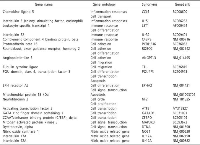
Table 2. Up-regulated genes expressed by over 50 percent between the KD patients Pre-IVIG treatment and the control group
Gene name Gene ontology Synonyms GeneBank
Chemokine ligand 5
Interleukin 5 (colony stimulating factor, eosinophil) Leukocyte specific transcript 1
Interleukin 32
Complement component 4 binding protein, beta Protocadherin beta 16
Roundabout, axon guidance receptor, homolog 2
Angiopoietin-like 3
Tubulin tyrosine ligase
POU domain, class 4, transcription factor 3
EPH receptor A2
Mitochondrial protein 18 kDa Neurofibromin 2
Activating transcription factor 3 GATA zinc finger domain containing 1 CCAAT/enhancer binding protein (C/EBP), delta Mitogen-activated protein kinase 3
Dystrobrevin, alpha Nitric oxide synthase 1 Interleukin 17A Interleukin 12A
Inflammation responses Cell transport
Inflammation responses Immune response Cell differentiation Immune response Immune response Cell adhesion Cell adhesion Cell differentiation Cell adhesion Cell migration Cell migration Cell differentiation Cell transcription Apoptosis Cell differentiation Cell signal transduction Apoptosis
Cell cycle Cell proliferation Cell transcription Cell transcription Cell transcription Cell signal transduction Cell signal transduction Nitric oxide related gene Nitric oxide related gene Nitric oxide related gene
CCL5
IL-5 LST1
IL-32 C4BPB PCDHB16 ROBO2
ANGPTL3
TTL POU4F3
EPHA2
NF2
ATF3 GATAD1 CEBPD MAP3K3 DTNA NOS1 IL-17A IL-12A
BC008600
BC066282 AF000424
BC009401 NM_000716 BC036062 NM_002942
NM_014495
BC036819 BC104923
NM_004431
NM_001003704 NM_181825
AY313927 BC031091 BC105109 BC093672 NM_001390 NM_000620 NM_002190 NM_000882 KD, Kawasaki disease; IVIG, intravenous immunoglobulin.
was used to compare pre- and post-IVIG patients with KD and the control patients.
Results
1. Comparison of microarray analysis between the KD group pre-IVIG treatment and the control group
Expressions of 393 genes in the KD group pre-IVIG treatment were significantly different to those of the control group. The KD group pre-IVIG treatment showed a 1.5-fold increase in the expression of 203 genes (two-fold increase in the expression of 88 genes) and 1.5-fold decrease in the expressions of 190 genes (two-fold decrease in the expression of 96 genes) com- pared to the control group (Table 1).
Among up-regulated genes (Table 2), three genes
(interkeukin-32, leukocyte specific transcript 1, com- plement component 4 binding protein) were related to immune responses and two genes (chemokine ligand 5, IL-5) were related to inflammation responses.
Fourteen genes (chemokine ligand 5, leukocyte specific transcript 1, protocadherin beta 16, roundabout, angio- poietin like 3, tubulin tyrosine ligase, POU domain class 4, transcription factor 3, EPH receptor A2, neuro-
Table 3. Down-regulated genes expressed by over 50 percent between Pre-IVIG in KD patients and the control group
Gene name Gene ontology Synonyms GeneBank
Major histocompatibility complex, class II, DP beta 1
Phosphoprotein associated with glycosphingolipid microdomains 1 Activation-induced cytidine deaminase
Proteasome (prosome, macropain) subunit, beta type, 8 (large multifunctional peptidase 7)
Integrin, alpha 7 Neurotrophin 3
Activation-induced cytidine deaminase
Protein disulfide isomerase family A, member 3
N-ethylmaleimide-sensitive factor attachment protein, beta Endosulfine alpha
Chloride intracellular channel 5 Polycomb group ring finger 5 Zinc finger protein 692 Zinc finger protein 10
DENN/MADD domain containing 4A c-myc binding protein
Oxoglutarate (alpha-ketoglutarate) receptor 1 Natriuretic peptide precursor C
Immune response Immune response Immune response Immune response
Cell adhesion Cell migration Cell differentiation Cell transduction Apoptosis Cell differentiation Apoptosis Transport Transport Transport Transport Transcription Transcription Transcription Transcription Transcription Signal transduction BNP related gene
HLA-DPB1 PAG1 AICDA PSMB8
ITGA7 NTF3
AICDA PDIA3
NAPB ENSA CLIC5 PCGF5 ZNF692 ZNF10 DENND4A MYCBP OXGR1 NPPC
BC007963 BC112159 NM_020661 NM_148919
NM_002206 NM_002527
NM_020661 BC014433
BC026310 BC069208 NM_016929 BC007377 CR595121 NM_015394 NM_005848 NM_012333 NM_080818 NM_024409 KD, Kawasaki disease; IVIG, intravenous immunoglobulin.
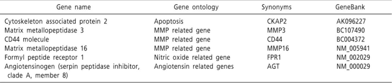
Table 4. Up-regulated genes expressed by over 50 percent between Pre- and Post-IVIG treatment in KD patients
Gene name Gene ontology Synonyms GeneBank
Cytoskeleton associated protein 2 Matrix metallopeptidase 3 CD44 molecule
Matrix metallopeptidase 16 Formyl peptide receptor 1
Angiotensinogen (serpin peptidase inhibitor, clade A, member 8)
Apoptosis MMP related gene MMP related gene MMP related gene Nitric oxide related gene Angiotensin related genes
CKAP2 MMP3 CD44 MMP16 FPR1 AGT
AK096227 BC107490 BC004372 NM_005941 NM_002029 NM_000029
KD, Kawasaki disease; IVIG, intravenous immunoglobulin.
fibromin 2, activating transcription factor 3, GATA zinc finger domain containing 1, CCAAT enhancer binding protein delta, mitogen activated protein kinase 3, dys- trobrevin, alpha) are related to cell proliferation process.
Among them, POU domain class 4, transcription factor 3 and mitochondrial protein 18 kDa are related to apoptosis. Three genes (nitric oxide synthase (NOS) 1, IL-17A, IL-12A) are nitric oxide (NO) related genes.
Eighteen genes are down-regulated compared with the control group (Table 3). Among down-regulated genes, four genes (major histocompatibility complex,
class II, DP beta 1, phosphoprotein associated with gly- cosphingolipid microdomains 1, activation-induced cy- tidine deaminase, proteasome subunit, beta type 8) are related to immune response. Neurotrophin 3 and protein disulfide isomerase family are related to apoptosis.
Thirteen genes (integrin alpha 7, neurotrophin 3, activa- tion-induced cytidine deaminase, protein disulfide iso- merase family A, member 3, N-ethylmaleimide-sensi- tive factor attachment protein, beta, endosulfine alpha, chloride intracellular channel 5, polycomb group ring finger 5, zinc finger protein 692, zinc finger protein
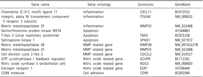
Table 5. Down-regulated genes expressed by over 50 percent between Pre- and Post-IVIG treatment in KD patients
Gene name Gene ontology Synonyms GeneBank
Chemokine (C-X-C motif) ligand 11 Integrin, alpha M (complement component 3 receptor 3 subunit)
Matrix metallopeptidase 25
Serine/threonine protein kinase MST4 T-box 3 (ulnar mammary syndrome) Sphingosine kinase 1
Matrix metallopeptidase 28 Matrix metallopeptidase 25 Cell division cycle 2-like 2
GTP cyclohydrolase I feedback regulator Nitric oxide synthase 3 (endothelial cell) Estrogen receptor 1
CD99 molecule
Inflammation Inflammation
Inflammation Apoptosis Apoptosis Apoptosis MMP related gene MMP related gene MMP related gene Nitric oxide related gene Nitric oxide related gene Nitric oxide related gene Cell adhesion
CXCL11 ITGAM
MMP25
TBX3 SPHK1 MMP28 MMP25 CDC2L2 GCHFR NOS3 ESR1 CD99
BC012532 NM_000632
NM_022468 AF344883 BC025258 NM_021972 NM_001032278 NM_022468 NM_033527 BC112262 NM_000603 AF258449 BC002584 KD, Kawasaki disease; IVIG, intravenous immunoglobulin.
Fig. 1. Gene heat map in KD patients Pre- and Post- IVIG treatment. The color red indicates over expre- ssion while green indicates down expression. KD, Ka- wasaki disease; IVIG, intra- venous immunoglobulin.
10, DENN/MADD domain containing 4A, c-myc bind- ing protein, oxoglutarate receptor 1) are related to cell proliferation process (Table 3).
2. Comparison of microarray analysis pre- and post-IVIG treatment in KD patients
In microarray analysis, 226 genes showed more than
two fold up expression and 117 genes were down-regu- lated in post-IVIG treatment group compared with the pre- IVIG treatment group (Table 1).
The expressed genes that had a 1.5-fold increase are summarized in Table 4. The genes that showed de- creased expressions are summarized in Table 5.
Six gene expressions (cytoskeleton associated protein 2, matrix metallopeptidase 3, matrix metallopeptidase 16, CD44 molecules, formyl peptide receptor receptor1 and angiotensinogen peptidase inhibitor) increased pre- and post-IVIG treatment in the KD patients (Table 4).
3. Total gene heat map
The genes that showed differential expressions by more than 1.5-fold (P<0.05) in at least one sample are shown in Fig. 1. The red color indicates an over expression while green indicates a down expression (Fig.
1). Through microarray analysis, the changes of gene expressions associated with proliferation, translation, inflammatory response, immune response, cell adhe- sion, cell migration, cell differentiation, apoptosis, cell growth, transport, cell cycle, transcription, signal trans- duction and metastasis were observed.
Discussion
Several studies have suggested that immune activa- tion and the secretion of cytokines contribute to the pathogenesis of KD. Although the etiology of KD re- mains unknown despite extensive investigations, the incidence of KD patients continues to increase in many countries [1,2,10,18].
There is no doubt that IVIG is a therapeutic utility in KD now. Infusion of high dose IVIG effectively re- duces systemic inflammation and prevents coronary ar- tery lesion in KD. Several mechanisms may explain the anti-inflammatory effects of IVIG in this disease [7, 8]. They include modification of the cytokine balance and alteration on both the differentiation and the func- tion of monocyte/macrophages, neutrophils and lym- phocytes [9,19,20]. However, the long term con- sequences of the cardiovascular sequelae in KD remain uncertain and therefore, KD is a leading cause of ac- quired heart disease in children [3,4,11].
To investigate the mechanisms underlying the ther- apeutic effects of IVIG, we examined gene expression profiles of fresh whole blood obtained in an acute stage before and in a subacute stage after IVIG therapy. The advantage of DNA microarrays is that it can evaluate changes in relative expression of thousands of genes simultaneously [15,21]. To gain further insight into the mechanism of KD related to immune processes and genetic factors, we investigated the difference of gene expression between KD patients and the control group.
Also, we compared the difference of gene expression levels after IVIG therapy to identify potential candidate genes that might link the systemic immune response to the development of vasculitis and coronary artery disease by examining the gene expression patterns be- tween acute and subacute stages in KD patients.
In the present study, many immunologic processes and genetic factors are attributed to the pathogenesis of KD. Immunologic abnormalities during the acute phase of KD reflect marked activation of the immune system leading to increased cytokine production.
Chemokine ligand 5 (CCL5) and IL-5, which are related to the inflammatory response are over expressed com- pared to the control group in the acute state of KD.
Also, over expressed CCL5, IL-32 protocadherin beta 16, angiopoietin like 3 are related to cell adhesion and migration. It has been reported that CCL5 is highly expressed in various tumors and stimulate tumor growth and metastasis by inducing tumor cell proliferation, angiogenesis, or the expression of MMPs [22,23]. We cannot find out a definite correlation between CCL5 and MMP but increased MMP genes, CCL5 and IL are potential candidates to understand this pathway.
After IVIG treatment, MMP related genes which be- longs to MMP-2, MMP-28, MMP-25, MMP-15, are decreased. MMPs especially MMP-2, and 9, have been considered to play pathophysiologic roles in the devel- opment of coronary artery lesions [24,25]. Many studies that find out MMP-28 are over expressed in several disease states. MMP-28, stimulated by TNF, is a potential novel therapeutic target for prevention and treatment of metastasis of gastric cancer. MMP-28 is frequently over expressed during the progression of gastric cancer and contributes to tumor cell invasion and metastasis
of tumor cells [26,27]. In this study, MMP-2 and 9 are insignificant compared to the control group.
However, the expression of MMP-28 decreased after IVIG treatment.
NO is secreted by immune and vascular endothelial cells, NO has several roles such as regulating vascular tone and the maintenance of the integrity of the vascula- ture [28]. In the KD group, NOS 1, IL-12A, IL-17A, which are related to NO related genes, are over expressed before IVIG treatment. These genes are not down regu- lated after IVIG infusion so further studies are needed.
Caspase (apoptosis related cysteine peptidase, CASP) 1 and cytoskeleton associated protein 2 (CKAP2) are also over expressed in this study. Endothelial cell dys- function and apoptosis are related to endothelial cell damage of the coronary artery. We can’t detect direct change after IVIG infusion among over expressed genes.
However, estrogen receptor 1, dimethylarginine dime- thylaminohydrolase 2 (DDAH2) GTP cyclohydrolase I feedback regulator, endothelial NOS decreased in ex- pression after IVIG compared to the control group. One study showed endothelial progenitor cell (EPC) partici- pated in the process of arterial repair. The number of EPC increased significantly in the subacute phase of KD. Especially, the number of circulating EPC positively correlated with the level of NO and negatively corre- lated with the levels of TNF-α and CRP [29]. IVIG suppresses induced NOS expression of mononuclear leu- kocytes in patients with KD, thus decreasing NO-medi- ated inflammatory responses and coronary artery dila- tion [28,30].
Severe vasculitis leading to coronary artery lesions are noted in the refractory KD group resistant to IVIG [31]. We expect down regulation of the up regulated genes among inflammatory related genes after IVIG in- fusion but we didn’t find different expression levels in this study. It seems this result is skewed because of small group size or IVIG resistant patients. TNF-α blockade (infliximab) has been reported to benefit KD patients with initial IVIG treatment failure. Further analysis of these genes in IVIG resistant group after infliximab is also attributed to understand pathogenesis of KD [31,32].
In conclusion, the data indicated that there are several genes which have different expressions in the KD group
than in the control group. We also confirmed that ex- pression levels change in several genes after an IVIG infusion. These genes are related to inflammatory re- sponse, immune response, cell adhesion, cell migration, cell differentiation, apoptosis, cell growth, transport, cell cycle, transcription, signal transduction, and metastasis. We found out a different expression pattern before and after IVIG treatment but there is lack of consistency in all KD patients. High dose IVIG is defi- nitely the gold standard treatment. There is no doubt that IVIG has a therapeutic utility in treating KD now.
However, many studies estimate that 10-20% of pa- tients do not respond to single dose IVIG, and the risk of aneurysms formation is higher in the unresponsive group than among patients who defervesce completely after a single dose of IVIG.
The limitation of our study is as follows. The sample size is small in number. Further analysis with larger samples of other independent set and specific sample such as peripheral blood T cell, monocytes/macrophages would be needed to find confirmative results in KD treatment.
References
1. Park AH, Batchra N, Rowley A, Hotaling A. Patterns of Kawasaki syndrome presentation. Int J Pediatr Otorhinolaryngol 1997;40:41-50.
2. Shulman ST, Rowley AH. Etiology and pathogenesis of Kawasaki disease. Prog Pediatr Cardiol 1997;6:187-192.
3. Fulton DR, Newburger JW. Long term cardiac sequelae of Kawasaki disease. Curr Rheumatol Rep 2000;2:324-329.
4. Crystal MA, Manhiot C, Yeung RSM, Smallhorn JF, McCrindle BW. Coronary artery dilation after Kawasa- ki disease for children within the normal range. Int J Cardiol 2009;136:27-32.
5. Newburger JW. Takahashi M, Beiser AS, Burns JC, Bastian J, Chung KJ, et al. Single infusion of intra- venous gamma globulin compared to four daily doses in the treatment of acute Kawasaki syndrome. N Engl J Med 1991;324:1633-1639.
6. Leung DY, Cortran RS, Kurt-Jones E, Burns JC, Newburger JW, Pober JS. Endothelial cell activation and high interleukin-1 secretion in the pathogenesis of acute Kawasaki disease. Lancet 1989;334:1298-1302.
7. Galeotti C, Bayry J, Kone-Paut I, Kaveri SV. Kawasaki disease: Aetiopathogenesis and therapeutic utility of
intravenous immunoglobulin. Autoimmun Rev 2010;9:
441-448.
8. Dajani AS, Taubert KA, Takahashi M, Bierman FZ, Freed MD, Ferrieri P, et al. Guidelines for long term management of patients with Kawasaki disease.
Circulation 1994;89:916-922.
9. Matsuda A, Morita H, Unno H, Saito H, Matsumoto K, Hirao Y, et al. Anti-inflammatory effects of high-dose IgG on TNF-α–activated human coronary artery endo- thelial cells. Eur J Immunol 2012;42:2121-2131.
10. Rowley AH, Shulman ST. Pathogenesis and manage- ment of Kawasaki disease. Expert Rev Anti Infect Ther 2010;8:197-203.
11. Cheung YF, Huang GY, Chen SB, Liu XQ, Xi L, Liang XC, et al. Inflammatory gene polymorphisms and sus- ceptibility to Kawasaki disease and its arterial sequelae.
Pediatrics 2008;122:e608-613.
12. Ikeda K, Ihara K, Yamaguchi K, Muneuchi J, Ohno T, Mizuno Y, et al. Genetic analysis of MMP gene poly- morphisms in patients with Kawasaki disease. Pediatr Res 2008;63:182-185.
13. Wang CL, Wu YT, Liu CA, Kuo HC, Yang KD. Kawasaki disease infection, immunity and genetics. Pediatr Infect Dis J 2005;24:998–1004.
14. Shimizu C, Matsubara T, Onouchi Y, Jain S, Sun S, Nievergelt CM, et al. Matrix metalloproteinase hap- lotypes associated with coronary artery aneurysm for- mation in patients with Kawasaki disease. J Hum Genet 2010;55:779-784.
15. Abe J, Hibiki T, Noma S, Nakajima T, Saito H, Terai M. Gene expression profiling of the effect of high dose intravenous Ig in patients with Kawasaki disease. J Immunol 2005;174:5837-5845.
16. Nomura I, Abe J, Noma S, Saito H, Gao B, Wheeler G, et al. Adrenomedullin is highly expressed in blood mono- cytes associated with acute Kawasaki disease: A micro- array gene expression study. Pediatr Res 2005;57:49-55.
17. Popper SJ, Shimizu C, Shike H, Kanegaye JT, Newburger JW, SundelRP, et al. Gene expression pat- terns reveal underlying biological processes in Kawasa- ki disease. Genome Bio 2007;8:R261.
18. Nakamura Y, Yashiro M, Uehara R, Sadakane A, Tsuboi S, Aoyama Y, et al. Epidemiologic features of Kawasaki disease in Japan: results of the 2009-2010 nationwide survey. J Epidemiol 2012;22:216-221.
19. Matsubara T, Ichiyma T, Furukawa S. Immunological profile of peripheral blood lymphocytes and mono- cytes/macrophages in Kawasaki disease. Clin Exp Immunol 2005;141:381-387.
20. Gupta M, Noel GJ, Schaefer M, Friedman D, Bussel J, Johann-Liang R. Cytokine modulation with immune
gamma-globulin in peripheral blood of normal children and its implications in Kawasaki disease treatment.
J Clin Immunol 2001;21:193-199.
21. Ikeda K, Yamaguchi K, Tanaka T, Mizuno Y, Hijikata A, Ohara O, et al. Unique activation status of peripheral blood mononuclear cells at acute phase of Kawasaki disease. Clin Exp Immunol 2009;160:246-255.
22. Chang LY, Lin YC, Mahalingam J, Huang CT, Chen TW, Kang CW, et al. Tumor derived chemokine CCL5 enhances TGF-β mediated killing of CD8+ T cells in colon cancer by T regulatory cells. Cancer Res 2012;72:
1092-1102.
23. Mamtani M, Matsubara T, Shimizu C,Furukawa S, Akagi T, Onouchi Y, et al. Association of CCR2-CCR5 haplotypes and CCL3L1 copy number with Kawasaki disease, coronary artery lesions, and IVIG responses in Japanese Children. PLoS One 2010;5:1-7.
24. FurunoK, Takada, H, Yamamoto K, Ikeda K, Ohno T, Khajoee V, et al. Tissue inhibitor of metalloproteinase 2 and coronary artery lesions in Kawasaki disease. J Pediatr 2007;151:155-160.
25. Lau AC, Duong TT, Ito S, Yeung RSM. Matrix metal- loproteinase 9 activity leads to elastin breakdown in an animal model of Kawasaki disease. Arthritis Rheum 2008;58:854-863.
26. Jian P, Yanfang T, Zhuan Z, Jian W, Xueming Z, Jian Ni. MMP 28(epilysin) as a novel promoter of invasion and metastasis in gastric cancer. BMC Caner 2011;11:
200-208.
27. Overall CM, Kleifeld O. Towards third generation ma- trix metalloproteinase inhibitors for cancer therapy. Br J Cancer 2006;94:941-946.
28. Khajoee V, Kariyazono H, Ohno T, Ihara K, Mizuno Y, Kusuhara K, et al. Inducible and endothelial con- stitutive nitric oxide synthase gene polymorphisms in Kawasaki disease. Pediatr Int 2003;45:130-134.
29. Xu MG, Men LN, Zhao CY, Zhao X, Wang UX, Meng XC, et al. The number and function of circulating endo- thelial progenitor cells in patients with Kawasaki disease. Eur J Pediatr 2010;169:289-296.
30. Wang CL, Wu YT, Lee CJ, Liu HC, Huang LT, Yang KD. Decreased nitric oxide production after intravenous immunoglobulin treatment in patients with Kawasaki disease. J Pediatr 2002;141:560-565.
31. Blaisdell LL, Hayman JA, Moran AM. Infliximab treat- ment for pediatric refractory Kawasaki disease. Pediatr Cardiol 2011;32:1023-1027.
32. Kuo HC, Yang KD, Chang WC, Ger LP. Hsieh KS.
Kawasaki disease: An update on diagnosis and treatment. Pediatr Neonatol 2012;53:4-11.