Many times head and neck lymphadenopathy may be caused by lymphomas.1 Extranodal lymphomas are seen almost exclusively as non-Hodgkin’s lymphoma (NHL) and these constitute 10-20% of all lymphomas.2Adult NHLs commonly arise from B cells. Swelling of unknown origin presenting as NHL of the head and neck may prove to be a challenge for diagnosis.1Salivary gland lympho- mas are quite rare and the majority of them originate from B cells.3This report described a case of 73-year-old female manifested as having both nodal and extranodal involve- ment, but on imaging it was shown to involve Waldeyer’s ring and the larynx, orbit, spleen, and liver.
Case Report
A 73-year-old Indian female patient presented with rapid- ly progressive painless swelling of face, mainly localized in the parotid and submandibular region bilaterally for the
duration of one year. The patient had a history of fever for the most recent two months, with unexplained weight loss (weight at the time of presentation was 35 kg). She had also noticed a recent change in her voice.
Physical examination revealed the patient to be moder- ately built and poorly nourished with signs of anemia.
Ocular examination showed bilateral subconjuctival hem- orrhage and puffiness of the eyelids. The spleen was also palpable 10 cm below the costal margin, whereas the liver was just barely palpable. Further systemic examination including that of the respiratory, cardiac, and central ner- vous systems was normal.
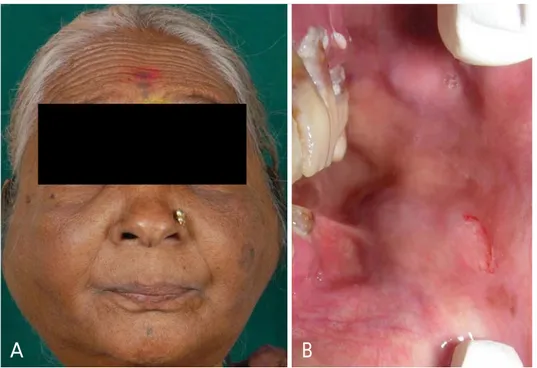
Extraorally, she had a diffuse firm swelling of both the parotid and submandibular glands (Fig. 1A), and a rubbery consistency of the cervical lymph nodes along with those of the submental, submandibular, and jugulodiagastric groups.
Intraorally, two soft, non-tender swellings of about 2×
3 cm were seen in the buccal vestibule in the region of the left upper and lower second molar. Multiple nodules were also palpable in the buccal mucosa bilaterally (Fig. 1B).
A routine hemogram showed pancytopenia with a hemo- globin level of 8.8 gm/dL, a total leukocyte count of 2.5
─ 59 ─
Disseminated non-Hodgkin’s lymphoma presenting as bilateral salivary gland enlargement:
a case report
Manjunatha M. Revanappa, Atul P. Sattur*, Venkatesh G. Naikmasur*, Arpita Rai Thakur**
Department of Oral Medicine and Radiology, College of Dental Sciences, Davangere, India
*Department of Oral Medicine and Radiology, SDM College of Dental Sciences and Hospital, Dharwad, India
**Department of Oral Medicine and Radiology, Jamia Milia Islamia University, New Delhi, India ABSTRACT
Non-Hodgkin’s lymphoma (NHL) constitutes a group of malignancies those arises from cellular components of lymphoid or extranodal tissues. The head and neck is the most common area for the presentation of these lympho- proliferative disorders. Primary involvement of salivary glands is uncommon. This report described a case of a 73- year-old female patient who presented with involvement of both nodal and extranodal sites, with predominant involvement of salivary glands. The tumor staging worked up along with imaging, histopathological, and immuno- histochemical findings were discussed. Computed tomographic images showed the involvement of Waldeyer’s ring, larynx, orbit, and spleen. This report described imaging and prognostic tumor markers in diagnosing, treatment planning, and prognosis. (Imaging Sci Dent 2013; 43 : 59-62)
KEY WORDS: Lymphoma, Non-Hodgkin; Diffuse, Large B-Cell, Lymphoma; Salivary Glands
Received July 23, 2012; Revised September 5, 2012; Accepted October 26, 2012 Correspondence to : Prof. Manjunatha M. Revanappa
Department of Oral Medicine and Radiology, College of Dental Sciences, Pavilion Road, Davangere, Karnataka-577004, India
Tel) 91-8192-231285, Fax) 91-8192-236493, E-mail) [email protected]
Imaging Science in Dentistry 2013; 43 : 59-62 http://dx.doi.org/10.5624/isd.2013.43.1.59
Copyright ⓒ 2013 by Korean Academy of Oral and Maxillofacial Radiology
This is an Open Access article distributed under the terms of the Creative Commons Attribution Non-Commercial License (http://creativecommons.org/licenses/by-nc/3.0) which permits unrestricted non-commercial use, distribution, and reproduction in any medium, provided the original work is properly cited.
Imaging Science in Dentistry∙pISSN 2233-7822 eISSN 2233-7830
×103/mm3and platelet count of 1.03×10³/μL. A lactate dehydrogenase level of 621 IU/L was noted and tests for human immunodeficiency virus (HIV) I and II were found to be non-reactive. Bone marrow cytology revealed an interstitial pattern of marrow involvement.
Panoramic and chest radiographs were unremarkable.
High resolution ultrasonography and color Doppler sonog- raphy studies confirmed the enlargement of the bilateral parotid and submandibular glands, and also the buccal and lingual lymph nodes (Figs. 2A and B). The cervical group of lymph nodes revealed similar enlargement of multiple
nodes belonging to IA, IB, IIA, and IIB levels along with the pre- and post-auricular and supraclavicular group of lymph nodes. Ultrasonography of the abdomen confirmed the enlargement of the spleen, which measured 17 cm. The porta hepatis lymph nodes were noted for enlargement and a hypoechogenic appearance.
Contrast-enhanced computed tomography confirmed the diffuse symmetrical enlargement of the parotid and submandibular glands (Figs. 3A and B), while asymmetric thickening of the subglottic larynx and right aryepiglottic fold were evident with narrowing of the subglottic lumen.
─ 60 ─
Disseminated non-Hodgkin’s lymphoma presenting as bilateral salivary gland enlargement: a case report
A B
Fig. 1.A. Bilateral enlargements of parotid and submandibular glands are seen. B. Two swellings measur- ing 2 cm×3 cm are seen in the buc- cal vestibule in the region of the upper and lower second molar.
Fig. 2.A. A high resolution ultrasonographic image shows the gross enlargement of the right parotid gland measuring 79 mm×31 mm and the involvement of both deep and superficial lobes by hypoechoic areas with a few echogenic septae traversing in between is evident. B. A high resolution ultrasonographic image shows the enlargement of the submandibular glands showing heterogeneous echogenicity with distorted architecture.
A B
Prominent pharyngeal tonsils confirmed the involvement of Waldeyer’s ring. The ocular adnexa of the left orbit were involved, presenting as a bulky lacrimal gland.
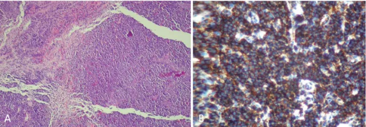
Fine needle aspiration cytology (FNAC) from the right parotid and submandibular glands, buccal vestibule, and tongue nodule showed a monomorphic population of lym- phoid cells with the absence of Reed-Sternberg cells, which was suggestive of non-Hodgkin’s lymphoma. Inci- sional biopsy of the right submandibular lymph node was performed, which revealed diffuse infiltration by sheets of a monomorphic population of moderately large cells, with moderate cytoplasm and large round vesicular nuclei having prominent nucleoli. Mitotic figures were frequently identified (Fig. 4A).
Immunohistochemistry was performed using standard
techniques. The tumor cells expressed CD20 (Fig. 4B) and were immunonegative for CD10, CD3, CD5, CD23, and Cyclin D1. The Mib-1 labeling index (for prolifera- tion marker Ki-67) was approximately 10-15%.
Diagnosis of NHL-diffuse large B-cell type was con- cluded based on FNAC and biopsies of the submandibu- lar glands, parotid glands, and lymph nodes, supported by immunohistochemical positivity for CD20 marker. The patient was referred to the oncology department regarding chemotherapy and radiotherapy.
Discussion
Non-Hodgkin’s lymphoma arises from a lymphocyte progenitor and comprises a heterogeneous group of highly
─ 61 ─
Manjunatha M. Revanappa et al
Fig. 3.A. Contrast-enhanced computed tomography shows diffuse symmetrical enlargement of the parotid gland with heterogeneous mod- erate enhancement. B. Contrast-enhanced computed tomography shows diffuse symmetrical enlargement of the submandibular gland with heterogeneous moderate enhancement.
A B
Fig. 4.A. Histopathological examination shows diffuse large B-cells with moderate cytoplasm and large round vesicular nuclei having prominent nucleoli (H&E stain, ×20). B. Immunohistochemical staining shows positive staining for CD20 (IHC, ×40).
A B
diverse malignancies.225-40% of NHLs are extranodal in origin4 and usually manifest in the gastrointestinal tract, followed by the head and neck region.5NHL mostly occurs in the pediatric age group in the head and neck region;6 however, it may be seen in an older age group, such as in the present case of a 73-year-old female.
The etiological factor for primary lymphoma of the sali- vary gland region is unclear.3Oral lymphomas are fre- quently seen with acquired immune deficiency syndrome (AIDS). In certain individuals, it might serve as the first presentation of AIDS.7The present case was seronegative for HIV.
The most common presentation of primary oral and paraoral lymphoma is a painless local mass with superfi- cial ulceration.7In the present case, patient presented with progressive painless swelling of the face involving mainly the parotid and submandibular glands. Lymphoma of the ocular adenexa is the most common in those aged over 60 years. Orbital lymphoma can involve the lacrimal gland, extraocular muscles, orbital fat, eyelids, and conjunctiva.8 In the present case, involvement of the larynx, orbit, and oropharynx were suspected based on the history and later confirmed by imaging. Based on the morphology, cell lineage, and immunohistochemical findings, the present case was categorized as aggressive NHL of diffuse large B-cell type.
To determine the prognosis and to guide therapy for NHL, staging is important. The Ann Arbor staging sys- tem9is one of the most widely used systems, and includes physical examination, hematological tests, imaging stud- ies, and selective biopsies.10According to the Ann Arbor system, stage IVEB (stage IV: diffuse or disseminated foci of involvement of one or more extralymphatic organs or tissues; E: extranodal organ involvement; B: presence of systemic symptoms like fever and loss of weight) was ascribed. All IPI parameters were found to be positive in this case, thus categorizing our patient as a high risk case.
Staging of salivary gland lymphoma plays a major role in management and enables more favorable prognosis.11 NHL is associated with significant morbidity. Early stages of NHL (minimal lymph node involvement) are more manageable with a greater prospect of long-term disease- free survival, whereas advanced stages (widespread in the lymph nodes) of the disease have a lower prognostic index.
Such patients are also vulnerable to infectious diseases that may involve multiple organ systems (e.g. central ner- vous system, liver).12
A diagnostic dilemma often occurs for oral physicians
when these lymphomas occur at extranodal sites.13 The present case is disseminated NHL with predominant in- volvement of the salivary glands. Though the involvement of both nodal and extranodal sites was present, the patient had complained mainly of enlargement of the salivary glands. Since NHL constitutes only 1.7% of salivary gland malignancies,14a clinical diagnosis of lymphoma is rarely suspected at initial presentation.
In conclusion, the present case emphasizes the impor- tance of imaging and prognostic markers for staging, which is essential for management.
References
1. Urquhart A, Berg R. Hodgkin’s and non-Hodgkin’s lymphoma of the head and neck. Laryngoscope 2001; 111 : 1565-9.
2. van der Waal RI, Huijgens PC, van der Valk P, van der Waal I. Characteristics of 40 primary extranodal non-Hodgkin lym- phomas of the oral cavity in perspective of the new WHO clas- sification and the International Prognostic Index. Int J Oral Maxillofac Surg 2005; 34 : 391-5.
3. Nadendla LK, Meduri V, Paramkusam G. Imaging character- istics of diffuse large cell extra nodal non-Hodgkin’s lymphoma involving the palate and maxillary sinus: a case report. Imag- ing Sci Dent 2012; 42 : 111-4.
4. Newton R, Ferlay J, Beral V, Devesa SS. The epidemiology of non-Hodgkin’s lymphoma: comparison of nodal and extra- nodal sites. Int J Cancer 1997; 72 : 923-30.
5. Teh CS, Chong SY. An unusual presentation of lymphoma of the head and neck region. Med J Malaysia 2011; 66 : 264-5.
6. Zagolski O, Dwivedi R, Kazi R, Subramanian S. Non-Hodg- kin’s lymphoma of the sino-nasal tract in children. J Cancer Res Ther 2010; 6 : 5-10.
7. Ioachim HL, Ryan JR, Blaugrund SM. Salivary gland lymph nodes. The site of lymphadenopathies and lymphomas asso- ciated with human immunodeficiency virus infection. Arch Pathol Lab Med 1988; 112 : 1224-8.
8. Aiken AH, Glastonbury C. Imaging Hodgkin and non-Hodg- kin lymphoma in the head and neck. Radiol Clin North Am 2008; 46 : 363-78.
9. Carbone PP, Kaplan HS, Musshoff K, Smithers DW, Tubiana M. Report of the committee on Hodgkin’s disease staging classification. Cancer Res 1971; 31 : 1860-1.
10. Burns FM, Parks S, Marley JJ. Primary non-Hodgkin’s lym- phoma of the mandible manifesting as a dentigerous cyst. Oral Surg 2011; 4 : 73-6.
11. Gleeson MJ, Bennett MH, Cawson RA. Lymphomas of sali- vary glands. Cancer 1986; 58 : 699-704.
12. DePeña CA, Van Tassel P, Lee YY. Lymphoma of the head and neck. Radiol Clin North Am 1990; 28 : 723-43.
13. Roh JL, Huh J, Suh C. Primary non-Hodgkin’s lymphomas of the major salivary glands. J Surg Oncol 2008; 97 : 35-9.
14. Batsakis JG. Primary lymphomas of the major salivary glands.
Ann Otol Rhinol Laryngol 1986; 95 : 107-8.
─ 62 ─
Disseminated non-Hodgkin’s lymphoma presenting as bilateral salivary gland enlargement: a case report