394 Primary colorectal lymphoma accounts for only 0.2∼
0.65% of large intestinal malignancies. Mucosa-associated lymphoid tissue lymphoma in the rectum is very rare. We report the case of a 73-year-old woman with mor- phologic and immunophenotypic findings consistent with mucosa-associated lymphoid tissue lymphoma of the rectum. The woman complained of consistent bloody diarrhea and recently developed abdominal pain, febrile and chilling sensation. Ultrasonography of upper abdomen detected multiple stones within the gallbladder and the gallbladder was resected using laparoscopic surgery. An ulcerative polypoid mass in the rectum was detected by colonoscopy and computerized tomography of abdomen.
Microscopic and immunohistochemical studies showed a diagnosis of mucosa-associated lymphoid tissue (MALT) lymphoma. This patient was consulted to the department of radiotherapy because of poor general condition and old age. Chemotherapy was not performed. She was followed up with no relapse of the lesions during 7 months after the diagnosis. J Korean Soc Coloproctol 2003;19:394-398 Key Words: Colorectal, Lymphoma, Mucosa-associated lymph-
oid tissue
직장결장, 점막 연관성 림프조직 림프종 ꠏꠏꠏꠏꠏꠏꠏꠏꠏꠏꠏꠏꠏꠏꠏꠏꠏꠏꠏꠏꠏꠏꠏꠏꠏꠏꠏꠏꠏꠏꠏꠏꠏꠏꠏꠏꠏꠏꠏꠏꠏꠏꠏꠏꠏꠏꠏꠏꠏ
In the revised European-American Lymphoma (REAL) classification, a MALT lymphoma is described as a subtype of a non-Hodgkin lymphoma derived from marginal-zone lymphocytes.1 It has been suggested that
‘acquired MALT' secondary to autoimmune disease or infection may form the substrate for lymphoma develop- ment. After Isaacson and Wright2 suggested the MALT lymphoma concept, 20 reported cases in the large intestine were found using MEDLINE. We report an additional case of MALT lymphoma arising in the rectum.
REPORT OF A CASE
A 73-year-old woman with diabetes had had bloody diarrhea 6 months before and had developed abdominal pain with a febrile and chilling sensation 5 days before.
Ultrasonography of the upper abdomen showed multiple stones within the gallbladder with wall thickening.
Cholelithiasis and acute cholecystitis were suspected. The gallbladder was resected using laparoscopic surgery.
Computerized tomography (CT) of the abdomen showed a diffuse thickening of the right lateral and posterior rectal wall with luminal narrowing (Fig. 1). Also, a hyperemic, easily bleeding, somewhat elevated and hard rectal mass was detected by colonoscopy. A biopsy was done. Micro- scopic and immunohistochemical studies provided a diagnosis of MALT lymphoma. A bone marrow biopsy showed no evidence of tumor involvement. Clinically, stage IE is curable by resection, but this patient was referred to the Department of Radiotherapy because of poor general condition and old age. 130 cGy five times per week for a total tumor dose of 4,520 cGy/5 wks was planned. Chemotherapy was not performed. She was followed-up closely with endoscopy and biopsy, and no relapse of the lesions was detected 7 months after the
Mucosa-associated Lymphoid Tissue Lymphoma of the Rectum -A case report-
Departments of Pathology, 1Internal Medicine and 2Diagnostic Radiology, Keimyung University School of Medicine and 3Department of Physical Medicine and Rehabilitation, Kyungpook University School of Medicine, Daegu, Korea
Yu Na Kang, M.D., Sun Young Kwon, M.D., Sang Pyo Kim, M.D., Kwan Kyu Park, M.D., Kun Young Kwon, M.D., Sang Sook Lee, M.D., Kyung Sik Park, M.D.
1, Jung Hyeok Kwon, M.D.
2and Chul Hyun Kim, M.D.
3Corresponding to: Yu Na Kang, Department of Pathology, Keimyung University School of Medicine, 194 Dongsan-dong Chung-gu, Daegu 700-712, Korea. Tel: 053-250-7481, Fax: 053-250-7852, E-mail: [email protected]
This study has been conducted by the Grant of Graduate School,
Keimyung University, Daegu, Korea in 2001.
diagnosis.
Microscopic findings showed a dense, diffuse lym- phocytic infiltration with lymphoepithelial lesions, charac- terized by infiltration of lymphoma cells around the epithe- lium (Figs. 2, 3). Nuclear features resembled those of small cleaved cells of a germinal center, so-called centrocyte-like cells. In immunohistochemical staining, lymphoma cells were positive for kappa light chain staining and negative for lambda light chain staining (Fig.
4), which demonstrates these lymphoma cells represent monoclonal neoplasms. In addition, lymphoma cells were stained with CD3-, CD20+, CD5-, CD10-, CD23-, Fig. 1. Computerized tomography of the abdomen showed
a diffuse thickening of the right lateral and posterior rectal wall with luminal narrowing.
Fig. 5. Lymphoma cells were stained with CD3- (A) and CD20+ (B) by immunohistochemical staining (IHC stain, × 200).
Fig. 3. Lymphoepithelial lesions, characterized by infiltra- tion of centrocyte-like lymphoma cells around the epithe- lium, are seen (H&E stain, ×400).
Fig. 4. In immunohistochemical staining, lymphoma cells were positive for kappa light chain staining (B) and negative for lambda light chain staining (A) (IHC stain, ×200).
Fig. 2. Microscopic findings of the nodular rectal mass showed a dense, diffuse lymphocytic infiltration with a de- crease in the mucosal glandular structure (H&E stain, ×40).
DISCUSSION
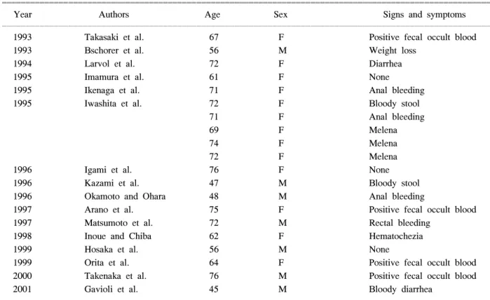
The concept of a MALT lymphoma was first described by Isaacson and Wright in 1983.2 In the 1990s, the distinct clinical-pathologic and molecular features of a MALT lymphoma became widely accepted.3 MALT lymphoma is now incorporated into the Revised European-American Lymphoma (REAL) and the World Health Organization (WHO) classification systems as an extranodal marginal zone B-cell lymphoma, MALT type.4,5 It accounts for 4∼13% of patients seen in individual cancer centers.6 Out of them, primary rectal MALT lymphoma is a rare neoplasm. After Isaacson and Wright2 suggested the MALT lymphoma concept, 20 reported cases of MALT lymphoma in the rectum were found by medline search (Table 1).1,3,7-20 In these 20 cases and our present case, the ages ranged from 45 to 76 (average 65.7 years) and the male : female ratio was 1:
2. Endoscopic findings revealed a submucosal tumor-like, a small nodular, or a polypoid mass. Especially, when
make a differential diagnosis between MALT lymphoma and mantle-cell lymphoma, which have different prognos- tic and therapeutic implications.21 In a large series by Shepherd et al.,22 MALT lymphoma was most common, accounting for 29 (64.5%) of 45 cases of primary colorectal lymphomas. A MALT-type of lymphoma usu- ally presents with a solitary lesion of polypoid appear- ance.23 In contrast, mantle-cell lymphoma (MCL) may also initially involve the gastrointestinal (GI) tract, although the most common presentation is lymphadeno- pathy. The GI involvement of MCL is characterized by multiple polypoid lesions, which is termed multiple lym- phomatous polyposis. The differential diagnosis is also difficult with a histological examination, especially on a small endoscopic biopsy specimen.23 The lymphoma cells of the two lesions, namely the centrocyte-like cells of MALT lymphoma and the so-called ‘centrocytes' of MCL, are similar in size and often closely resemble each other morphologically. The immunohistochemical detec- tion of cyclin D1 overexpression is a most important tool for distinguishing between an MCL and an MALT lym-
Table 1. Reported cases diagnosed as mucosa-associated lymphoid tissue lymphoma of the rectum
ꠚꠚꠚꠚꠚꠚꠚꠚꠚꠚꠚꠚꠚꠚꠚꠚꠚꠚꠚꠚꠚꠚꠚꠚꠚꠚꠚꠚꠚꠚꠚꠚꠚꠚꠚꠚꠚꠚꠚꠚꠚꠚꠚꠚꠚꠚꠚꠚꠚꠚꠚꠚꠚꠚꠚꠚꠚꠚꠚꠚꠚꠚꠚꠚꠚꠚꠚꠚꠚꠚꠚꠚꠚꠚꠚꠚꠚꠚꠚꠚꠚꠚꠚꠚꠚꠚꠚꠚꠚꠚꠚꠚꠚꠚꠚꠚꠚꠚꠚꠚꠚꠚꠚꠚ
Year Authors Age Sex Signs and symptoms
ꠏꠏꠏꠏꠏꠏꠏꠏꠏꠏꠏꠏꠏꠏꠏꠏꠏꠏꠏꠏꠏꠏꠏꠏꠏꠏꠏꠏꠏꠏꠏꠏꠏꠏꠏꠏꠏꠏꠏꠏꠏꠏꠏꠏꠏꠏꠏꠏꠏꠏꠏꠏꠏꠏꠏꠏꠏꠏꠏꠏꠏꠏꠏꠏꠏꠏꠏꠏꠏꠏꠏꠏꠏꠏꠏꠏꠏꠏꠏꠏꠏꠏꠏꠏꠏꠏꠏꠏꠏꠏꠏꠏꠏꠏꠏꠏꠏꠏꠏꠏꠏꠏꠏꠏ
1993 Takasaki et al. 67 F Positive fecal occult blood
1993 Bschorer et al. 56 M Weight loss
1994 Larvol et al. 72 F Diarrhea
1995 Imamura et al. 61 F None
1995 Ikenaga et al. 71 F Anal bleeding
1995 Iwashita et al. 72 F Bloody stool
71 F Anal bleeding
69 F Melena
74 F Melena
72 F Melena
1996 Igami et al. 76 F None
1996 Kazami et al. 47 M Bloody stool
1996 Okamoto and Ohara 48 M Anal bleeding
1997 Arano et al. 75 F Positive fecal occult blood
1997 Matsumoto et al. 72 M Rectal bleeding
1998 Inoue and Chiba 62 F Hematochezia
1999 Hosaka et al. 56 M None
1999 Orita et al. 64 F Positive fecal occult blood
2000 Takenaka et al. 76 M Positive fecal occult blood
2001 Gavioli et al. 45 M Bloody diarrhea
ꠏꠏꠏꠏꠏꠏꠏꠏꠏꠏꠏꠏꠏꠏꠏꠏꠏꠏꠏꠏꠏꠏꠏꠏꠏꠏꠏꠏꠏꠏꠏꠏꠏꠏꠏꠏꠏꠏꠏꠏꠏꠏꠏꠏꠏꠏꠏꠏꠏꠏꠏꠏꠏꠏꠏꠏꠏꠏꠏꠏꠏꠏꠏꠏꠏꠏꠏꠏꠏꠏꠏꠏꠏꠏꠏꠏꠏꠏꠏꠏꠏꠏꠏꠏꠏꠏꠏꠏꠏꠏꠏꠏꠏꠏꠏꠏꠏꠏꠏꠏꠏꠏꠏꠏ
phoma. Also, rare lymphoid polyps of the rectum should be differentiated from malignant lymphomas by using cytomorphology and the immunohistochemical findings of the lymphatic infiltrate.24 In our case, the immunohis- tochemical stains with CD5-, CD10-, CD23-, and cyclin D1- confirmed an MALT lymphoma, not an MCL.
Surgery, radiation therapy, and chemotherapy have been used in the treatment of GI lymphomas. Especially, the therapy for MALT lymphomas has included antibiotic regimens that can eliminate Helicobacter pylori in the rectum,17 as well as in the stomach. Also, radiation therapy or surgery has been used when the disease is of limited extent.
REFERENCES
1. Takenaka R, Tomoda J, Sakata T, Ichiba T, Motoi M, Mizuno M, et al. Mucosa-associated lymphoid tissue lymphoma of the rectum that regressed spontaneously. J Gastroenterol Hepatol 2000;15:331-5.
2. Isaacson P, Wright DH. Malignant lymphoma of mucosa- associated lymphoid tissue. A distinctive type of B-cell lymphoma. Cancer 1983;52:1410-6.
3. Larvol L, Cervoni JP, Hagiage M, Barge J, Soule JC.
Colonic lymphoma simulating cryptogenetic colitis asso- ciated with common variable hypogammaglobulinemia.
Gastroenterol Clin Biol 1994;18:779-81.
4. Harris NL, Jaffe ES, Stein H, Banks PM, Chan JK, Cleary ML, et al. A revised European-American clas- sification of lymphoid neoplasms: a proposal from the International Lymphoma Study Group. Blood 1994;84:
1361-92.
5. Harris NL, Jaffe ES, Diebold J, Flandrin G, Muller- Hermelink HK, Vardiman J, et al. World Health Organi- zation classification of neoplastic diseases of the hema- topoietic and lymphoid tissues: report of the Clinical Advisory Committee meeting-Airlie House, Virginia, November 1997. J Clin Oncol 1999;17:3835-49.
6. Armitage JO, Weisenburger DD. New approach to clas- sifying non-Hodgkins lymphomas: Clinical features of the major histologic subtypes. Non-Hodgkins Lympho- ma Classification Project. J Clin Oncol 1998;16:2780-95.
7. Takasaki M, Yorimitsu Y, Eguchi T. Primary malignant lymphoma of mucosa-associated lymphoid tissue arising from the rectum. Endosc Dig 1993;5:113-8.
8. Bschorer R, Lingenfelser T, Kaiserling E, Schwenzer N.
Malignant lymphoma of the mucosa-associated lymphoid tissue (MALT)--consecutive unusual manifestation in the rectum and gingiva. J Oral Pathol Med 1993;22:190-2.
9. Iwashita A, Takeshita M, Takemura S. Clinicopatho- logical study concerning primary malignant lymphoma of the large intestine. Stomach Intestine 1995;30:869-86.
10. Imamura A, Ambo T, Murashima Y, Sato T. Primary malignant lymphoma of the rectum, measuring 16 mm in radiologic size. Stomach Intestine 1995;30:951-4.
11. Ikenaga M, Takano Y, Nishi Y, Tateishi M, Hiki Y, Kakita A. An operated case of MALT (mucosa-associ- ated lymphoid tissue) lymphoma of the rectum. J Jpn Soc Clin Surg 1995;56:2143-8.
12. Igami T, Yamaguchi A, Hori I. A case of primary rectal mucosa-associated lymphoid tissue (MALT) lymphoma.
Jpn J Cancer Clin 1996;42:748-52.
13. Kazami A, Nakamura T, Nobayashi S. A case of low- grade B-cell lymphoma of mucosa-associated lymphoid tissue (MALT) of the rectum appearing as type IIa+IIc early cancer. Gastroenterol Endosc 1996;38:2209-22.
14. Okamoto T, Ohara N. A case of MALT-lymphoma of the rectum combined with reactive lymphoreticular hyper- plasia (RLH): Special reference to its biopsy and patho- logical diagnosis. Jpn J Natl Med Services 1996;50:784-7.
15. Arano Y, Hirano M, Murakami N, Nagao S, Kikuchi T, Kurokawa M, et al. Primary mucosa-associated lymphoid tissue lymphoma of the rectum. Jpn J Gastroenterol Surg 1997;30:1814-8.
16. Matsumoto T, Iida M, Shimizu. Regression of mucosa- associated lymphoid tissue lymphoma of rectum after eradication of Helicobacter pylori. Lancet 1997;350:115-6.
17. Inoue F, Chiba T. Regression of MALT lymphoma of the rectum after anti-H. pylori therapy in a patient nega- tive for H. pylori. Gastroenterology 1999;117:514-5.
18. Orita M, Yamashita K, Okino M, Enoki T, Noshima S, Morita N, et al. A case of MALT (mucosa-associated lymphoid tissue) lymphoma occurring in the rectum.
Hepatogastroenterology 1999;46:2352-4.
19. Hosaka S, Akamatsu T, Nakamura S, Kaneko T, Kitano K, Kiyosawa K, et al. Mucosa-associated lymphoid tissue (MALT) lymphoma of the rectum with chromosomal translocation of the t (11;18)(q21;q21) and an additional aberration of trisomy 3. Am J Gastroenterol 1999;94:
1951-4.
20. Gavioli M, Bagni A, Santacroce G, Piccagli I, Natalini G. Endorectal sonographic appearances of rectal MALT lymphoma, its response to therapy, and local recurrence.
J Clin Ultrasound 2001;29:401-5.
21. Yatabe Y, Nakamura S, Nakamura T, Seto M, Ogura M, Kimura M, et al. Multiple polypoid lesions of primary mucosa-associated lymphoid-tissue lymphoma of colon.
Histopathology 1998;32:116-25.
22. Shepherd NA, Hall PA, Coates PJ, Levison DA. Primary malignant lymphoma of the colon and rectum. A histo- pathological and immunohistochemical analysis of 45
logy 1988;12:235-52.
23. Schmid C, Vazquez JJ, Diss TC, Isaacson PG. Primary B-cell mucosa-associated lymphoid tissue lymphoma pre- senting as a solitary colorectal polyp. Histopathology 1994;4:357-62.
24. Rutsch F, Henker J, Fischer R, Gobel P. Gastrointestinal lymphonodular hyperplasia and lymphoid polyps of the rectum-a rare coincidence. Z Gastroenterol 1997;35: 271-5.
국문초록
직장의 점막 연관성 림프조직 림프종
계명의대 병리학교실, 1내과학교실, 2진단방사선과학교실,
경북의대 3재활의학과
강유나․권선영․김상표․박관규․권건영․이상숙 박경식1․권중혁2․김철현3
원발성 직장결장 림프종은 대장 악성 암종의 0.2∼
0.65%에 이른다. 그 중에서도 직장의 점막 연관성 림 프조직 림프종은 매우 드물다. 저자는 73세 여성의 점 막 연관성 림프조직 림프종 한 예를 경험하였기에 조 직학적 그리고 면역조직화학 염색의 특징을 보고하고 자 한다. 환자는 지속되는 혈성 설사와 최근에 발생한 복통, 열 그리고 오한감을 주소로 내원 하였다. 상복부 의 초음파촬영술에서 담낭 내 다수의 담석을 발견하 여 담낭절제술을 시행하였다. 결장내시경술과 전산화 단층촬영술로 직장에서 궤양을 동반한 용종성 종물을 발견하였다. 현미경 검사와 면역조직화학염색 결과를 토대로 점막 연관성 림프조직 림프종으로 진단하였다.
이 환자는 전신 쇠약이 심하고 나이가 많아서 수술이 나 화학요법은 실시하지 않았고, 5주간 직장에 4,520 cGy의 방사선 치료를 받았다. 환자는 진단 후 7개월 동안 재발없이 지내고 있다.