48 J Cerebrovasc Endovasc Neurosurg
Treatment of Cerebral Vasospasm in an Infant Using a Modified Dotter Technique
Brian M. Snelling, Samir Sur, Sumedh S. Shah, Eric C. Peterson
Department of Neurological Surgery, University of Miami Miller School of Medicine, Miami, FL, USA
An 8-month old female presented with spontaneous subarachnoid hemor- rhage and was treated successfully with endovascular coil embolization of the ruptured aneurysm. Transcranial Doppler ultrasound performed four days later demonstrated middle cerebral artery (MCA) velocities greater than 350 cm/sec on the right and greater than 200 cm/sec on the left, despite medical management. The patient demonstrated no focal neuro- logical deficits, though examination was limited by our patient's sedation and intubation. Angiography revealed severe vasospasm of the supra- clinoid internal carotid and MCA territories, bilaterally. The vasospasm was refractory to the administration of intra-arterial verapamil. Balloon angio- plasty was attempted, but the device could not be advanced safely due to the small size of the patient's vessels and the stiffness of the device.
A microcatheter (0.0165'' diameter) was advanced over a J-shaped soft microwire (0.014'' diameter) to perform mechanical angioplasty in the in- ternal carotid artery and MCA vessels bilaterally. Dramatic improvement was seen angiographically and on transcranial Doppler, and no complica- tions were seen.
J Cerebrovasc Endovasc Neurosurg.
2017 March;19(1):48-51 Received : 18 August 2016 Revised : 8 October 2016 Accepted : 30 November 2016 Correspondence to Eric C. Peterson Departments of Neurosurgery, Endovascular Neurosurgery, University of Miami Miller School of Medicine, 1095 NW 14th Terrace, Miami, FL 33136, USA
Tel : 1-305-355-1101 Fax : 1-305-355-1102
E-mail : [email protected]
ORCID : http://orcid.org/0000-0002-1420-2346
This is an Open Access article distributed under the terms of the Creative Commons Attribution Non- Commercial License (http://creativecommons.org/li- censes/by-nc/3.0) which permits unrestricted non- commercial use, distribution, and reproduction in any medium, provided the original work is properly cited.
Keywords Aneurysm, Angioplasty, Dotter technique, Pediatric, Cerebral vasospasm Journal of Cerebrovascular and Endovascular Neurosurgery
pISSN 2234-8565, eISSN 2287-3139, http://dx.doi.org/10.7461/jcen.2017.19.1.48
Case Report
INTRODUCTION
Aneurysmal subarachnoid hemorrhage is rare in the pediatric population.5) Cerebral vasospasm (CV) can be seen in 50-80% of these patients, however, delayed ischemia is rarely seen due to robust collateral circulation.3)4) In patients with severe or symptomatic CV despite medical management, intra-arterial vaso- spasm therapy or balloon angioplasty can be performed safely.3) We present an infant with CV, treated with a modified Dotter technique to perform mechanical an- gioplasty of the intracranial arteries refractory to in- tra-arterial verapamil and in whom balloon angio- plasty could not be safely performed.2)
CASE REPORT
An 8-month old female presented with acute sub- arachnoid hemorrhage due to a four-millimeter para- clinoid ruptured aneurysm of the right internal car- otid artery (ICA). She was treated successfully with endovascular coiling of the aneurysm. Trans-cranial Doppler ultrasound (TCD) performed in four days following the rupture demonstrated mean middle cer- ebral artery (MCA) velocities greater than 330 cm/sec on the right and greater than 160 cm/sec on the left.
The patient showed no focal neurological deficit, though she was sedated using fentanyl in the in- tensive care unit during this time. On the following day, mean velocities of all intracranial vessels on TCD
BRIAN M. SNELLING ET AL
Volume 19 · Number 1 · March 2017 49 Fig. 2. The supraclinoid ICA and MCA were mechanically di- lated bilaterally using a MarathonTM microcatheter (0.0165'' di- ameter), which advanced over a J-shaped 0.014'' soft MirageTM microwire (0.014'' diameter). ICA = internal carotid artery; MCA
= middle cerebral artery.
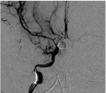
Fig. 1. Angiography three days post aneurysm coiling revealed bilateral vasospasm of the supraclinoid ICA, ACA, and MCA territories. The vasospasm was greater than 60%, when com- pared to her initial angiogram. ICA = internal carotid artery;
ACA = anterior cerebral artery; MCA = middle cerebral artery.
increased despite medical management, with mean velocities greater than 350 cm/sec in all intracranial arteries on the right and greater than 200 cm/sec in arteries on the left. The patient was taken to the an- giography suite for cerebral angiogram and possible angioplasty. Vascular access was obtained, and the patient was heparinized with a dose of 60 units/kg.
The angiogram revealed vasospasm of the supraclinoid ICA, MCA, and anterior cerebral artery (ACA) terri- tories bilaterally of greater than 60% in diameter if all intracranial arteries when compared to her initial an- giogram (Fig. 1). The proximal segments of right and left MCA segments, in particular, were 0.6 and 0.5 mm in diameter, respectively (compared to roughly 1.5 mm on original angiogram in both MCAs). Intra-arte- rial verapamil was administered, however, after 15 minutes, follow-up angiogram revealed persistent vasospasm. Balloon angioplasty was then attempted using a 1.5 mm "over-the-wire" balloon through a 5F guide catheter, but the device could not be advanced safely through the ICA due to the small size of the patient's intracranial vessels.
Therefore, a microcatheter (MarathonTM Microcatheter [Covidien, Irvine, CA, USA]; 0.0165'' diameter) was advanced over a J-shaped 0.014'' soft microwire (MirageTM Microwire, Covidien, Irvine, CA, USA) under road- map guidance to mechanically dilate the supraclinoid ICA and MCA bilaterally (Fig. 2). The microwire was formed into a tight J-shape in order to decrease the risk of vessel perforation and was tracked into the MCA without need for excessive forward tension. The microcatheter was then brought into the MCA vessels over the wire without incident. By passing the micro- wire and subsequently the microcatheter, sequential mechanical angioplasty of the vessel was performed.
Dramatic improvement was seen angiographically in that the proximal MCA M1 segment diameters in- creased to 1.0 and 1.2 mm, right and left (Fig. 3).
Mean TCD velocities decreased to below 200 cm/sec in all vessels the following day, and continued to trend down thereafter. Follow-up angiography demonstrated a 66% and 140% improvement in the degree of steno- sis in the right and left MCA, respectively (2/3 of their initial diameter). However, angioplasty of the ACA A1 segments, though also in severe angiographic vasospasm, was not attempted due to the increased
TREATING CEREBRAL VASOSPASM USING THE DOTTER TECHNIQUE
50 J Cerebrovasc Endovasc Neurosurg
Fig. 3. Post-procedural angiography revealed dramatic improve- ment as the proximal MCA M1 segment diameters increased to 1.0 in the right and 1.2 mm in the left. MCA = middle cere- bral artery.
risk of vessel perforation during catheterization. ACA TCD mean velocities decreased following the procedure.
There were no complications from the procedure and follow-up magnetic resonance imaging the fol- lowing day demonstrated no ischemic injury and no apparent vasospasm.
DISCUSSION
Dotter and Judkins successfully performed a cathe- ter angioplasty of an occluded popliteal artery in an elderly patient, achieving revascularization via a se- quential system of coaxial catheters.2) Although this procedure may seem elementary when compared to modern neurointerventional techniques and devices, it remains an effective tool in the neurointerventionalist's armamentarium when other treatments fail. This is es- pecially true in the pediatric population, where small vessel diameters often preclude the use of most guide catheters and many transluminal devices. Our patient did not appear to have symptomatic vasospasm, but demonstrated severe CV in all anterior circulation vessels using commonly accepted criteria for adults (mean velocities > 120 cm/sec).1) The pre-vasospasm
MCA M1 diameter was at the lower size limit of cur- rently available percutaneous transluminal angio- plasty balloons, and as we anticipated, they could not be delivered safely to the vessels of interest.
Therefore, we believed that employing a similar tech- nique, as the one performed by Dotter and Judkins2) was inevitable in our patient, despite the high risk of dissection or vasospasm aggravations.
In our practice with adult patients and older chil- dren, verapamil and nitroglycerine are our agents of choice due to drug response and our experience in dosing these drugs. Because the patient in this case was so small, we elected to proceed with mechanical angioplasty given the lack of response to verapamil and concerns regarding systemic effects on such a small child with increased use of calcium channel blockers or other agents. Initially, we attempted in- tra-arterial verapamil to chemically treat our patient's vasospasm; however, after 15 minutes, follow-up an- giogram showed that the vasospasm persisted. By this time, vasodilation due to calcium channel blocker ef- fect should have been observed based on considerable prior experience in adult patients. Although we can- not state with certainty that a delayed response to ve- rapamil had no role in the observed improvement in vasospasm, we believe this to be highly likely based on our experience with adults and older children.
As previously mentioned, by passing the microwire and subsequently the microcatheter, sequential me- chanical angioplasty of the vessel was performed.
This technique was highly successful, and the pa- tient's vasospasm resolved on follow-up angiogram, mean TCD velocities in all anterior circulation arteries improved immediately, and follow-up imaging re- vealed no ischemia. While the MCA was dilated us- ing the modified Dotter technique, there is a possi- bility that the associated ACA velocities would have stayed elevated. Had this occurred, we would have revisited the risks and benefits of administering addi- tional vasodilators or attempting mechanical angio- plasty of the ACA. However, we believe that navigat- ing the microwire and microcatheter into the MCA
BRIAN M. SNELLING ET AL
Volume 19 · Number 1 · March 2017 51 from the ICA is substantially less risky as compared
to ACA catheterization for two reasons: (1) the natu- ral curvature of the ICA terminus is directed toward the larger caliber MCA which allows, and (2) main- tenance of a "J" curvature on the distal wire while ad- vancing it into the MCA. To access the ACA, the wire tip must be used to direct the wire into the A1 segment from the ICA, which can lead to vessel perforation.
CONCLUSION
Overall, this case report highlights a novel mod- ification of an existing technique to safely treat CV in an infant due to aneurysmal subarachnoid hemorrhage becauae balloon angioplasty was not available and chemical angioplasty was ineffective. The use of me- chanical angioplasty-a modified Dotter technique-with modern microcatheters and microwires can provide excellent angiographic and clinical outcomes in this setting.
Disclosure
The authors report no conflict of interest concerning the materials or methods used in this study or the findings specified in this paper.
REFERENCES
1. Aaslid R, Huber P, Nornes H. Evaluation of cerebrovascular spasm with transcranial Doppler ultrasound. J Neurosurg.
1984 Jan;60(1):37-41.
2. Dotter CT, Judkins MP. Percutaneous transluminal treatment of arteriosclerotic obstruction. Radiology. 1965 Apr;84:631-43.
3. Moftakhar P, Cooke DL, Fullerton HJ, Ko NU, Amans MR, Narvid JA, et al. Extent of collateralization predicting symptomatic cerebral vasospasm among pediatric patients:
correlations among angiography, transcranial Doppler ul- trasonography, and clinical findings. J Neurosurg Pediatr.
2015 Mar;15(3):282-90.
4. Ostergaard JR, Voldby B. Intracranial arterial aneurysms in children and adolescents. J Neurosurg. 1983 Jun;58(6):832-7.
5. Proust F, Toussaint P, Garniéri J, Hannequin D, Legars D, Houtteville JP, et al. Pediatric cerebral aneurysms. J Neurosurg. 2001 May;94(5):733-9.