Index terms
Papillary Thyroid Microcarcinoma Papillary Thyroid Macrocarcinoma Lateral Metastatic Lymph Node Central Metastatic Lymph Node Round Shape
INTRODUCTION
A papillary thyroid microcarcinoma (PTMC) is defined by the World Health Organization as a carcinoma measured at 1.0 cm or less in its greatest dimension (1). There is an increase in the proportion of PTMC identified among all differentiated thy- roid carcinomas mainly due to the improvement and increased in use of ultrasound (US) examination, fine-needle aspiration biopsy, and other diagnostic procedures (2). It is estimated that PTMC accounts for up to 30% of all papillary thyroid cancers, although marked geographic differences in incidence rates have been noted (3). In South Korea, PTMC was reported in up to 50.2% of thyroid carcinomas (4). Currently, there is controversy
on the extent of surgery necessary for PTMC. In published clini- cal guidelines of organizations including the American Thyroid Association, the National Comprehensive Cancer Network, and the British Thyroid Association, the general recommendation in cases of lymphadenopathy is to perform lymph node dissection of the affected compartments. In patients with biopsy-proven metastatic lateral cervical lymphadenopathy, therapeutic lateral neck compartmental lymph node dissection was recommended with total thyroidectomy (TT) to provide clearance of disease (5-8). Patients with preoperatively detected lateral neck node metastasis are more likely to develop recurrence in the lymph nodes (9). Thus, preoperative US examination is important for evaluating lateral metastatic lymph nodes to determine the ex-
J Korean Soc Radiol 2015;72(2):92-100 http://dx.doi.org/10.3348/jksr.2015.72.2.92
Received August 5, 2014; Accepted September 29, 2014 Corresponding author: Hee Kang, MD
Department of Radiology, Kosin University Gospel Hospital, 262 Gamcheon-ro, Seo-gu, Busan 602-702, Korea.
Tel. 82-51-990-6341 Fax. 82-51-255-2764 E-mail: [email protected]
This is an Open Access article distributed under the terms of the Creative Commons Attribution Non-Commercial License (http://creativecommons.org/licenses/by-nc/3.0) which permits unrestricted non-commercial use, distri- bution, and reproduction in any medium, provided the original work is properly cited.
Purpose: To analyze ultrasonographic (US) features of metastatic lymph nodes (LNs) in papillary thyroid microcarcinomas (PTMC) and in papillary thyroid macro- carcinomas.
Materials and Methods: The study reviewed US findings of 273 patients with pathologically confirmed papillary thyroid carcinoma (PTC) and metastatic LNs based on the US examination. Patients were divided into two groups: PTMC and papillary thyroid macrocarcinomas.
Results: The 273 patients with PTC included 87 with PTMC and 186 with papillary thyroid macrocarcinoma. No significant difference of US features in patients with lateral neck node metastasis was found between PTMC (n = 96) and macrocarcino- ma (n = 29). In central neck node metastasis, round shape was the most frequent findings in both groups (p < 0.001).
Conclusion: There was no significant difference in US features of metastatic LNs between PTMC and papillary thyroid macrocarcinomas. Therefore, careful evalua- tion of the whole neck should be made.
Ultrasonographic Features of Metastatic Lymph Nodes in Papillary Thyroid Microcarcinomas and Macrocarcinomas
미세 유두상 갑상선암에서 전이 림프절의 초음파 소견 분석과 일반 유두상 갑상선암 전이 림프절 소견과의 비교 연구
Young Gyung Shin, MD, Hee Kang, MD, Young Doc Joh, MD, Kyung Soon Jeong, MD, Beom Su Kim, MD
Department of Radiology, Kosin University Gospel Hospital, Busan, Korea
lary thyroid macrocarcinoma group were selected because they were suspected of metastatic lymph nodes based on US exami- nation.
Image Evaluation
The US features of metastatic lymph nodes of the total 273 pa- tients were reviewed by two radiologists. The following US crite- ria were used to define lymph node metastasis: round shape (minor axis greater than 50% of major axis), loss of fatty hilum, focal or diffuse hyperechogenicity, cystic change, and microcal- cification (12-14). Initially, we compared US features of meta- static lymph nodes in the lateral neck of patients with PTMC and those of patients with papillary thyroid macrocarcinoma.
Subsequently, the US features of metastatic lymph nodes in the central neck were reviewed.
Lymph Nodes
Based on findings of US examinations, patients were classified into two groups: the first group had only central neck node (lev- el VI) metastasis; the second group had metastases extended to the lateral neck node (levels II–V). The definition of cervical compartment used in this study was based on the rules outlined by the Head and Neck Service of the Memorial Sloan-Kettering Cancer Center (15, 16).
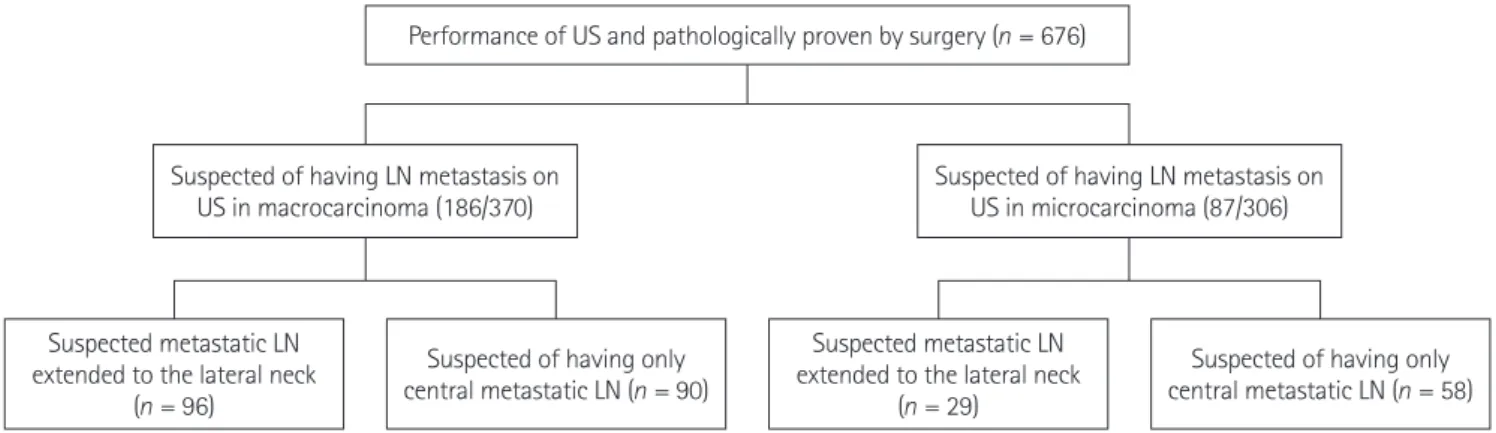
Of the 186 patients with papillary thyroid macrocarcinoma, 90 were suspected of only central neck node metastasis, whereas 96 were suspected of central and lateral neck node metastasis.
Of the 87 patients with PTMC, 58 were suspected of only cen- tral neck node metastasis, while 29 were suspected of central and lateral neck node metastasis.
Cytopathologic Evaluation
Lateral neck node metastasis was confirmed by Nodal fine- needle aspiration biopsy (FNAB) or Nodal FNAB needle-wash thyroglobulin (Tg). Nodal FNAB needle-wash Tg measure- ments could complement cytology in thyroid cancer because it has been reported that these measurements could be used as di- agnostic substitute (17, 18). All thyroid tumors and lymph node metastases were confirmed as PTC by pathologic examination.
In addition, lymph node metastases were identified by levels.
Radiologic findings of suspected lymph node metastasis were correlated with the pathologic evaluation findings.
tent of operation in potentially aggressive PTMC.
Previous studies have reported the usefulness of preoperative US in detecting the presence of clinically apparent cervical lymph node metastasis (9-11). Ito et al. (9) reported that preoperative US could detect 39% of metastases among patients with patho- logically confirmed lateral lymph node metastasis. However, no report has described whether there are any characteristic or dominant imaging features of metastatic lymph nodes in PTMC in comparison with those of papillary thyroid macrocarcino- mas. Therefore, this study retrospectively reviewed the sono- graphic features of metastatic lymph nodes in PTMC compared to papillary thyroid macrocarcinomas and determined whether there were any dominant findings of lateral metastatic lymph nodes in PTMC. Additionally, this study investigated the sono- graphic features of metastatic lymph nodes limited to the central neck in PTMC and papillary thyroid macrocarcinoma.
MATERIALS AND METHODS
Subjects
From January 2009 through May 2012, 676 patients with pathologically confirmed papillary thyroid carcinoma (PTC) were identified by the Institutional Review Board. None of these patients had undergone previous operation on the head or neck.
All patients underwent US before surgery.
Ultrasound Examination
Thyroid US was performed using a 5- to 12-MHz linear transducer (iU22; Philips Healthcare, Bothell, WA, USA) by one radiologist with ten years of experience. When US examination was performed for detecting metastatic lymph nodes, the radi- ologist was unaware of cytologic confirmation of malignancy in the thyroid masses. The US examinations included both thyroid lobes and all neck levels (I–VI).
Tumor Size
Patients were placed into two groups depending on the size of the tumor. Patients placed in the PTMC group (n = 306) had tu- mors with a maximum diameter of 10 mm, and those placed in the papillary thyroid macrocarcinoma group (n = 370) had tu- mors greater than a minimum diameter of 1 cm. A total of 87 patients from the PTMC group and 186 patients from the papil-
were men. The mean age was 45 years (range, 19–81 years).
Ultrasound Features of Metastatic Lymph Nodes The US features of metastatic lymph nodes in PTMC and pap- illary thyroid macrocarcinoma are summarized in Table 1. The following diagnostic results were found in the papillary thyroid macrocarcinoma group: round shape (central vs. lateral neck node metastasis: n = 86, 95.6% vs. n = 70, 72.9%), loss of hilum (n = 29, 32.2% vs. n = 84, 87.5%), hyperechogenicity (n = 14, 15.6% vs. n = 87, 90.6%), cystic change (n = 12, 13.3% vs. n = 32, 33.3%), and calcification (n = 2, 2.2% vs. n = 20, 20.8%). For the PTMC group, the following results were obtained: round shape (central vs. lateral neck node metastasis: n = 54, 93.1% vs. n = 19, 65.5%), loss of hilum (n = 12, 20.7% vs. n = 26, 89.7%), hyper- echogenicity (n = 8, 13.8% vs. n = 27, 93.1%), cystic change (n = 4, 6.9% vs. n = 9, 31%), and calcification (n = 1, 1.7% vs. n = 7, 24.1%) (Figs. 2-5).
Of the 125 patients suspected of lateral neck node metastasis, no significant difference of US features was found between the microcarcinoma (n = 96) and macrocarcinoma (n = 29) groups Surgical Procedure
Patient flow diagram is shown in Fig. 1. All patients under- went routinely TT with central compartment neck dissection.
Modified radical neck dissection (MRND) of the affected side was performed for patients diagnosed with extended lateral neck node metastasis (PTMC, n = 29; papillary thyroid macro- carcinoma, n = 96).
Statistical Analysis
Statistical analysis was performed using the chi-square test and a Fisher’s exact test. Statistical calculations were performed using the statistical package SPSS for Windows, released in 20.0.0 (SPSS Inc., Chicago, IL, USA). Statistical significance was considered when p value was less than 0.05.
RESULTS
Patients
Of the 273 patients suspected of metastatic lymph nodes based on the US examination, 231 (84.6%) were women, 42 (15.4%)
Suspected metastatic LN extended to the lateral neck
(n = 96)
Suspected of having only central metastatic LN (n = 90)
Suspected metastatic LN extended to the lateral neck
(n = 29)
Suspected of having only central metastatic LN (n = 58) Suspected of having LN metastasis on
US in macrocarcinoma (186/370)
Performance of US and pathologically proven by surgery (n = 676)
Suspected of having LN metastasis on US in microcarcinoma (87/306)
Fig. 1. Patient flow diagram. Flowchart shows the study population and included patients. Of 676 patients with pathologically confirmed papil- lary thyroid carcinoma, 186 patients from the papillary thyroid macrocarcinoma group and 87 patients from the PTMC group were selected be- cause they were suspected of having metastatic lymph nodes based on the US examination.
Note.—LN = lymph node, PTMC = papillary thyroid microcarcinoma, US = ultrasonographic Table 1. US Features of Metastatic LNs of 273 Patients
US Imaging Features
Location of Metastatic LN
Macrocarcinoma (Tumor Size > 1 cm) Microcarcinoma (Tumor Size ≤ 1 cm) Central (n = 90) Lateral (n = 96) Total (n = 186) Central (n = 58) Lateral (n = 29) Total (n = 87)
Round shape 86 70 156 54 19 73
Loss of fatty hilum 29 84 113 12 26 38
Hyperechogenicity 14 87 101 8 27 35
Cystic change 12 32 44 4 9 13
Presence of calcification 2 20 22 1 7 8
Note.-LN = lymph node, US = ultrasonographic
histological subtype, accounting for 80% of all cases. Increasing global interest in this disease partially stems from the increasing number of its diagnoses. Several authors have demonstrated that the incidence of PTC has almost doubled over the last three de- cades, mainly due to the higher incidence of PTMC (19, 20).
This increase is attributable to the widespread use of cervical ul- trasound and ultrasound-guided fine-needle aspiration biopsies of thyroid nodules as well as more accurate histopathological search for small PTMC (19, 21).
In spite of the overall excellent prognosis for patients with PTMC, there is a 1.0% disease-related mortality rate, 5.0% lymph node recurrence rate, and 2.5% distant metastasis rate associat- ed with PTMC. In addition, PTMC has been highly associated with lymph node metastasis at the time of diagnosis, with inci- dence rate up to 26.3% (22). The presence of lymph node metas- tasis is an important prognostic factor that indicates the poten- tial for an increasing rate of distant metastasis and the risk of cervical lymph node recurrence (3). Many factors such as tumor mutifocality, bilaterality, capsule invasion, and size (> 5 mm) have been proposed as risk factors of cervical lymph node me- tastasis in PTMC. These factors are the same for individuals with papillary thyroid macrocarcinoma (22-24). Some studies have demonstrated that distinguishing PTMC from convention- al PTC based on size alone may be erroneous because the recur- rence rates for PTMC and conventional PTC are not significant- as shown in Figs. 4 and 5. The following analysis was made based
on image findings: round shape (microcarcinoma vs. macrocar- cinoma: n = 19, 65.5% vs. n = 70, 72.9%, p = 0.221), loss of hilum (n = 26, 89.7% vs. n = 84, 87.5%, p = 0.556), hyperechogenicity (n
= 27, 93.1% vs. n = 87, 90.6%, p = 0.555), cystic change (n = 9, 31.0% vs. n = 32, 33.3%, p = 0.533), calcification (n = 7, 24.1% vs.
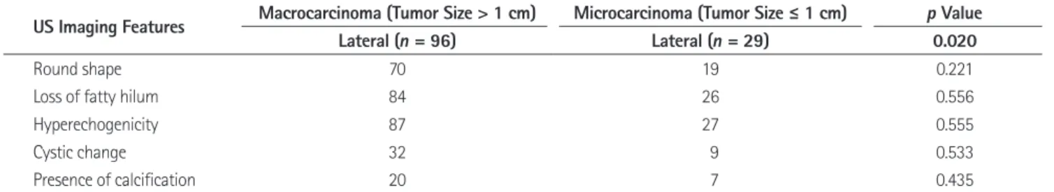
n = 20, 20.8%, p = 0.435). The frequency of lateral neck node me- tastasis in papillary thyroid macrocarcinoma (52%) was signifi- cantly (p = 0.020) higher than that in PTMC (33%) (Table 2).
There was significant difference of frequency in PTMC between central neck node (n = 58) and lateral neck node (n = 29) me- tastases, with p < 0.001 for loss of hilum, hyperechogenicity, cys- tic change, and calcification. Similar results were obtained for the papillary thyroid macrocarcinoma group with p < 0.001 for loss of hilum, hyperechogenicity, cystic change, and calcifica- tion. The round shape was the more frequent finding than other four variables in central neck node metastasis in both papillary thyroid macrocarcinoma group and PTMC group, with p <
0.001 for loss of hilum, hyperechogenicity, cystic change, and calcification (Table 3).
DISCUSSION
Currently research has indicated that thyroid cancer is the most common endocrine malignancy. PTC is the most frequent
Table 2. US Features of Lateral Metastatic Lymph Nodes in Papillary Thyroid Macrocarcinoma and PTMC
US Imaging Features Macrocarcinoma (Tumor Size > 1 cm) Microcarcinoma (Tumor Size ≤ 1 cm) p Value
Lateral (n = 96) Lateral (n = 29) 0.020
Round shape 70 19 0.221
Loss of fatty hilum 84 26 0.556
Hyperechogenicity 87 27 0.555
Cystic change 32 9 0.533
Presence of calcification 20 7 0.435
Note.-PTMC = papillary thyroid microcarcinomas, US = ultrasonographic
Table 3. US Features of Central Metastatic Lymph Nodes in Papillary Thyroid Macrocarcinoma and PTMC
US Imaging Features Macrocarcinoma (Tumor Size > 1 cm) Microcarcinoma (Tumor Size ≤ 1 cm)
p Value
Central (n = 90) Central (n = 58)
Round shape 86 54 < 0.001
Loss of fatty hilum 29 12 < 0.001
Hyperechogenicity 14 8 < 0.001
Cystic change 12 4 < 0.001
Presence of calcification 2 1 < 0.001
Note.-PTMC = papillary thyroid microcarcinomas, US = ultrasonographic
Fig. 2. Central neck node metastasis in 56-year-old man.
A. Ultrasonographic image shows ill-defined hypoechoic papillary thyroid microcarcinoma with calcifications in the right thy- roid gland.
B. An oval shaped lymph node at left level VI shows focal hyperechogenicity (arrow) with loss of hilum in the left central neck on ultrasonography.
Fig. 3. Central neck node metastasis in 35-year-old woman.
A. Ultrasonographic image shows a lobulated margin, marked hypoechoic papillary thyroid macrocarcinoma with extracapsular extension in the right thyroid gland.
B. A round shaped lymph node (arrow) shows internal cystic change in the left central neck on ultrasonography.
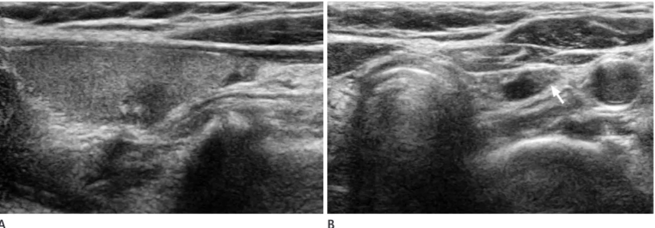
Fig. 4. Lateral neck node metastasis in 56-year-old man.
A. Ultrasonographic image shows a spiculated margin, marked hypoechoic papillary thyroid microcarcinoma in the right thyroid gland.
B. An oval shaped lymph node (arrow) shows loss of hilum, hyperechogenicity, and microcalcification in the left lateral neck on ultrasonography.
A
A
A
B
B
B
used it to predict and detect central metastasis. However, the characteristic US features of central metastatic lymph nodes have not yet been demonstrated. Therefore, the US features of metastatic central neck lymph nodes were reviewed in this study to determine if there were any dominant findings that could be used clinically for central neck lymph node examinations. Our results indicated that round shape was the most frequent find- ing in central neck node metastasis of both PTMC and papillary thyroid macrocarcinomas. This might be due to the relatively low sensitivity of US in evaluating potential metastatic lymph nodes in the central neck. A considerably high percentage of small lesions might have led to the difficulty in identifying inner structures and any changes of lesions. Our findings also indicat- ed that the possibility of metastasis might be suspected when the central lymph node exhibited only round shape.
This study had several limitations. First, only patients suspect- ed of lateral neck node metastasis based on US and those who had undergone surgery with pathologically confirmed metastasis were included, which might have selection bias. However, based on research findings, MRND is not necessary nor recommended for patients without definite lateral neck node metastasis detect- ed on preoperative US. This was because lymph node recur- rence-free survival rates did not differ between patients who un- derwent MRND and patients who did not undergo MRND if there was no preoperative detection of lateral neck node metas- tasis. In addition, research has shown that, in more than 60% of patients, latent lymph node metastasis only occasionally be- comes clinically apparent that requires treatment (9). Finally, this study analyzed only central neck nodes detected with US, ly different. In fact, both tumors behave similarly (25). Some
authors have demonstrated that the ipsilateral lateral compart- ment was involved almost as often as the central compartment (24, 26-28). However, currently no report has compared any characteristic or dominant imaging features of metastatic lymph nodes in PTMC with papillary thyroid macrocarcinomas. The results from this study indicated that there were no significant difference in US features of metastatic lymph nodes between PTMC and papillary thyroid macrocarcinomas. Not only rela- tively high incidence of lymph node metastasis, but also all fea- tures of metastatic lymph nodes were well presented in PTMC examination. Treatment of patients with PTMC has been sug- gested to be similar to treatment of patients with conventional PTC (3, 29). Given the relatively low sensitivity of US in evaluat- ing potential metastatic lymph nodes in the central neck, it is reasonable to consider a prophylactic central neck dissection with a total thyroidectomy or lobectomy, even when clear US evidence for local metastatic disease is absent. It is reasonable to consider a therapeutic central neck dissection based on intraop- erative findings of positive nodal metastases in this region. Cer- vical nodal metastases are quite common in PTC. Initial nodal metastasis in PTC usually occurs in the paratracheal and pretra- cheal nodes in level VI of the central compartment of the ipsilat- eral neck (26). Preoperative B-Raf proto-oncogene analysis by FNAB based on US may assist the prediction of occult central neck lymph node metastasis in patients with PTC. Previous studies have used indications of clinically node-negative neck and tumor size > 5 mm as predictive factors for subclinical cen- tral lymph node metastasis in PTMC (30, 31). Many trials have Fig. 5. Lateral neck node metastasis in 61-year-old woman.
A. Ultrasonographic image shows a spiculated margin, marked hypoechoic papillary thyroid macrocarcinoma with extracapsular extension in the right thyroid gland.
B, C. The right level III (B) and level IV (C) metastatic lymph nodes show microcalcification (white arrow), loss of hilum, and hyperechogenicity (black arrow) on ultrasonography.
B
A C
guidelines for patients with thyroid nodules and differen- tiated thyroid cancer. Thyroid 2009;19:1167-1214
9. Ito Y, Tomoda C, Uruno T, Takamura Y, Miya A, Kobayashi K, et al. Preoperative ultrasonographic examination for lymph node metastasis: usefulness when designing lymph node dissection for papillary microcarcinoma of the thy- roid. World J Surg 2004;28:498-501
10. Hwang HS, Orloff LA. Efficacy of preoperative neck ultra- sound in the detection of cervical lymph node metastasis from thyroid cancer. Laryngoscope 2011;121:487-491 11. Boland GW, Lee MJ, Mueller PR, Mayo-Smith W, Dawson
SL, Simeone JF. Efficacy of sonographically guided biopsy of thyroid masses and cervical lymph nodes. AJR Am J Roentgenol 1993;161:1053-1056
12. Ying M, Ahuja A, Metreweli C. Diagnostic accuracy of so- nographic criteria for evaluation of cervical lymphade- nopathy. J Ultrasound Med 1998;17:437-445
13. Rosário PW, de Faria S, Bicalho L, Alves MF, Borges MA, Purisch S, et al. Ultrasonographic differentiation between metastatic and benign lymph nodes in patients with pap- illary thyroid carcinoma. J Ultrasound Med 2005;24:1385- 1389
14. Vassallo P, Wernecke K, Roos N, Peters PE. Differentiation of benign from malignant superficial lymphadenopathy:
the role of high-resolution US. Radiology 1992;183:215- 220
15. Shah JP. Cervical lymph node metastases--diagnostic, therapeutic, and prognostic implications. Oncology (Wil- liston Park) 1990;4:61-69; discussion 72, 76
16. Som PM, Curtin HD, Mancuso AA. An imaging-based clas- sification for the cervical nodes designed as an adjunct to recent clinically based nodal classifications. Arch Otolar- yngol Head Neck Surg 1999;125:388-396
17. Snozek CL, Chambers EP, Reading CC, Sebo TJ, Sistrunk JW, Singh RJ, et al. Serum thyroglobulin, high-resolution ul- trasound, and lymph node thyroglobulin in diagnosis of differentiated thyroid carcinoma nodal metastases. J Clin Endocrinol Metab 2007;92:4278-4281
18. Uruno T, Miyauchi A, Shimizu K, Tomoda C, Takamura Y, Ito Y, et al. Usefulness of thyroglobulin measurement in fine-needle aspiration biopsy specimens for diagnosing cervical lymph node metastasis in patients with papillary which may cause selection bias. To overcome these limitations,
further studies are needed to assess the findings using large study groups.
In conclusion, there was no significant difference in US find- ings of metastatic LNs between the PTMC and the papillary thyroid macrocarcinoma groups, although the frequency of lat- eral neck node metastasis in the macrocarcinoma group was higher than in the PTMC group. Therefore, it is recommended that careful evaluation of metastatic lymph nodes should be conducted, even if only microcarcinoma is present in the thy- roid gland.
REFERENCES
1. Hedinger CE, Williams ED, Sobin LH. Histologic typing of thyroid tumours, 2nd ed. Berlin: Springer-Verlag Berlin Heidelberg, 1988:9-10
2. Baudin E, Travagli JP, Ropers J, Mancusi F, Bruno-Bossio G, Caillou B, et al. Microcarcinoma of the thyroid gland: the Gustave-Roussy Institute experience. Cancer 1998;83:553- 559
3. Chow SM, Law SC, Chan JK, Au SK, Yau S, Lau WH. Papil- lary microcarcinoma of the thyroid-Prognostic signifi- cance of lymph node metastasis and multifocality. Cancer 2003;98:31-40
4. Lee J, Rhee Y, Lee S, Ahn CW, Cha BS, Kim KR, et al. Fre- quent, aggressive behaviors of thyroid microcarcinomas in Korean patients. Endocr J 2006;53:627-632
5. Hartl DM, Travagli JP. The updated American Thyroid As- sociation Guidelines for management of thyroid nodules and differentiated thyroid cancer: a surgical perspective.
Thyroid 2009;19:1149-1151
6. Tuttle RM, Ball DW, Byrd D, Dilawari RA, Doherty GM, Duh QY, et al. Thyroid carcinoma. J Natl Compr Canc Netw 2010;
8:1228-1274
7. British Thyroid Association, Royal College of Physicians Lodon. Guidelines for the management of thyroid cancer, 2nd ed. London: The Lavenham Press, 2007:13-15
8. American Thyroid Association (ATA) Guidelines Taskforce on Thyroid Nodules and Differentiated Thyroid Cancer, Cooper DS, Doherty GM, Haugen BR, Kloos RT, Lee SL, et al. Revised American Thyroid Association management
25. Arora N, Turbendian HK, Kato MA, Moo TA, Zarnegar R, Fahey TJ 3rd. Papillary thyroid carcinoma and microcarci- noma: is there a need to distinguish the two? Thyroid 2009;19:473-477
26. Machens A, Hinze R, Thomusch O, Dralle H. Pattern of nodal metastasis for primary and reoperative thyroid can- cer. World J Surg 2002;26:22-28
27. Grebe SK, Hay ID. Thyroid cancer nodal metastases: bio- logic significance and therapeutic considerations. Surg Oncol Clin N Am 1996;5:43-63
28. Gimm O, Rath FW, Dralle H. Pattern of lymph node metas- tases in papillary thyroid carcinoma. Br J Surg 1998;85:
252-254
29. Page C, Biet A, Boute P, Cuvelier P, Strunski V. ‘Aggressive papillary’ thyroid microcarcinoma. Eur Arch Otorhinolar- yngol 2009;266:1959-1963
30. Joo JY, Park JY, Yoon YH, Choi B, Kim JM, Jo YS, et al. Pre- diction of occult central lymph node metastasis in papil- lary thyroid carcinoma by preoperative BRAF analysis us- ing fine-needle aspiration biopsy: a prospective study. J Clin Endocrinol Metab 2012;97:3996-4003
31. Kim BY, Jung CH, Kim JW, Lee SW, Kim CH, Kang SK, et al.
Impact of clinicopathologic factors on subclinical central lymph node metastasis in papillary thyroid microcarcino- ma. Yonsei Med J 2012;53:924-930
thyroid cancer. World J Surg 2005;29:483-485
19. Grodski S, Brown T, Sidhu S, Gill A, Robinson B, Learoyd D, et al. Increasing incidence of thyroid cancer is due to in- creased pathologic detection. Surgery 2008;144:1038- 1043; discussion 1043
20. Pazaitou-Panayiotou K, Capezzone M, Pacini F. Clinical features and therapeutic implication of papillary thyroid microcarcinoma. Thyroid 2007;17:1085-1092
21. Mazzaferri EL. Managing small thyroid cancers. JAMA 2006;295:2179-2182
22. Vasileiadis I, Karakostas E, Charitoudis G, Stavrianaki A, Ka- petanakis S, Kouraklis G, et al. Papillary thyroid microcarci- noma: clinicopathological characteristics and implications for treatment in 276 patients. Eur J Clin Invest 2012;42:
657-664
23. Scheumann GF, Gimm O, Wegener G, Hundeshagen H, Dralle H. Prognostic significance and surgical manage- ment of locoregional lymph node metastases in papillary thyroid cancer. World J Surg 1994;18:559-567; discussion 567-568
24. Wada N, Duh QY, Sugino K, Iwasaki H, Kameyama K, Mimura T, et al. Lymph node metastasis from 259 papillary thyroid microcarcinomas: frequency, pattern of occur- rence and recurrence, and optimal strategy for neck dis- section. Ann Surg 2003;237:399-407
미세 유두상 갑상선암에서 전이 림프절의 초음파 소견 분석과 일반 유두상 갑상선암 전이 림프절 소견과의 비교 연구
신영경 · 강 희 · 조영덕 · 정경순 · 김범수
목적: 미세 유두상 갑상선암에서 전이 림프절의 초음파 소견을 분석하고 이를 일반 유두상 갑상선암에서 전이 림프절의 초음파 소견과 비교하여 유의한 차이점을 알아보고자 한다.
대상과 방법: 유두상 갑상선암으로 수술을 시행하고 림프절 전이가 확진된 환자 중 술전 초음파 검사상 경부 림프절 전이 가 의심되었던 273명의 환자를 대상으로, 1 cm 이하의 미세 유두상 갑상선암과 1 cm를 초과하는 일반 유두상 갑상선암 으로 나누어 전이 림프절의 초음파 소견을 후향적으로 비교분석하였다.
결과: 총 273명의 환자 중 87명은 미세 유두상 갑상선암, 186명은 일반 유두상 갑상선암으로 진단되었다. 미세 유두상 갑상선암 환자 중 29명, 일반 유두상 갑상선암 환자 중 96명에서 외측 경부 림프절 전이가 있었다. 두 그룹 간 전이 림프 절의 초음파 소견에서 통계적으로 유의한 차이는 없었다. 외측 경부 전이 림프절과 비교하여 중앙 경부 전이 림프절에 둥 근 모양이 우세하게 나타났다(p < 0.001).
결론: 미세 유두상 갑상선암과 일반 유두상 갑상선암에서 전이 림프절의 초음파 소견에 유의한 차이는 없었다. 미세 유두 상 갑상선암 환자에서 술전 초음파 검사를 시행할 때에도 일반 유두상 갑상선암에서처럼 범위와 초음파 소견의 제한 없이 발생할 수 있는 림프절 전이의 가능성을 명심하고 전 경부를 면밀히 검사하여야 한다.
고신대학교 복음병원 영상의학과