Primary Cutaneous Adenoid Cystic Carcinoma
Vol. 31, No. 6, 2019 669
Received February 26, 2018, Revised August 24, 2018, Accepted for publication October 17, 2018
Corresponding author: Kee-Yang Chung, Department of Dermatology, Severance Hospital, Cutaneous Biology Research Institute, Yonsei Uni- versity College of Medicine, 50-1 Yonsei-ro, Seodaemun-gu, Seoul 03722, Korea. Tel: 82-2-2228-2080, Fax: 82-2-393-9157, E-mail: [email protected] ORCID: https://orcid.org/0000-0003-3257-0297
This is an Open Access article distributed under the terms of the Creative Commons Attribution Non-Commercial License (http://creativecommons.
org/licenses/by-nc/4.0) which permits unrestricted non-commercial use, distribution, and reproduction in any medium, provided the original work is properly cited.
Copyright © The Korean Dermatological Association and The Korean Society for Investigative Dermatology
pISSN 1013-9087ㆍeISSN 2005-3894
Ann Dermatol Vol. 31, No. 6, 2019 https://doi.org/10.5021/ad.2019.31.6.669
CASE REPORT
Immediate Umbilical Reconstruction after a Mohs Micrographic Surgery for Primary Cutaneous Adenoid Cystic Carcinoma Arising in the Umbilicus
Jee Eun Kim, Mi Ryung Roh, Kee-Yang Chung1
Department of Dermatology, Gangnam Severance Hospital, Cutaneous Biology Research Institute, Yonsei University College of Medicine,
1Department of Dermatology, Severance Hospital, Cutaneous Biology Research Institute, Yonsei University College of Medicine, Seoul, Korea
Adenoid cystic carcinoma (ACC) is a malignant neoplasm of glands commonly occurs in salivary glands. Primary cuta- neous adenoid cystic carcinoma (PCACC) is a rare form of ACC that primarily presents on the skin. Herein, we repre- sent a rare case of PCACC occurred in the umbilicus in a 66-year-old Korean male patient. The patient visited our cen- ter with erythematous indurated patch on the umbilicus diag- nosed as ACC by incisional biopsy at another center. The di- agnosis of PCACC was confirmed by additional histopatho- logic examination and imaging study. We proceeded Mohs micrographic surgery and reconstructed umbilicus with tacked purse string suture. Local recurrence and distant metastasis were not observed during 30-month follow-up.
We report this rare case of PCACC on the umbilicus so that dermatologist can aware of the rare disease. Furthermore, we recommend MMS and tacked purse string suture as effective methods for treatment of PCACC and immediate umbilical reconstruction. (Ann Dermatol 31(6) 669∼672, 2019) -Keywords-
Immediate umbilical reconstruction, Mohs micrographic
surgery, Primary cutaneous adenoid cystic carcinoma
INTRODUCTION
Adenoid cystic carcinoma (ACC) is a malignant neoplasm of glands commonly occurs in salivary glands. Primary cu- taneous adenoid cystic carcinoma (PCACC) is a rare form of ACC that primarily presents on the skin showing in- dolent course with low rate of distant metastasis. To date, less than 70 cases have been reported in the English liter- ature. Herein, we report a case of PCACC of the umbi- licus. To the best of author’s knowledge, this is he first re- ported case of PCACC in the umbilicus.
The patient was diagnosed as PCACC upon histopatho- logical examination and the lesion was removed by Mohs micrographic surgery (MMS). Since umbilicus is an aes- thetically important body structure located at the center of the abdomen, the sacrificed umbilicus in the process of the surgery was reconstructed with special technique of purse string sutures.
CASE REPORT
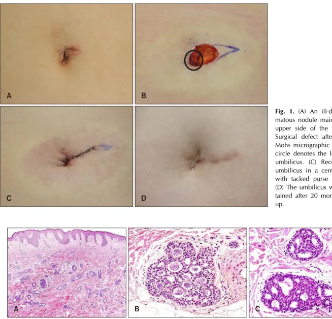
A 66-year-old Korean male patient presented with an ill- defined erythematous indurated patch on the umbilicus (Fig. 1A). The patient had not received any treatment for 1 year as the lesion was asymptomatic. Recently, the patient was referred to our center after an incisional biopsy. The tumor was cleared after three stages of MMS. We received the patient’s consent form about publishing all photo- graphic materials.
Histopathologic examination revealed basaloid tumor cells forming nests and cords in the dermis and subcutaneous
JE Kim, et al
670 Ann Dermatol
Fig. 1. (A) An ill-defined erythe- matous nodule mainly on the left upper side of the umbilicus. (B) Surgical defect after 3 stages of Mohs micrographic surgery. Black circle denotes the location of the umbilicus. (C) Reconstruction of umbilicus in a centralized defect with tacked purse string sutures.
(D) The umbilicus was well main- tained after 20 months of follow- up.
Fig. 2. Histopathologic features of the excised tumor. (A) Basaloid tumor cells forming nests and cords in the dermis and subcutaneous tissue without connection to the epidermis (H&E, original magnification ×100). (B) Cribriform or tubular structure of the basaloid cells with abundant mucin inside (H&E, ×400). (C) Tumor cells had hyperchromatic nuclei but did not demonstrate distinct cellular atypia (H&E, ×400).
tissue (Fig. 2A). The tumor showed cribriform or tubular structure without connection to the epidermis. Abundant mucin was observed inside the tubular structure (Fig. 2B).
Tumor cells had hyperchromatic nuclei but did not dem- onstrate distinct cellular atypia (Fig. 2C). Immunohistoche- mical study against receptor tyrosine kinase (cKIT) and ep- ithelial membrane antigen (EMA) showed strong positive for tumor cells. The tumor was diagnosed as ACC upon histopathologic examination and immunohistochemical study.
Physical examination and whole-body positron emission tomography fused with computed tomography (PET/CT) performed to exclude the possibility of metastasis from other original lesion showed no evidence of additional
lesion. Finally, the patient was diagnosed as PCACC.
In the process of removing the tumor, the umbilicus was completely eliminated with an asymmetrical defect that extended into the left upper quadrant, which required ela- borate reconstruction. The size of the postoperative defect was 2.0×1.8 cm (Fig. 1B). An elliptical repair was done at the left side of the defect to close the paramedian defect as well as to remove the dogear. To close the remaining cen- tral defect and to recreate the concavity of the umbilicus, about 0.5 cm of the soft tissue was removed from the sur- gical margin, deeper layered purse string sutures were placed to adjust the size of the umbilicus, and the super- ficial purse string suture involving the dermal layer was
Primary Cutaneous Adenoid Cystic Carcinoma
Vol. 31, No. 6, 2019 671 Fig. 3. (A) Deeper layered purse string sutures were placed to adjust the size of the umbilicus. (B) The superficial purse string suture involving the dermal layer was tacked at 3 areas at 12, 4, and 8 o’clock directions to the underlying linea alba. (C) Concave and normal looking umbilicus was recreated after tightening the purse string suture.
tacked at 3 areas at 12, 4, and 8 o’clock directions to the underlying linea alba (Fig. 3). Concave structure of the umbilicus was recreated at the center of the abdomen (Fig.
1C). Follow-up at six months showed a well-healed surgi- cal defect with recreated concave and normal looking um- bilicus and its shape was well maintained at 20-month fol- low-up (Fig. 1D).
The patient showed no local recurrence or distant meta- stasis at 30-month follow-up.
DISCUSSION
ACC is a rare tumor which occurs most commonly in the head and neck, especially in the major and minor salivary glands. It can arise from a variety of primary sites includ- ing the breast, trachea, external auditory canal, uterus and prostate1. It also occurs on the skin primarily or metasta- tically. Cutaneous ACC usually presents as a solitary, flesh-colored or erythematous, ill-defined, slow-growing mass1. Patients are usually asymptomatic or may present with tenderness. PCACC is diagnosed upon histopatho- logic examination, which shows basaloid cells with hyper- chromatic nuclei arranged in a tubular or cribriform archi- tecture with abundant mucin in the cystic spaces2. cKIT and EMA are known to be strongly reactive in the PCACC2. For confirming the diagnosis of PCACC, since PCACC is not distinguishable from other ACCs by histopathologic examination, the possibility of metastasis should be ex- cluded by imaging studies. Our case was confirmed as PCACC after excluding the possibility of metastatic lesion with whole-body PET/CT.
The standard treatment for PCACC is wide local excision.
PCACC shows high tendency of local recurrence, though overall disease course is indolent with low rate of distant metastasis2. Thus, it is important to secure enough safety margin to reduce the opportunity of local recurrence.
Recently, MMS is considered as the adequate treatment for PCACC, as MMS shows better outcome with lower rate of local recurrence and minimal skin defect in other
skin cancers, such as squamous cell carcinoma or basal cell carcinoma. Among reported 7 cases of cutaneous ACC treated with MMS, there has been no local re- currence with a follow-up range of 10 to 28 months3. Although significant conclusion cannot be made due to the short observation period and the insufficient number of cases, MMS is well worth to be considered as the treat- ment for PCACC. Our case was treated with MMS and did not show recurrence at 30-month follow-up.
In the process of abdominal surgery including urachal cyst repair, umbilical herniorrhaphy and tumor excision, it is often unavoidable to sacrifice the umbilicus. Various meth- ods for umbilical reconstruction have been developed in- cluding an inverted C-V flap4 or a lunchbox approach5. However, these methods leave extensive scars around the reconstructed umbilicus and the three-dimensional struc- ture of the reconstructed umbilicus may become flattened with passing of time. The purse string suture has been re- ported to be a method that allows immediate recon- struction of umbilicus with minimizing the scars6-8. This method also allows formation of the three-dimensional structure of the umbilicus and is suitable for long-term maintenance of the umbilical shape. In addition, in some cases of tumor surgery involving the umbilicus, as in our case, the surgeon has to deal with an asymmetric skin defect. In this situation, initial procedure to centralize the defect is necessary. After centralization of the defect, de- fatting of the surgical margin followed by double-layered purse string suture with the superficial suture being tacked down at three locations will successfully recreate a cos- metically acceptable umbilicus.
In summary, we reported a case of a rare cutaneous tu- mor, PCACC occurred on the umbilicus. The histopatho- logic features and the results of immunohistochemical study were suitable for diagnosis of ACC. The exclusion of other possible origin for the lesion was made before the fi- nal diagnosis of PCACC. The lesion was removed by MMS and the umbilicus was reconstructed with tacked purse string suture. The patient is maintaining three-dimensional
JE Kim, et al
672 Ann Dermatol
umbilicus with no local recurrence or distant metastasis for 30-month follow-up. In our experience, MMS and tacked purse string suture were effective methods for treatment of PCACC and immediate umbilical reconstruction.
CONFLICTS OF INTEREST
The authors declare no conflicts of interest.
ORCID
Jee Eun Kim, https://orcid.org/0000-0003-0795-5742 Mi Ryung Roh, https://orcid.org/0000-0002-6285-2490 Kee-Yang Chung, https://orcid.org/0000-0003-3257-0297
REFERENCES
1. Ramakrishnan R, Chaudhry IH, Ramdial P, Lazar AJ, McMenamin ME, Kazakov D, et al. Primary cutaneous adenoid cystic carcinoma: a clinicopathologic and immuno- histochemical study of 27 cases. Am J Surg Pathol 2013;37:
1603-1611.
2. Naylor E, Sarkar P, Perlis CS, Giri D, Gnepp DR, Robinson- Bostom L. Primary cutaneous adenoid cystic carcinoma. J Am Acad Dermatol 2008;58:636-641.
3. Xu YG, Hinshaw M, Longley BJ, Ilyas H, Snow SN. Cutaneous adenoid cystic carcinoma with perineural invasion treated by mohs micrographic surgery-a case report with literature review. J Oncol 2010;2010:469049.
4. Shinohara H, Matsuo K, Kikuchi N. Umbilical reconstruction with an inverted C-V flap. Plast Reconstr Surg 2000;105:
703-705.
5. Onishi K, Yang YL, Maruyama Y. A new lunch box-type method in umbilical reconstruction. Ann Plast Surg 1995;
35:654-656.
6. Bartsich SA, Schwartz MH. Purse-string method for im- mediate umbilical reconstruction. Plast Reconstr Surg 2003;
112:1652-1655.
7. Schoeller T, Rainer C, Wechselberger G, Piza-Katzer H. Im- mediate navel reconstruction after total excision: a simple three-suture technique. Surgery 2002;131:105-107.
8. Meirer R, Wechselberger G, Schoeller T. Purse-string method for immediate umbilical reconstruction. Plast Reconstr Surg 2004;114:831-832.