J Korean Soc Radiol 2017;77(3):192-196 https://doi.org/10.3348/jksr.2017.77.3.192
INTRODUCTION
Currently, the World Health Organization (WHO) classifies uterine smooth muscle neoplasms that cannot be clearly diag- nosed as benign or malignant based on generally applied histo- pathologic parameters as “smooth muscle tumors of uncertain malignant potential (STUMP)” (1). Extrauterine STUMPs are very rare; only one metastasizing case, in the retroperitoneum, has been reported (2). Herein, we report on the first case of a STUMP that originated from the superior mesenteric vein (SMV), with emphasis on the radiologic findings.
CASE REPORT
A 59-year-old woman was referred to our hospital for a pan- creatic mass incidentally found on abdominal ultrasonography, which had been performed as part of her general health screen- ing. She had an unremarkable medical history and no symptoms except mild, intermittent epigastric pain; her laboratory findings were also not remarkable. As for a tumor marker, the result of a carbohydrate antigen 19-9 test was also within the normal range.
The patient underwent computed tomography (CT) and mag- netic resonance imaging (MRI) for further evaluation. The con- trast-enhanced CT images showed a 4 × 3 × 3 cm sized smooth- margined mass compressing the pancreas head, with a sharp
Smooth Muscle Tumor of Uncertain Malignant Potential Originating from the Superior Mesenteric Vein: A Case Report
상장간막정맥에서 유래된 Smooth Muscle Tumor of Uncertain Malignant Potential: 증례 보고
Kyung Ho Kim, MD
1, Kyung A Kang, MD
2*, Ji Yeon Park, MD
1, Seon-Jeong Kim, MD
1, Jeong-Ju Lee, MD
3Departments of 1Radiology, 3Pathology, Myongji Hospital, Seonam University College of Medicine, Goyang, Korea
2Department of Radiology, Kangbuk Samsung Hospital, Sungkyunkwan University School of Medicine, Seoul, Korea
Uterine smooth muscle neoplasms that cannot be clearly diagnosed as benign or malignant based on generally applied histopathologic parameters are classified as
“smooth muscle tumors of uncertain malignant potential (STUMP)”. Herein, we report on the first case of a STUMP that originated from the superior mesenteric vein (SMV), with emphasis on the radiologic findings. Magnetic resonance imaging showed a well-defined mass encasing the SMV with progressive and homogeneous enhance- ment in the arterial and portal venous phases. The lesion showed mild hyperintensity on T2-weighted images when compared with skeletal muscle, which was quite dif- ferent from typical leiomyomas. The lesion also showed hyperintensity on the diffu- sion-weighted images, and hypointensity on the apparent diffusion coefficient map.
These results may reflect the high cellularity of the mass.
Index terms
Smooth Muscle Tumor Mesenteric Veins
Magnetic Resonance Imaging Tomography, X-Ray Computed
Received December 16, 2016 Revised January 10, 2017 Accepted February 25, 2017
*Corresponding author: Kyung A Kang, MD Department of Radiology, Kangbuk Samsung Hospital, Sungkyunkwan University School of Medicine, 29 Saemunan-ro, Jongno-gu, Seoul 03181, Korea.
Tel. 82-2-2001-1029 Fax. 82-2-2001-1030 E-mail: [email protected]
This is an Open Access article distributed under the terms of the Creative Commons Attribution Non-Commercial License (http://creativecommons.org/licenses/by-nc/4.0) which permits unrestricted non-commercial use, distri- bution, and reproduction in any medium, provided the original work is properly cited.
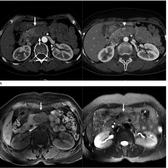
beak-shaped interface (Fig. 1A). The mass was encasing the entire circumference of the SMV (Fig. 1A). Progressive homogeneous enhancement was seen on the arterial and portal venous phases (Fig. 1A). Similarly, gadoxetic acid-enhanced MR images re- vealed progressive homogeneous enhancement, and the lesion on MRI showed mild hypointensity on a T1-weighted image (Fig.
1B) and mild hyperintensity on a T2-weighted image when com-
pared with the skeletal muscle (Fig. 1B). The lesion also showed hyperintensity on a diffusion-weighted image (DWI) obtained with a b value of 600 sec/mm2 and hypointensity on an apparent diffusion coefficient (ADC) map (Fig. 1C). The main pancreatic duct was not dilated. Based on these imaging findings, the mass was first considered to be a SMV-origin tumor, such as leiomyo- ma. The differential diagnosis was a nonepithelial tumor of the
Fig. 1. Smooth muscle tumor of uncertain malignant potential originating from the SMV in a 59-year-old woman.
A. On axial contrast-enhanced CT images, an approximately 4 × 3 × 3 cm sized, well-defined mass showed progressively homogeneous enhancement during arterial (left) and portal phases (right). The pancreas head appears compressed by the lesion with a sharp, beak-shaped interface (arrows). The mass is encasing the entire circumference of the SMV (arrowhead).
B. On MR images, the mass shows slightly low signal intensity (arrow) on a T1-weighted image (left) and slightly high signal intensity (arrow) on a T2-weighted image (right) compared with that of skeletal muscle. Gadoxetic acid-enhanced MR images revealed progressive homogeneous en- hancement (not shown).
SMV = superior mesenteric vein B
A
pancreas with SMV invasion.
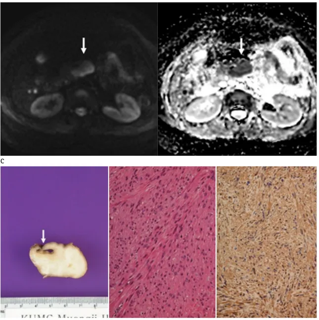
During an exploratory laparotomy a round encapsulated tu- mor firmly attached to the wall of the SMV was identified in the pancreas head. En bloc resection, with an involved part of the vein, was performed and the tumor was easily dissected from the pancreas. Grossly, the excised mass was 4.4 × 4.3 × 2.5 cm in size, and the cut surface revealed a firm, circumscribed grayish
white mass encircling the SMV (Fig. 1D). Histologically, the tu- mor was characterized by interlacing bundles of compact uni- form spindled cells with little collagen. The tumor showed diffuse moderate cytologic atypia, four mitoses events per 50 high-pow- er fields, and no tumor cell necrosis (Fig. 1D). Immunohisto- chemical staining for smooth muscle actin (SMA), S-100, CD117, and Ki-67 was performed, and the results were positive for SMA
Fig. 1. Smooth muscle tumor of uncertain malignant potential originating from the SMV in a 59-year-old woman.
C. A diffusion-weighted image obtained with a b value of 600 sec/mm2 (left) shows hyperintensity (arrow), and the apparent diffusion coefficient map (right) shows hypointensity of the mass (arrow).
D. Grossly (left), the cut surface reveals a firm and circumscribed grayish white mass encircling the SMV (arrow). Microscopically, spindle cell pro- liferation with moderate cytologic atypia and no tumor necrosis (hematoxylin and eosin stain, × 200, middle) was shown. Immunohistochemical staining for SMA (× 200, right) shows diffuse positivity.
SMA = smooth muscle actin, SMV = superior mesenteric vein D
C
(Fig. 1D). Additionally, 30% of tumor cells were positive for Ki- 67. The final pathological diagnosis was a STUMP.
DISCUSSION
There is an ambiguous group of uterine smooth muscle neo- plasms, with overlapping features between leiomyoma and leio- myosarcoma, that can be challenging for even the most experi- enced pathologist to diagnose. Therefore, the current WHO guide- lines indicate that uterine smooth muscle neoplasms, that do not fit the definitions for any other categories, should be classified as STUMPs (1).
A number of investigators have studied parameters for assess- ing the diagnosis and prognosis of uterine smooth muscle neo- plasms. One of the most widely used classification systems was proposed by Bell et al. (3). The histologic distinctions between the five subgroups are based on assessing a combination of fea- tures including cytologic atypia, mitotic index, and coagulative tumor cell necrosis. STUMPs are defined as tumors with the fol- lowing features: 1) no, or no more than, mild cytologic atypia; no tumor cell necrosis; and a mitotic index of 5 ≤ mitoses < 20 per 10 high-power fields; 2) diffuse moderate to severe cytologic atypia, no tumor cell necrosis, and a mitotic index of < 10 mito- ses per 10 high-power fields; 3) focal or multifocal moderate to severe cytologic atypia, no tumor cell necrosis, and a mitotic in- dex of 1 < mitoses < 20 per 10 high-power fields; 4) no, to mild, cytologic atypia; tumor cell necrosis; and a mitotic index < 10 mitoses per 10 high-power fields.
In this case, the tumor showed diffuse moderate cytologic atypia, four mitoses events per 50 high-power fields, and no tu- mor cell necrosis; the histologic appearance of the smooth mus- cle neoplasm in our case fulfilled the Bell’s criteria for atypical leiomyoma with a low risk of recurrence. For this reason, our fi- nal diagnosis was a STUMP, even though STUMP is typically a term used only with uterine neoplasms. We found only one case report of an extrauterine STUMP that arose from the retroperito- neum with lung metastasis after a hysterectomy (4). To the best of our knowledge, this is the first case report of a STUMP that originated from the SMV, without a primary uterine STUMP.
Regardless of their anatomic locations, classic leiomyomas have signal intensities similar to those of skeletal and smooth muscles on T1- and T2-weighted MR images. Upon histologic
examination, non-degenerated leiomyomas are composed of whorls of uniform smooth muscle cells with varying amounts of intervening collagen. Cellular leiomyoma is a specific subtype of leiomyoma composed of compact smooth muscles cells with lit- tle or no collagen that can have relatively increased T2-weighted signal intensity and homogeneous enhancement on contrast- enhanced images. On the other hand, high signal intensity on T2-weighted images can also be caused by myxoid or cystic de- generation (5). However, mild hyperintense tumors on T2- weighted images were more likely to be highly cellular tumors, whereas markedly hyperintense tumors with heterogeneous ar- chitecture tended to be degenerated tumors (6). Because our case showed mild hyperintensity on T2-weighted images com- pared with the skeletal muscle and homogeneous enhancement on contrast-enhanced T1-weighted images, the cause of the in- creased signal intensity, unlike with a usual leiomyoma, was likely high cellularity rather than myxoid or cystic degeneration. Patho- logic findings also revealed a uniform cellular mass composed of compact smooth muscle cells with little collagen. Only one study described the MR characteristics of three uterine STUMPs (7);
one of the three STUMPs resembled the MR findings of ordi- nary leiomyoma, but, in the other two cases, more than half of the mass showed hyperintensity on T2-weighted images.
In this case, the mass showed high signal intensity on a DWI and low signal intensity on an ADC map. Because previous in- vestigations have shown that ADC values of the tissues are de- pendent on the cellularity and amount of tissue fibrosis (8), we presumed that high cellularity was responsible for the low ADC value in this case.
Prognostic significance is not well-known because of insuffi- cient cases of extrauterine STUMPs. A number of studies have shown that uterine STUMPs are usually clinically benign, but that they can occasionally recur or metastasize to distant sites (9).
Therefore, they should be considered tumors with low malignant potential, but patients who are diagnosed with STUMPs require close, long-term follow-up. Additionally, radiologic clues for sus- pected STUMPs might be important.
In conclusion, we report on the first case report of an extra- uterine STUMP, that originated from the SMV, without a pri- mary uterine STUMP. Because STUMPs can occasionally recur or metastasize, radiologic clues about the suspect STUMP might be important. Imaging findings of STUMPs are not well estab-
lished, but a STUMP can show slightly high signal intensity on T2-weighted imaging with homogeneous or heterogeneous contrast-enhancement. Additionally, diffusion restriction could be seen in the STUMP due to its high cellularity.
REfERENCES
1. Fletcher CDM, Unni KK, Mertens F. World Health Organiza- tion classification of tumors: pathology and genetics of tu- mors of soft tissue and bone. Lyon: IARC Press, 2002 2. Nishino M, Hayakawa K, Minami M, Yamamoto A, Ueda H,
Takasu K. Primary retroperitoneal neoplasms: CT and MR imaging findings with anatomic and pathologic diagnostic clues. Radiographics 2003;23:45-57
3. Bell SW, Kempson RL, Hendrickson MR. Problematic uterine smooth muscle neoplasms. A clinicopathologic study of 213 cases. Am J Surg Pathol 1994;18:535-558
4. Won HS, Chun HG, Lee K. Retroperitoneal smooth muscle tumor of uncertain malignant potential after hysterectomy:
a case report. J Med Case Rep 2011;5:214
5. Ueda H, Togashi K, Konishi I, Kataoka ML, Koyama T, Fuji- wara T, et al. Unusual appearances of uterine leiomyomas:
MR imaging findings and their histopathologic backgrounds.
Radiographics 1999;19 Spec No:S131-S145
6. Yamashita Y, Torashima M, Takahashi M, Tanaka N, Katabu- chi H, Miyazaki K, et al. Hyperintense uterine leiomyoma at T2-weighted MR imaging: differentiation with dynamic en- hanced MR imaging and clinical implications. Radiology 1993;
189:721-725
7. Tanaka YO, Nishida M, Tsunoda H, Okamoto Y, Yoshikawa H.
Smooth muscle tumors of uncertain malignant potential and leiomyosarcomas of the uterus: MR findings. J Magn Reson Imaging 2004;20:998-1007
8. Koh DM, Collins DJ. Diffusion-weighted MRI in the body:
applications and challenges in oncology. AJR Am J Roent- genol 2007;188:1622-1635
9. Ip PP, Cheung AN, Clement PB. Uterine smooth muscle tu- mors of uncertain malignant potential (STUMP): a clinico- pathologic analysis of 16 cases. Am J Surg Pathol 2009;33:
992-1005
상장간막정맥에서 유래된 Smooth Muscle Tumor of Uncertain Malignant Potential: 증례 보고
김경호
1· 강경아
2* · 박지연
1· 김선정
1· 이정주
3자궁평활근종 중 조직병리학적으로 양성 또는 악성으로 명확하게 진단할 수 없는 그룹을 smooth muscle tumors of un-
certain malignant potential (이하 STUMP)로 정의한다. 저자들은 상장간막정맥에서 유래된 STUMP의 첫 증례에 대해 방사선학적 소견에 중점을 두어 보고하고자 한다. 자기공명영상에서 상장간막정맥을 감싸는 종괴는 점진적이고 균질한 조영증강을 보이고, T2 강조영상에서 골격근과 비교하여 경미한 고신호를 보였는데 이는 전형적인 평활근종의 자기공명영 상 소견과는 차이가 있다. 그리고 확산강조영상에서 고신호 강도를 보였고 겉보기확산계수에서 저신호 강도를 보였는데, 이러한 소견들은 종괴의 높은 세포질을 반영한 것이라고 생각된다.
서남대학교 의과대학 명지병원 1영상의학과학교실, 3병리학교실
2성균관대학교 의과대학 강북삼성병원 영상의학과